
Click image to see more details
-

-
-
-
-
+7
Product Info Summary
| SKU: | M00344 |
|---|---|
| Size: | 100 μl |
| Reactive Species: | Human |
| Host: | Rabbit |
| Application: | Flow Cytometry, IF, IHC, ICC, WB |
Customers Who Bought This Also Bought

Product info
Product Name
Anti-CD4 Rabbit Monoclonal Antibody
SKU/Catalog Number
M00344
BM4263 is an alternative SKU for this antibody, used in previous lots.
Size
100 μl
Form
Liquid
Description
Boster Bio Anti-CD4 Rabbit Monoclonal Antibody catalog # M00344. Tested in WB, IHC, ICC/IF, Flow Cytometry applications. This antibody reacts with Human.
Storage & Handling
Store at -20°C for one year. For short term storage and frequent use, store at 4°C for up to one month. Avoid repeated freeze-thaw cycles.
Cite This Product
Anti-CD4 Rabbit Monoclonal Antibody (Boster Biological Technology, Pleasanton CA, USA, Catalog # M00344)
Host
Rabbit
Contents
Rabbit IgG in stabilizing components, phosphate buffered saline, pH 7.4, 150mM NaCl, 0.02% sodium azide and 50% glycerol.
*This antibody is supplied in a stabilized formulation.
Compatibility with conjugation reactions depends on the chemistry of the conjugation method used.
For conjugation methods that are not compatible with the stabilizing components present in this formulation, a carrier-free antibody format is required.
Clonality
Monoclonal
Clone Number
DOE-3
Isotype
Rabbit IgG
Immunogen
A synthesized peptide derived from human CD4
Reactive Species
M00344 is reactive to CD4 in Human
Observed Molecular Weight
55 kDa
Calculated molecular weight
51.1 kDa
Antibody Validation
Boster validates all antibodies on WB, IHC, ICC, Immunofluorescence, and ELISA with known positive control and negative samples to ensure specificity and high affinity, including thorough antibody incubations.
Application & Images
Applications
M00344 is guaranteed for Flow Cytometry, IF, IHC, ICC, WB Boster Guarantee
Assay Dilutions Recommendation
The recommendations below provide a starting point for assay optimization. The actual working concentration varies and should be decided by the user.
WB 1:500-2000
IHC 1:50-200
ICC/IF 1:50-200
FC 1:40
Positive Control
IHC: human appendix tissue, human appendix tissue, human appendix tissue, human appendix tissue, human tonsil tissue, human tonsil tissue
Validation Images & Assay Conditions
Click image to see more details
All lanes use the Antibody at 1:2K dilution for 1 hour at room temperature.

Click image to see more details
All lanes use the Antibody at 1:2K dilution for 1 hour at room temperature.

Click image to see more details
Western blot analysis of CD4 expression in THP-1 cell lysate.

Click image to see more details
IHC analysis of CD4 using anti-CD4 antibody (M00344).
CD4 was detected in a paraffin-embedded section of human appendix tissue. Heat mediated antigen retrieval was performed in EDTA buffer (pH 8.0, epitope retrieval solution). The tissue section was blocked with 10% goat serum. The tissue section was then incubated with 1:50 rabbit anti-CD4 Antibody (M00344) overnight at 4°C. Peroxidase Conjugated Goat Anti-rabbit IgG was used as secondary antibody and incubated for 30 minutes at 37°C. The tissue section was developed using HRP Conjugated Rabbit IgG Super Vision Assay Kit (Catalog # SV0002) with DAB as the chromogen.

Click image to see more details
IHC analysis of CD4 using anti-CD4 antibody (M00344).
CD4 was detected in a paraffin-embedded section of human tonsil tissue. Heat mediated antigen retrieval was performed in EDTA buffer (pH 8.0, epitope retrieval solution). The tissue section was blocked with 10% goat serum. The tissue section was then incubated with 1:50 rabbit anti-CD4 Antibody (M00344) overnight at 4°C. Peroxidase Conjugated Goat Anti-rabbit IgG was used as secondary antibody and incubated for 30 minutes at 37°C. The tissue section was developed using HRP Conjugated Rabbit IgG Super Vision Assay Kit (Catalog # SV0002) with DAB as the chromogen.

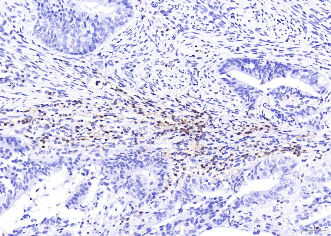
Click image to see more details
IHC analysis of CD4 using anti-CD4 antibody (M00344).
CD4 was detected in a paraffin-embedded section of human cervical cance tissue. Heat mediated antigen retrieval was performed in EDTA buffer (pH 8.0, epitope retrieval solution). The tissue section was blocked with 10% goat serum. The tissue section was then incubated with 1:200 rabbit anti-CD4 Antibody (M00344) overnight at 4°C. Two-step IHC detection kit was used as secondary antibody and incubated for 30 minutes at 37°C. The tissue section was developed using HRP Conjugated Rabbit IgG Super Vision Assay Kit (Catalog # SV0002) with DAB as the chromogen.

Click image to see more details
Interaction between GZMK + CD8 + T cells and fibroblasts contributes to neutrophilic inflammation in nasal polyps. a Representative immunofluorescence staining of collagen I (COL1A1, green), CD8 (red), and GZMK (yellow) in NPs. The right image shows a greater magnification of the outlined area. b Spatial distribution analysis of GZMK + CD8 + T and COL1A1 + cells in the same tissue field demonstrated in ( a ) using HALO software. c–d The number of COL1A1 + fibroblasts within a radius of 25 μm from the nuclear center of GZMK + CD8 + T, GZMB + CD8 + T, CD4 + T, or CD19 + B cells in CIT group (left, n = 10 samples) and NP group (right, n = 10 samples) ( c ). Average distance from the indicated cell types to the closest COL1A1 + fibroblasts in CIT group (left, n = 10 samples) and NP group (right, n = 10 samples) ( d ). e DEGs between NP-derived primary fibroblasts (NPDF) treated with and without recombinant human GZMK ( n = 4). Two-sided Wald test (default for DESeq2 r-package) was used for differential expression analysis utilizing standard cutoffs of
Specific Publications For Anti-CD4 Rabbit Monoclonal Antibody (M00344)
Loading publications
Recommended Resources
Here are featured tools and databases that you might find useful.
- Boster's Pathways Library
- Protein Databases
- Bioscience Research Protocol Resources
- Data Processing & Analysis Software
- Photo Editing Software
- Scientific Literature Resources
- Research Paper Management Tools
- Molecular Biology Software
- Primer Design Tools
- Bioinformatics Tools
- Phylogenetic Tree Analysis
Customer Reviews
Have you used Anti-CD4 Rabbit Monoclonal Antibody?
Share your experimental results or join a short interview to earn up to $1,000 in product credits or other rewards.
1 Reviews For Anti-CD4 Rabbit Monoclonal Antibody
In this IHC experiment using Anti-CD4 antibody (Cat# M00344) on human cervical cancer paraffin sections, CD4-positive helper T cells were clearly and specifically stained with minimal background, demonstrating excellent antibody performance.
Excellent
| SKU | M00344 |
|---|---|
| Application | Western blot |
| Sample | human cervical cancer paraffin sections |
| Sample Processing Description | Cervical cancer tissue collected from clinical surgery, fixed in formalin and paraffin-embedded |
| Other Reagents | Goat serum, DAB substrate solution |
| Primary Antibody | CD4 Rabbit Monoclonal Antibody |
| Primary Incubation | 1:200, overnight at 4 ℃ |
| Secondary Antibody | Two-step IHC detection kit |
| Secondary Incubation | 30 min at 37℃ |
| Detection | Substrate: ECL substrate, Imaging system:ChemiDoc MP |
| Results Summary | CD4 is a marker of helper T cells; in this IHC experiment, CD4 staining clearly identified helper T cells in human cervical cancer samples, showing excellent specificity and performance of the antibody. |
Yanan Peng, The First Affiliated Hospital of Shihezi University
Verified customer
Submitted 2026-02-25
Customer Q&As
Have a question?
Find answers in Q&As, reviews.
Can't find your answer?
Submit your question
4 Customer Q&As for Anti-CD4 Rabbit Monoclonal Antibody
Question
We are currently using anti-CD4 Rabbit Monoclonal antibody M00344 for human tissue, and we are content with the IF results. The species of reactivity given in the datasheet says human. Is it true that the antibody can work on pig tissues as well?
Verified Customer
Verified customer
Asked: 2020-05-08
Answer
The anti-CD4 Rabbit Monoclonal antibody (M00344) has not been tested for cross reactivity specifically with pig tissues, but there is a good chance of cross reactivity. We have an innovator award program that if you test this antibody and show it works in pig you can get your next antibody for free. Please contact me if I can help you with anything.
Boster Scientific Support
Answered: 2020-05-08
Question
We ordered your anti-CD4 Rabbit Monoclonal antibody for WB on thymus in a previous experiment. I am using human, and I plan to use the antibody for IF next. My question regards examining thymus as well as blood in our next experiment. Do you have any suggestion on which antibody would work the best for IF?
Verified Customer
Verified customer
Asked: 2019-09-03
Answer
I looked at the website and datasheets of our anti-CD4 Rabbit Monoclonal antibody and it seems that M00344 has been tested on human in both WB and IF. Thus M00344 should work for your application. Our Boster satisfaction guarantee will cover this product for IF in human even if the specific tissue type has not been validated. We do have a comprehensive range of products for IF detection and you can check out our website bosterbio.com to find out more information about them.
Boster Scientific Support
Answered: 2019-09-03
Question
Our team were happy with the WB result of your anti-CD4 Rabbit Monoclonal antibody. However we have seen positive staining in brain cell membrane using this antibody. Is that expected? Could you tell me where is CD4 supposed to be expressed?
Verified Customer
Verified customer
Asked: 2019-08-22
Answer
Based on literature, brain does express CD4. Generally CD4 expresses in cell membrane. Regarding which tissues have CD4 expression, here are a few articles citing expression in various tissues:
Blood, Pubmed ID: 1425921
Brain, Pubmed ID: 9074930
Liver, Pubmed ID: 19159218
Pancreas, Pubmed ID: 15489334
Thymus, Pubmed ID: 14702039
Boster Scientific Support
Answered: 2019-08-22
Question
We have been able to see staining in human blood. Any tips? Is anti-CD4 Rabbit Monoclonal antibody supposed to stain blood positively?
Verified Customer
Verified customer
Asked: 2017-07-28
Answer
From literature blood does express CD4. From Uniprot.org, CD4 is expressed in leukocyte, brain, thymus, pancreas, blood, liver, among other tissues. Regarding which tissues have CD4 expression, here are a few articles citing expression in various tissues:
Blood, Pubmed ID: 1425921
Brain, Pubmed ID: 9074930
Liver, Pubmed ID: 19159218
Pancreas, Pubmed ID: 15489334
Thymus, Pubmed ID: 14702039
Boster Scientific Support
Answered: 2017-07-28
