Click image to see more details
-

-
-
-
-
+1
Product Info Summary
| SKU: | A01114-2 |
|---|---|
| Size: | 100 μg/vial |
| Reactive Species: | Human, Mouse, Rat |
| Host: | Rabbit |
| Application: | ELISA, IHC, WB |
Customers Who Bought This Also Bought

Product info
Product Name
Anti-CD40L/Cd40lg Antibody Picoband®
SKU/Catalog Number
A01114-2
Size
100 μg/vial
Form
Lyophilized
Description
Boster Bio Anti-CD40L/Cd40lg Antibody Picoband® catalog # A01114-2. Tested in ELISA, IHC, WB applications. This antibody reacts with Human, Mouse, Rat. The brand Picoband indicates this is a premium antibody that guarantees superior quality, high affinity, and strong signals with minimal background in Western blot applications. Only our best-performing antibodies are designated as Picoband, ensuring unmatched performance.
Storage & Handling
Store at -20˚C for one year from date of receipt. After reconstitution, at 4˚C for one month. It can also be aliquotted and stored frozen at -20˚C for six months. Avoid repeated freeze-thaw cycles.
Cite This Product
Anti-CD40L/Cd40lg Antibody Picoband® (Boster Biological Technology, Pleasanton CA, USA, Catalog # A01114-2)
Host
Rabbit
Contents
Each vial contains 4mg Trehalose, 0.9mg NaCl, 0.2mg Na2HPO4, 0.05mg NaN3.
Clonality
Polyclonal
Isotype
Rabbit IgG
Immunogen
E. coli-derived mouse CD40L recombinant protein (Position: E107-L260).
Cross-reactivity
No cross-reactivity with other proteins.
Reactive Species
A01114-2 is reactive to Cd40lg in Human, Mouse, Rat
Observed Molecular Weight
36 kDa
Calculated molecular weight
29.4 kDa
Background of Cd40lg
CD40 ligand (CD40L) is a type II membrane protein of 261 amino acids on activated T cells that induces B cell proliferation and immunoglobulin secretion. It has homology with tumour necrosis factor-alpha and -beta, and has important functions in B-cell activation and differentiation. Human CD40L with 5 exons, is mapped to the proximal region of the mouse X chromosome on Xq26.3-27.1, and can be detected on T cells but is absent from B cells and monocytes. Since CD40L is expressed on platelets and released from them on activation, its predictive value as a marker for clinical outcome and the therapeutic effect of inhibition of glycoprotein IIb /IIIa receptor in patients with acute coronary syndromes was investigated. The soluble CD40L may be involved in the process of restenosis and that it exerts its effect by triggering a complex group of inflammatory reactions on endothelial and mononuclear cells.CD40L plays a central role in the pathophysiology of acute coronary syndromes, and has a role in the pathogenesis of coronary artery lesions.
Antibody Validation
Boster validates all antibodies on WB, IHC, ICC, Immunofluorescence, and ELISA with known positive control and negative samples to ensure specificity and high affinity, including thorough antibody incubations.
Application & Images
Applications
A01114-2 is guaranteed for ELISA, IHC, WB Boster Guarantee
Recommend Dilution
| Application | Dilution | Species |
|---|---|---|
| Western blot | 0.1-0.5μg/ml | |
| Immunohistochemistry (Paraffin-embedded Section) | 0.5-1μg/ml | |
| ELISA | 0.1-0.5μg/ml |
Tested application
Suggested blocking solution with 5% non-fat milk or BSA; (*)Recommended protein loading: 20-40 µg per lane
Use TE buffer pH 9.0 for antigen retrieval; (*) citrate buffer pH 6.0 is an alternative.
Validation Images & Assay Conditions
Click image to see more details
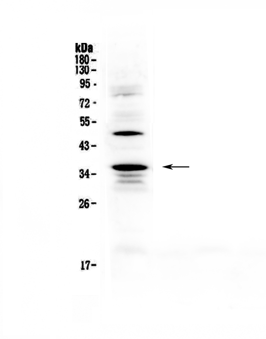
Western blot analysis of CD40L using anti-CD40L antibody (A01114-2).
Electrophoresis was performed on a 5-20% SDS-PAGE gel at 70V (Stacking gel) / 90V (Resolving gel) for 2-3 hours. The sample well of each lane was loaded with 50ug of sample under reducing conditions.
Lane 1: rat spleen tissue lysates.
After Electrophoresis, proteins were transferred to a Nitrocellulose membrane at 150mA for 50-90 minutes. Blocked the membrane with 5% Non-fat Milk/ TBS for 1.5 hour at RT. The membrane was incubated with rabbit anti-CD40L antigen affinity purified polyclonal antibody (Catalog # A01114-2) at 0.5 μg/mL overnight at 4°C, then washed with TBS-0.1%Tween 3 times with 5 minutes each and probed with a goat anti-rabbit IgG-HRP secondary antibody at a dilution of 1:10000 for 1.5 hour at RT. The signal is developed using an Enhanced Chemiluminescent detection (ECL) kit (Catalog # EK1002) with Tanon 5200 system. A specific band was detected for CD40L at approximately 36KD. The expected band size for CD40L is at 29KD.

Click image to see more details
IHC analysis of CD154/CD40LG using anti-CD154/CD40LG antibody (A01114-2).
CD154/CD40LG was detected in a paraffin-embedded section of human tonsil tissue. Heat mediated antigen retrieval was performed in EDTA buffer (pH 8.0, epitope retrieval solution). The tissue section was blocked with 10% goat serum. The tissue section was then incubated with 2 μg/ml rabbit anti-CD154/CD40LG Antibody (A01114-2) overnight at 4°C. Peroxidase Conjugated Goat Anti-rabbit IgG was used as secondary antibody and incubated for 30 minutes at 37°C. The tissue section was developed using HRP Conjugated Rabbit IgG Super Vision Assay Kit (Catalog # SV0002) with DAB as the chromogen.

Click image to see more details
IHC analysis of CD40L using anti-CD40L antibody (A01114-2).
CD40L was detected in paraffin-embedded section of mouse spleen tissue. Heat mediated antigen retrieval was performed in citrate buffer (pH6, epitope retrieval solution) for 20 mins. The tissue section was blocked with 10% goat serum. The tissue section was then incubated with 1μg/ml rabbit anti-CD40L Antibody (A01114-2) overnight at 4°C. Biotinylated goat anti-rabbit IgG was used as secondary antibody and incubated for 30 minutes at 37°C. The tissue section was developed using Strepavidin-Biotin-Complex (SABC)(Catalog # SA1022) with DAB as the chromogen.

Click image to see more details
IHC analysis of CD40L using anti-CD40L antibody (A01114-2).
CD40L was detected in paraffin-embedded section of rat spleen tissue. Heat mediated antigen retrieval was performed in citrate buffer (pH6, epitope retrieval solution) for 20 mins. The tissue section was blocked with 10% goat serum. The tissue section was then incubated with 1μg/ml rabbit anti-CD40L Antibody (A01114-2) overnight at 4°C. Biotinylated goat anti-rabbit IgG was used as secondary antibody and incubated for 30 minutes at 37°C. The tissue section was developed using Strepavidin-Biotin-Complex (SABC)(Catalog # SA1022) with DAB as the chromogen.

Click image to see more details
Western blot analysis of CD40L using anti-CD40L antibody (A01114-2).
Electrophoresis was performed on a 5-20% SDS-PAGE gel at 70V (Stacking gel) / 90V (Resolving gel) for 2-3 hours. The sample well of each lane was loaded with 50ug of sample under reducing conditions.
Lane 1: human MCF-7 whole cell lysates.
After Electrophoresis, proteins were transferred to a Nitrocellulose membrane at 150mA for 50-90 minutes. Blocked the membrane with 5% Non-fat Milk/ TBS for 1.5 hour at RT. The membrane was incubated with rabbit anti-CD40L antigen affinity purified polyclonal antibody (Catalog # A01114-2) at 0.5 μg/mL overnight at 4°C, then washed with TBS-0.1%Tween 3 times with 5 minutes each and probed with a goat anti-rabbit IgG-HRP secondary antibody at a dilution of 1:10000 for 1.5 hour at RT. The signal is developed using an Enhanced Chemiluminescent detection (ECL) kit (Catalog # EK1002) with Tanon 5200 system. A specific band was detected for CD40L at approximately 36KD. The expected band size for CD40L is at 29KD.
Specific Publications For Anti-CD40L/Cd40lg Antibody Picoband® (A01114-2)
Loading publications
Recommended Resources
Here are featured tools and databases that you might find useful.
- Boster's Pathways Library
- Protein Databases
- Bioscience Research Protocol Resources
- Data Processing & Analysis Software
- Photo Editing Software
- Scientific Literature Resources
- Research Paper Management Tools
- Molecular Biology Software
- Primer Design Tools
- Bioinformatics Tools
- Phylogenetic Tree Analysis
Customer Reviews
Have you used Anti-CD40L/Cd40lg Antibody Picoband®?
Share your experimental results or join a short interview to earn up to $1,000 in product credits or other rewards.
0 Reviews For Anti-CD40L/Cd40lg Antibody Picoband®
Customer Q&As
Have a question?
Find answers in Q&As, reviews.
Can't find your answer?
Submit your question
5 Customer Q&As for Anti-CD40L/Cd40lg Antibody Picoband®
Question
Our lab were content with the WB result of your anti-CD40L/Cd40lg antibody. However we have observed positive staining in blood cell membrane using this antibody. Is that expected? Could you tell me where is CD40LG supposed to be expressed?
Verified Customer
Verified customer
Asked: 2019-11-26
Answer
From what I have seen in literature, blood does express CD40LG. Generally CD40LG expresses in cell membrane. Regarding which tissues have CD40LG expression, here are a few articles citing expression in various tissues:
Blood, Pubmed ID: 15489334
Boster Scientific Support
Answered: 2019-11-26
Question
I was wanting to use using your anti-CD40L/Cd40lg antibody for r-mmu-198933; immunoregulatory interactions between a lymphoid and a non-lymphoid cell studies. Has this antibody been tested with western blotting on mouse spleen tissue? We would like to see some validation images before ordering.
Verified Customer
Verified customer
Asked: 2019-10-17
Answer
We appreciate your inquiry. This A01114-2 anti-CD40L/Cd40lg antibody is validated on rat spleen tissue, mouse spleen tissue. It is guaranteed to work for ELISA, IHC, WB in human, mouse, rat. Our Boster guarantee will cover your intended experiment even if the sample type has not been be directly tested.
Boster Scientific Support
Answered: 2019-10-17
Question
We bought anti-CD40L/Cd40lg antibody for WB on blood a few years ago. I am using rat, and We want to use the antibody for IHC next. Our lab want to know about examining blood as well as leukocyte in our next experiment. Could you please give me some suggestion on which antibody would work the best for IHC?
B. Williams
Verified customer
Asked: 2015-02-06
Answer
I have checked the website and datasheets of our anti-CD40L/Cd40lg antibody and I see that A01114-2 has been tested on rat in both WB and IHC. Thus A01114-2 should work for your application. Our Boster satisfaction guarantee will cover this product for IHC in rat even if the specific tissue type has not been validated. We do have a comprehensive range of products for IHC detection and you can check out our website bosterbio.com to find out more information about them.
Boster Scientific Support
Answered: 2015-02-06
Question
We have observed staining in human blood. Are there any suggestions? Is anti-CD40L/Cd40lg antibody supposed to stain blood positively?
A. Singh
Verified customer
Asked: 2013-05-30
Answer
From what I have seen in literature blood does express CD40LG. From what I have seen in Uniprot.org, CD40LG is expressed in leukocyte, blood, among other tissues. Regarding which tissues have CD40LG expression, here are a few articles citing expression in various tissues:
Blood, Pubmed ID: 15489334
Boster Scientific Support
Answered: 2013-05-30
Question
We are currently using anti-CD40L/Cd40lg antibody A01114-2 for rat tissue, and we are content with the WB results. The species of reactivity given in the datasheet says human, mouse, rat. Is it likely that the antibody can work on monkey tissues as well?
B. Jones
Verified customer
Asked: 2013-03-08
Answer
The anti-CD40L/Cd40lg antibody (A01114-2) has not been tested for cross reactivity specifically with monkey tissues, but there is a good chance of cross reactivity. We have an innovator award program that if you test this antibody and show it works in monkey you can get your next antibody for free. Please contact me if I can help you with anything.
Boster Scientific Support
Answered: 2013-03-08
