Click image to see more details
Product Info Summary
| SKU: | A01047-2 |
|---|---|
| Size: | 100ul |
| Reactive Species: | Human |
| Host: | Rabbit |
| Application: | Flow Cytometry, IF, IHC, WB |
Customers Who Bought This Also Bought

Product info
Product Name
Anti-CD79a Antibody
SKU/Catalog Number
A01047-2
Size
100ul
Form
Liquid
Description
Boster Bio Anti-CD79a Antibody catalog # A01047-2. Tested in WB,IF,IHC,Flow Cytometry applications. This antibody reacts with Human.
Storage & Handling
Store at -20°C for one year. For short term storage and frequent use, store at 4°C for up to one month. Avoid repeated freeze-thaw cycles.
Cite This Product
Anti-CD79a Antibody (Boster Biological Technology, Pleasanton CA, USA, Catalog # A01047-2)
Host
Rabbit
Contents
Rabbit IgG, 1mg/ml in PBS with 0.02% sodium azide, 50% glycerol, pH7.2
Clonality
Polyclonal
Isotype
IgG
Immunogen
Recombinant protein within human CD79a aa 33-143.
Cross-reactivity
No cross reactivity with other proteins.
Reactive Species
A01047-2 is reactive to CD79A in Human
Calculated molecular weight
25.0 kDa
Antibody Validation
Boster validates all antibodies on WB, IHC, ICC, Immunofluorescence, and ELISA with known positive control and negative samples to ensure specificity and high affinity, including thorough antibody incubations.
Application & Images
Applications
A01047-2 is guaranteed for Flow Cytometry, IF, IHC, WB Boster Guarantee
Recommend Dilution
| Application | Dilution | Species |
|---|---|---|
| WB: 1:500-1:1 | 000 |
Validation Images & Assay Conditions
Click image to see more details
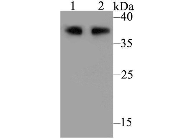
Western blot analysis of CD79a on different lysates. Proteins were transferred to a PVDF membrane and blocked with 5% BSA in PBS for 1 hour at room temperature. The primary antibody was used at a 1:500 dilution in 5% BSA at room temperature for 2 hours. Goat Anti-Rabbit IgG - HRP Secondary Antibody (HA1001) at 1:5,000 dilution was used for 1 hour at room temperature.Positive control: Lane 1: Daudi cell lysateLane 2: Raji cell lysate

Click image to see more details
Immunohistochemical analysis of paraffin-embedded human tonsil tissue using anti-CD79a antibody. The section was pre-treated using heat mediated antigen retrieval with Tris-EDTA buffer (pH 8.0-8.4) for 20 minutes. The tissues were blocked in 5% BSA for 30 minutes at room temperature, washed with ddH2O and PBS, and then probed with ET7109-33 at 1/100 dilution, for 30 minutes at room temperature and detected using an HRP conjugated compact polymer system. DAB was used as the chrogen. Counter stained with hematoxylin and mounted with DPX.
Specific Publications For Anti-CD79a Antibody (A01047-2)
Loading publications
Recommended Resources
Here are featured tools and databases that you might find useful.
- Boster's Pathways Library
- Protein Databases
- Bioscience Research Protocol Resources
- Data Processing & Analysis Software
- Photo Editing Software
- Scientific Literature Resources
- Research Paper Management Tools
- Molecular Biology Software
- Primer Design Tools
- Bioinformatics Tools
- Phylogenetic Tree Analysis
Customer Reviews
Have you used Anti-CD79a Antibody?
Share your experimental results or join a short interview to earn up to $1,000 in product credits or other rewards.
0 Reviews For Anti-CD79a Antibody
Customer Q&As
Have a question?
Find answers in Q&As, reviews.
Can't find your answer?
Submit your question
