Click image to see more details
-

-
-
-
-
+5
Product Info Summary
| SKU: | A00196 |
|---|---|
| Size: | 0.1 mg |
| Reactive Species: | Human, Mouse, Rat |
| Host: | Rabbit |
| Application: | ELISA, IF, WB |
Customers Who Bought This Also Bought

Product info
Product Name
Anti-CD80 Antibody
SKU/Catalog Number
A00196
Size
0.1 mg
Form
Liquid
Description
Boster Bio Anti-CD80 Antibody (Catalog # A00196). Tested in ELISA, WB, IF applications. This antibody reacts with Human, Mouse, Rat.
Storage & Handling
CD80 antibody can be stored at 4°C for three months and -20°C, stable for up to one year. Avoid repeated freeze-thaw cycles. Antibodies should not be exposed to prolonged high temperatures.
Cite This Product
Anti-CD80 Antibody (Boster Biological Technology, Pleasanton CA, USA, Catalog # A00196)
Host
Rabbit
Contents
CD80 Antibody is supplied in PBS containing 0.02% sodium azide.
Clonality
Polyclonal
Isotype
IgG
Immunogen
CD80 antibody was raised against a peptide corresponding to 17 amino acids near the amino terminus of human CD80. The immunogen is located within amino acids 60 - 110 of CD80.
Reactive Species
A00196 is reactive to CD80 in Human, Mouse, Rat
Observed Molecular Weight
60 kDa
Calculated molecular weight
33.0 kDa
Background of CD80
CD80, also known as B7-1, is a type I membrane protein that is a member of the immunoglobulin superfamily. Like the related protein CD86, this protein is expressed by antigen-presenting cells, and is the ligand for two proteins at the cell surface of T cells, CD28 and the cytotoxic T-lymphocyte-associated protein 4 (CTLA-4). Binding of this protein with CD28 antigen is a costimulatory signal for activation of the T-cell and induces T-cell proliferation and cytokine production. CTLA-4 binding negatively regulates T-cell activation and diminishes the immune response (1). Blocking the CTLA-4-CD80/CD86 interaction has been shown to enhance T-cell functions in acute lymphoblastomic leukemia (ALL), suggesting that this pathway may be an attractive target for future cancer immunotherapy (2).
Antibody Validation
Boster validates all antibodies on WB, IHC, ICC, Immunofluorescence, and ELISA with known positive control and negative samples to ensure specificity and high affinity, including thorough antibody incubations.
Application & Images
Applications
A00196 is guaranteed for ELISA, IF, WB Boster Guarantee
Assay Dilutions Recommendation
The recommendations below provide a starting point for assay optimization. The actual working concentration varies and should be decided by the user.
WB: 1-4 μg/mL; IHC-P/IF: 5-20 μg/mL.
Antibody validated: Western Blot in human, mouse and rat samples; Immunofluorescence in the human, mouse and rat samples. All other applications and species not yet tested.
Validation Images & Assay Conditions
Click image to see more details
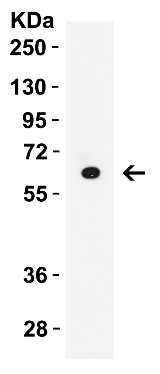
WB Validation in Human Raji Cells Loading: 15 μg of lysate Antibodies: CD80, A00196, 2 μ g/mL , 1 h incubation at RT in 5% NFDM/TBST. Secondary: Goat Anti-Rabbit IgG HRP conjugate at 1:10000 dilution.

Click image to see more details
WB Validation in Human Thymus Loading: 15 μg of lysate Antibodies: CD80, A00196, 2 μ g/mL , 1 h incubation at RT in 5% NFDM/TBST. Secondary: Goat Anti-Rabbit IgG HRP conjugate at 1:10000

Click image to see more details
WB Validation in Human Tonsil Loading: 15 μg of lysate Antibodies: CD80, A00196, 2 μg/mL , 1 h incubation at RT in 5% NFDM/TBST. Secondary: Goat Anti-Rabbit IgG HRP conjugate at 1:10000 dilution.

Click image to see more details
WB Validation in Mouse Tissues Loading: 15 μg of lysate Antibodies: CD80, A00196, 2 μg/mL , 1 h incubation at RT in 5% NFDM/TBST. Secondary: Goat Anti-Rabbit IgG HRP conjugate at 1:10000 dilution.

Click image to see more details
WB Validation in Rat Thymus Loading: 15 μg of lysate Antibodies: CD80, A00196, 2 μg/mL , 1 h incubation at RT in 5% NFDM/TBST. Secondary: Goat Anti-Rabbit IgG HRP conjugate at 1:10000 dilution.

Click image to see more details
Immunofluorescence Validation of CD80 in Human Tonsil Immunofluorescent analysis of 4% paraformaldehyde-fixed human tonsil tissue labeling CD80 with A00196 at 20 μg/mL, followed by goat anti-rabbit IgG secondary antibody at 1/500 dilution (red) and DAPI staining (blue).

Click image to see more details
Immunofluorescence Validation of CD80 in Mouse Spleen Immunofluorescent analysis of 4% paraformaldehyde-fixed mouse spleen tissue labeling CD80 with A00196 at 20 μg/mL, followed by goat anti-rabbit IgG secondary antibody at 1/500 dilution (red) and DAPI staining (blue).

Click image to see more details
Immunofluorescence Validation of CD80 in Human Tonsil Immunofluorescent analysis of 4% paraformaldehyde-fixed human tonsil tissue labeling CD80 with A00196 at 20 μg/mL, followed by goat anti-rabbit IgG secondary antibody at 1/500 dilution (red) and DAPI staining (blue).

Click image to see more details
Immunofluorescence Validation of CD80 in Rat Spleen Immunofluorescent analysis of 4% paraformaldehyde-fixed rat spleen tissue labeling CD80 with A00196 at 20 μg/mL, followed by goat anti-rabbit IgG secondary antibody at 1/500 dilution (red) and DAPI staining (blue).
Specific Publications For Anti-CD80 Antibody (A00196)
Loading publications
Recommended Resources
Here are featured tools and databases that you might find useful.
- Boster's Pathways Library
- Protein Databases
- Bioscience Research Protocol Resources
- Data Processing & Analysis Software
- Photo Editing Software
- Scientific Literature Resources
- Research Paper Management Tools
- Molecular Biology Software
- Primer Design Tools
- Bioinformatics Tools
- Phylogenetic Tree Analysis
Customer Reviews
Have you used Anti-CD80 Antibody?
Share your experimental results or join a short interview to earn up to $1,000 in product credits or other rewards.
0 Reviews For Anti-CD80 Antibody
Customer Q&As
Have a question?
Find answers in Q&As, reviews.
Can't find your answer?
Submit your question
