Click image to see more details
-

-
-
-
-
+3
Product Info Summary
| SKU: | PB9546 |
|---|---|
| Size: | 100 μg/vial |
| Reactive Species: | Human, Mouse, Rat |
| Host: | Rabbit |
| Application: | Flow Cytometry, IHC, WB |
Customers Who Bought This Also Bought

Product info
Product Name
Anti-CYP1B1 Antibody Picoband®
SKU/Catalog Number
PB9546
Size
100 μg/vial
Form
Lyophilized
Description
Boster Bio Anti-CYP1B1 Antibody Picoband® catalog # PB9546. Tested in Flow Cytometry, IHC, WB applications. This antibody reacts with Human, Mouse, Rat. The brand Picoband indicates this is a premium antibody that guarantees superior quality, high affinity, and strong signals with minimal background in Western blot applications. Only our best-performing antibodies are designated as Picoband, ensuring unmatched performance.
Storage & Handling
Store at -20˚C for one year from date of receipt. After reconstitution, at 4˚C for one month. It can also be aliquotted and stored frozen at -20˚C for six months. Avoid repeated freeze-thaw cycles.
Cite This Product
Anti-CYP1B1 Antibody Picoband® (Boster Biological Technology, Pleasanton CA, USA, Catalog # PB9546)
Host
Rabbit
Contents
Each vial contains antibody formulated with stabilizing components, 0.9 mg NaCl, 0.2 mg Na2HPO4, and 0.05 mg NaN3.
*This antibody is supplied in a stabilized formulation.
Compatibility with conjugation reactions depends on the chemistry of the conjugation method used.
For conjugation methods that are not compatible with the stabilizing components present in this formulation, a carrier-free antibody format is required.
Clonality
Polyclonal
Isotype
Rabbit IgG
Immunogen
E.coli-derived human CYP1B1 recombinant protein (Position: R255-L480). Human CYP1B1 shares 85.4% and 84.5% amino acid (aa) sequence identity with mouse and rat CYP1B1, respectively.
Cross-reactivity
No cross-reactivity with other proteins
Reactive Species
PB9546 is reactive to CYP1B1 in Human, Mouse, Rat
Observed Molecular Weight
61 kDa
Calculated molecular weight
60.8 kDa
Background of CYP1B1
Cytochrome P450 1B1 is an enzyme that in humans is encoded by the CYP1B1 gene. CYP1B1 belongs to the the cytochrome P450 superfamily of enzymes. The cytochrome P450 proteins are monooxygenases which catalyze many reactions involved in drug metabolism and synthesis of cholesterol, steroids and other lipids. The enzyme encoded by this gene localizes to the endoplasmic reticulum and metabolizes procarcinogens such as polycyclic aromatic hydrocarbons and 17beta-estradiol. Mutations in this gene have been associated with primary congenital glaucoma; therefore it is thought that the enzyme also metabolizes a signaling molecule involved in eye development, possibly a steroid.
Antibody Validation
Boster validates all antibodies on WB, IHC, ICC, Immunofluorescence, and ELISA with known positive control and negative samples to ensure specificity and high affinity, including thorough antibody incubations.
Application & Images
Applications
PB9546 is guaranteed for Flow Cytometry, IHC, WB Boster Guarantee
Recommend Dilution
| Application | Dilution | Species |
|---|---|---|
| Western blot | 0.1-0.5μg/ml | Human, Mouse, Rat |
| Immunohistochemistry (Paraffin-embedded Section) | 0.5-1μg/ml | Human, Mouse, Rat |
| Flow Cytometry (Fixed) | 1-3μg/1x106 cells | Human |
Tested application
Suggested blocking solution with 5% non-fat milk or BSA; (*)Recommended protein loading: 20-40 µg per lane
Use TE buffer pH 9.0 for antigen retrieval; (*) citrate buffer pH 6.0 is an alternative.
Validation Images & Assay Conditions
Click image to see more details
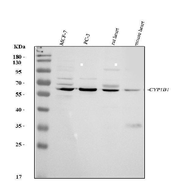
Western blot analysis of CYP1B1 using anti-CYP1B1 antibody (PB9546).
Electrophoresis was performed on a 5-20% SDS-PAGE gel at 70V (Stacking gel) / 90V (Resolving gel) for 2-3 hours. The sample well of each lane was loaded with 30 ug of sample under reducing conditions.
Lane 1: human MCF-7 whole cell lysates,
Lane 2: human PC-3 whole cell lysates,
Lane 3: rat heart tissue lysates,
Lane 4: mouse heart tissue lysates.
After electrophoresis, proteins were transferred to a nitrocellulose membrane at 150 mA for 50-90 minutes. Blocked the membrane with 5% non-fat milk/TBS for 1.5 hour at RT. The membrane was incubated with rabbit anti-CYP1B1 antigen affinity purified polyclonal antibody (Catalog # PB9546) at 0.5 μg/mL overnight at 4°C, then washed with TBS-0.1%Tween 3 times with 5 minutes each and probed with a goat anti-rabbit IgG-HRP secondary antibody at a dilution of 1:5000 for 1.5 hour at RT. The signal is developed using an Enhanced Chemiluminescent detection (ECL) kit (Catalog # EK1002) with Tanon 5200 system. A specific band was detected for CYP1B1 at approximately 61 kDa. The expected band size for CYP1B1 is at 61 kDa.

Click image to see more details
aHSC release LTB4R2 ligands in a manner dependent on CYP1B1. a TEAD-luciferase activity in Huh7 cells treated with conditioned medium (CM) from LX2 cells transduced with scrambled shRNA (SCRsh-CM) vs. SCD-shRNA (SCDsh-CM) as compared to the media without Huh7 cells (Media). * p < 0.05 by two-sided t-test. Data presented as means ± SEM ( n = 4 separate experiments). b TEAD-luciferase activity in Huh7 cells exposed to SCRsh-CM vs. SCDsh-CM in the presence of the TBXAS1 inhibitor Ozagarel or vehicle DMSO. * p < 0.001 vs. Media, # p < 0.001 vs. DMSO by two-sided t-test. Data presented as means ± SEM ( n = 3 experiments). c A scRNA - seq t-SNE plot showing Cyp1b1 + cells and violin plots revealing selective Cyp1b1 expression by Fbln2 + cells. Cell numbers for different cell type groups are provided in Supplementary Data . d Contour FACS plots of liver mesenchymal cells isolated from control vs. DEN + WAD treated Rosa26mTmG;Col1a1-Cre; ( mTmG;CC ) mouse, gated by DAPI ( Y -axis) for Vit A fluorescence and FITC (X-axis) for Col1a1-GFP, revealing VitA + GFP - quiescent HSC (blue), VitA + GFP + aHSC (red), and VitA − GFP + cells (green). FACS gating strategies are provided in . e scRNA-seq analysis for expression of 12-HHTrE biosynthetic genes in VitA + GFP + (top) and VitA − GFP + (bottom) subpopulations from DEN+WAD mouse (DEN) vs. normal (Cont.) livers. f Violin plots for Cyp1b1 expression by subpopulations based on Lrat , Thy1 , and Fbln2 expression in VitA + GFP + cells from the DEN mouse and g in VitA − GFP + cells. (See Supplementary Data for parameter values for violin plots and cell numbers for different subpopulations). h CRISPR/Cas9 ablation of CYP1B1 in LX2 cells using the guide RNA-A (sgRNA-A) or -B (sgRNA-B) (top), represses CYP1B1 mRNA. * p < 0.05 vs. control by two-sided t-test. Data presented as means ± SEM ( n = 3 separate samples). i Oxylipin concentrations in CM from LX2 cells with CYP1B1 KD are described above. * p < 0.05 vs. control by two-sided t-test. Data presented as means ± SEM ( n = 4 separate samples). (Raw data provided in Supplementary Data in Supplementary File). j Reduced stimulatory effects of CM from CYP1B1 ablated LX2 cells on the TEAD promoter activity in Huh7 cells (right). *** p < 0.001 vs. Control CM by two-sided t-test. Data presented as means ± SEM ( n = 3 experiments). For all relevant figures, source data and exact p values are provided in the file.
Index in PubMed under a CC BY license. PMID: 37156770

Click image to see more details
HCC growth is dependent on LTB4R2. a IB analysis of LTB4R2 and NaKATPase (membrane), pYAP1, pGSK3β, GSK3β, HuR, GAPDH (cytosolic), YAP1 and CTNNB1 (nuclear) proteins from B6 mice subjected to the DEN + WAD regimen and injected with AAV vector (4 × 10 11 GC per mouse) expressing SCR-shRNA (SCR-sh) vs. LTB4R2-shRNA (LTB4R2-sh) one month prior to the end of experiment ( n = 6 mice per group). b Liver tumor development in the mice with SCR-sh vs. LTB4R2-sh treatment depicted by representative images and total tumor volume and multiplicity. * p < 0.05 vs. SCR-sh mice by two-sided t-test. Data presented as means ± SEM ( n = 9 mice each). c qPCR data for oxylipin synthetic genes, LTB4R2 , and YAP1 in patient HCC ( n = 6) vs. normal subject livers ( n = 6). * p < 0.05, ** p < 0.01 vs. normal liver by two-sided t-test. Data presented as means ± SEM. d Representative IHC-HRP staining for CYP1B1 and LTB4R2 of patient HCC liver sections (×200) from four patient samples examined. Areas demarked by broken lines are HCC. e Top: Growth of patient HCC organoid (model-1 and model-2) in the presence of DMSO (vehicle) or the LTB4R2 antagonist LY255283. * p < 0.05, ** p < 0.01, *** p < 0.005 vs. DMSO by two-way ANOVA with post hoc test. Data presented as means ± SEM ( n = 3 experiments). Bottom: Growth of patient HCC organoid model-1 and model-2 without (CTRL) or with infection with adenovirus expressing scrambled shRNA ( shCTRL ) or LTB4R2 shRNA (shLTB4R2). * p < 0.05, *** p < 0.005 vs. DMSO ( n = 3 experiments). f Schematic diagram of HCC promotion initiated by SCD-CYP1B1-dependent release of 12-HHTrE by Lart + Fbln2 + aHSC, activating LTB4R2-CTNNB1-YAP1 pathway in HCC cell. For all relevant figures , source data and exact p values are provided in the file.
Index in PubMed under a CC BY license. PMID: 37156770

Click image to see more details
IHC analysis of CYP1B1 using anti-CYP1B1 antibody (PB9546).
CYP1B1 was detected in paraffin-embedded section of Mouse Kidney Tissue. Heat mediated antigen retrieval was performed in citrate buffer (pH6, epitope retrieval solution) for 20 mins. The tissue section was blocked with 10% goat serum. The tissue section was then incubated with 1μg/ml rabbit anti-CYP1B1 Antibody (PB9546) overnight at 4°C. Biotinylated goat anti-rabbit IgG was used as secondary antibody and incubated for 30 minutes at 37°C. The tissue section was developed using Strepavidin-Biotin-Complex (SABC)(Catalog # SA1022) with DAB as the chromogen.

Click image to see more details
IHC analysis of CYP1B1 using anti-CYP1B1 antibody (PB9546).
CYP1B1 was detected in paraffin-embedded section of Rat Kidney Tissue. Heat mediated antigen retrieval was performed in citrate buffer (pH6, epitope retrieval solution) for 20 mins. The tissue section was blocked with 10% goat serum. The tissue section was then incubated with 1μg/ml rabbit anti-CYP1B1 Antibody (PB9546) overnight at 4°C. Biotinylated goat anti-rabbit IgG was used as secondary antibody and incubated for 30 minutes at 37°C. The tissue section was developed using Strepavidin-Biotin-Complex (SABC)(Catalog # SA1022) with DAB as the chromogen.

Click image to see more details
IHC analysis of CYP1B1 using anti-CYP1B1 antibody (PB9546).
CYP1B1 was detected in paraffin-embedded section of Human Liver Cancer Tissue. Heat mediated antigen retrieval was performed in citrate buffer (pH6, epitope retrieval solution) for 20 mins. The tissue section was blocked with 10% goat serum. The tissue section was then incubated with 1μg/ml rabbit anti-CYP1B1 Antibody (PB9546) overnight at 4°C. Biotinylated goat anti-rabbit IgG was used as secondary antibody and incubated for 30 minutes at 37°C. The tissue section was developed using Strepavidin-Biotin-Complex (SABC)(Catalog # SA1022) with DAB as the chromogen.

Click image to see more details
Flow Cytometry analysis of SiHa cells using anti-CYP1B1 antibody (PB9546).
Overlay histogram showing SiHa cells stained with PB9546 (Blue line). To facilitate intracellular staining, cells were fixed with 4% paraformaldehyde and permeabilized with permeabilization buffer. The cells were blocked with 10% normal goat serum. And then incubated with rabbit anti-CYP1B1 Antibody (PB9546,1μg/1x106 cells) for 30 min at 20°C. DyLight®488 conjugated goat anti-rabbit IgG (BA1127, 5-10μg/1x106 cells) was used as secondary antibody for 30 minutes at 20°C. Isotype control antibody (Green line) was rabbit IgG (1μg/1x106) used under the same conditions. Unlabelled sample without incubation with primary antibody and secondary antibody (Red line) was used as a blank control.
Specific Publications For Anti-CYP1B1 Antibody Picoband® (PB9546)
Loading publications
Recommended Resources
Here are featured tools and databases that you might find useful.
- Boster's Pathways Library
- Protein Databases
- Bioscience Research Protocol Resources
- Data Processing & Analysis Software
- Photo Editing Software
- Scientific Literature Resources
- Research Paper Management Tools
- Molecular Biology Software
- Primer Design Tools
- Bioinformatics Tools
- Phylogenetic Tree Analysis
Customer Reviews
Have you used Anti-CYP1B1 Antibody Picoband®?
Share your experimental results or join a short interview to earn up to $1,000 in product credits or other rewards.
0 Reviews For Anti-CYP1B1 Antibody Picoband®
Customer Q&As
Have a question?
Find answers in Q&As, reviews.
Can't find your answer?
Submit your question
2 Customer Q&As for Anti-CYP1B1 Antibody Picoband®
Question
We are currently using anti-CYP1B1 antibody PB9546 for human tissue, and we are happy with the Flow Cytometry results. The species of reactivity given in the datasheet says human, mouse, rat. Is it likely that the antibody can work on pig tissues as well?
Verified Customer
Verified customer
Asked: 2019-05-30
Answer
The anti-CYP1B1 antibody (PB9546) has not been tested for cross reactivity specifically with pig tissues, but there is a good chance of cross reactivity. We have an innovator award program that if you test this antibody and show it works in pig you can get your next antibody for free. Please contact me if I can help you with anything.
Boster Scientific Support
Answered: 2019-05-30
Question
Has PB9546 been tested on IF application?
Verified customer
Asked: 2019-03-11
Answer
Unfortunately, our lab failed to get positive staining for IF applicaiton using Anti-CYP1B1 Antibody Picoband™ (PB9546).
Boster Scientific Support
Answered: 2019-03-19
