Click image to see more details
Product Info Summary
| SKU: | A00339-2 |
|---|---|
| Size: | 100 μg/vial |
| Reactive Species: | Human |
| Host: | Rabbit |
| Application: | ELISA, Flow Cytometry, WB |
Customers Who Bought This Also Bought

Product info
Product Name
Anti-Cytochrome P450 3A4/CYP3A4 Antibody Picoband®
SKU/Catalog Number
A00339-2
Size
100 μg/vial
Form
Lyophilized
Description
Boster Bio Anti-Cytochrome P450 3A4/CYP3A4 Antibody Picoband® catalog # A00339-2. Tested in ELISA, Flow Cytometry, WB applications. This antibody reacts with Human. The brand Picoband indicates this is a premium antibody that guarantees superior quality, high affinity, and strong signals with minimal background in Western blot applications. Only our best-performing antibodies are designated as Picoband, ensuring unmatched performance.
Storage & Handling
Store at -20˚C for one year from date of receipt. After reconstitution, at 4˚C for one month. It can also be aliquotted and stored frozen at -20˚C for six months. Avoid repeated freeze-thaw cycles.
Cite This Product
Anti-Cytochrome P450 3A4/CYP3A4 Antibody Picoband® (Boster Biological Technology, Pleasanton CA, USA, Catalog # A00339-2)
Host
Rabbit
Contents
Each vial contains 4mg Trehalose, 0.9mg NaCl, 0.2mg Na2HPO4.
Clonality
Polyclonal
Isotype
Rabbit IgG
Immunogen
E.coli-derived human CYP3A4 recombinant protein (Position: L44-H267).
Cross-reactivity
No cross-reactivity with other proteins
Reactive Species
A00339-2 is reactive to CYP3A4 in Human
Observed Molecular Weight
57 kDa
Calculated molecular weight
57.3 kDa
Background of CYP3A4
Cytochrome P450 3A4 (abbreviated CYP3A4), is an important enzyme in the body, mainly found in the liver and in the intestine. This gene encodes a member of the cytochrome P450 superfamily of enzymes. The cytochrome P450 proteins are monooxygenases that catalyze many reactions involved in drug metabolism and synthesis of cholesterol, steroids and other lipids. This protein localizes to the endoplasmic reticulum and its expression is induced by glucocorticoids and some pharmacological agents. This enzyme is involved in the metabolism of approximately half the drugs in use today, including acetaminophen, codeine, cyclosporin A, diazepam and erythromycin. The enzyme also metabolizes some steroids and carcinogens. This gene is part of a cluster of cytochrome P450 genes on chromosome 7q21.1. Previously another CYP3A gene, CYP3A3, was thought to exist; however, it is now thought that this sequence represents a transcript variant of CYP3A4. Alternatively spliced transcript variants encoding different isoforms have been identified.
Antibody Validation
Boster validates all antibodies on WB, IHC, ICC, Immunofluorescence, and ELISA with known positive control and negative samples to ensure specificity and high affinity, including thorough antibody incubations.
Application & Images
Applications
A00339-2 is guaranteed for ELISA, Flow Cytometry, WB Boster Guarantee
Recommend Dilution
| Application | Dilution | Species |
|---|---|---|
| Western blot | 0.25-0.5μg/ml | Human |
| Flow Cytometry (Fixed) | 1-3μg/1x106 cells | Human |
| ELISA | 0.1-0.5μg/ml | - |
Tested application
Suggested blocking solution with 5% non-fat milk or BSA; (*)Recommended protein loading: 20-40 µg per lane
Validation Images & Assay Conditions
Click image to see more details
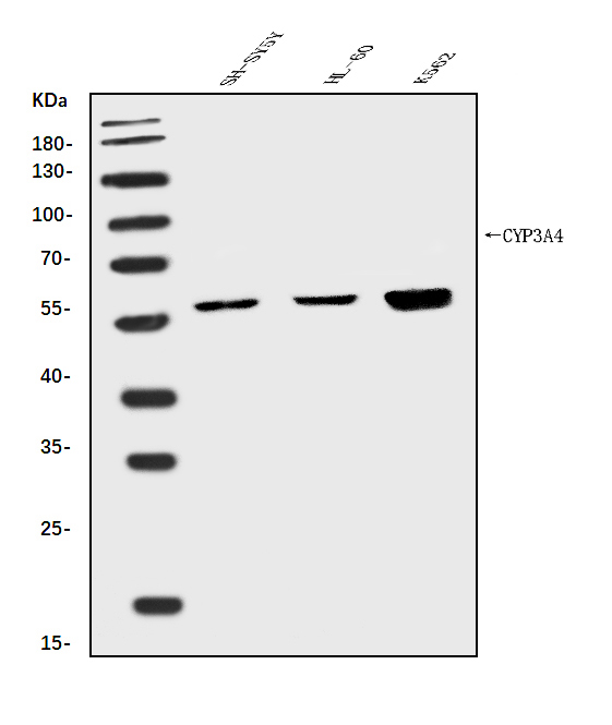
Western blot analysis of Cytochrome P450 3A4/CYP3A4 using anti-Cytochrome P450 3A4/CYP3A4 antibody (A00339-2).
Electrophoresis was performed on a 5-20% SDS-PAGE gel at 70V (Stacking gel) / 90V (Resolving gel) for 2-3 hours. The sample well of each lane was loaded with 30 ug of sample under reducing conditions.
Lane 1: human SH-SY5Y whole cell lysates,
Lane 2: human HL-60 whole cell lysates,
Lane 3: human K562 whole cell lysates.
After electrophoresis, proteins were transferred to a nitrocellulose membrane at 150 mA for 50-90 minutes. Blocked the membrane with 5% non-fat milk/TBS for 1.5 hour at RT. The membrane was incubated with rabbit anti-Cytochrome P450 3A4/CYP3A4 antigen affinity purified polyclonal antibody (Catalog # A00339-2) at 0.5 μg/mL overnight at 4°C, then washed with TBS-0.1%Tween 3 times with 5 minutes each and probed with a goat anti-rabbit IgG-HRP secondary antibody at a dilution of 1:5000 for 1.5 hour at RT. The signal is developed using an Enhanced Chemiluminescent detection (ECL) kit (Catalog # EK1002) with Tanon 5200 system. A specific band was detected for Cytochrome P450 3A4/CYP3A4 at approximately 57 kDa. The expected band size for Cytochrome P450 3A4/CYP3A4 is at 57 kDa.

Click image to see more details
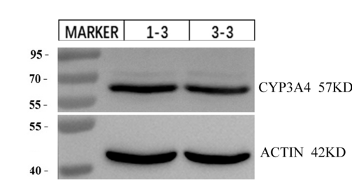
Western blot analysis of Cytochrome P450 3A4/CYP3A4 using anti-Cytochrome P450 3A4/CYP3A4 antibody (A00339-2).
Electrophoresis was performed on a 5-20% SDS-PAGE gel at 70V (Stacking gel) / 90V (Resolving gel) for 2-3 hours. The sample well of each lane was loaded with 30 ug of sample under reducing conditions.
Lane 1-2: Human keratinocytes isolated from skin.
After electrophoresis, proteins were transferred to a nitrocellulose membrane at 150 mA for 50-90 minutes. Blocked the membrane with 5% non-fat milk/TBS for 1.5 hour at RT. The membrane was incubated with rabbit anti-Cytochrome P450 3A4/CYP3A4 antigen affinity purified polyclonal antibody (Catalog # A00339-2) at 0.5 μg/mL overnight at 4°C, then washed with TBS-0.1%Tween 3 times with 5 minutes each and probed with a goat anti-rabbit IgG-HRP secondary antibody at a dilution of 1:5000 for 1 hour at RT. The signal is developed using an Enhanced Chemiluminescent detection (ECL) kit with ChemiDoc MP system. A specific band was detected for Cytochrome P450 3A4/CYP3A4 at approximately 57 kDa. The expected band size for Cytochrome P450 3A4/CYP3A4 is at 57 kDa.

Click image to see more details
Flow Cytometry analysis of HL-60 cells using anti-Cytochrome P450 3A4/CYP3A4 antibody (A00339-2).
Overlay histogram showing HL-60 cells stained with A00339-2 (Blue line). To facilitate intracellular staining, cells were fixed with 4% paraformaldehyde and permeabilized with permeabilization buffer. The cells were blocked with 10% normal goat serum. And then incubated with rabbit anti-Cytochrome P450 3A4/CYP3A4 Antibody (A00339-2, 1 μg/1x106 cells) for 30 min at 20°C. DyLight®488 conjugated goat anti-rabbit IgG (BA1127, 5-10 μg/1x106 cells) was used as secondary antibody for 30 minutes at 20°C. Isotype control antibody (Green line) was rabbit IgG (1 μg/1x106) used under the same conditions. Unlabelled sample without incubation with primary antibody and secondary antibody (Red line) was used as a blank control.

Click image to see more details
Flow Cytometry analysis of U87 cells using anti-Cytochrome P450 3A4/CYP3A4 antibody (A00339-2).
Overlay histogram showing U87 cells stained with A00339-2 (Blue line). To facilitate intracellular staining, cells were fixed with 4% paraformaldehyde and permeabilized with permeabilization buffer. The cells were blocked with 10% normal goat serum. And then incubated with rabbit anti-Cytochrome P450 3A4/CYP3A4 Antibody (A00339-2, 1 μg/1x106 cells) for 30 min at 20°C. DyLight®488 conjugated goat anti-rabbit IgG (BA1127, 5-10 μg/1x106 cells) was used as secondary antibody for 30 minutes at 20°C. Isotype control antibody (Green line) was rabbit IgG (1 μg/1x106) used under the same conditions. Unlabelled sample without incubation with primary antibody and secondary antibody (Red line) was used as a blank control.
Specific Publications For Anti-Cytochrome P450 3A4/CYP3A4 Antibody Picoband® (A00339-2)
Loading publications
Recommended Resources
Here are featured tools and databases that you might find useful.
- Boster's Pathways Library
- Protein Databases
- Bioscience Research Protocol Resources
- Data Processing & Analysis Software
- Photo Editing Software
- Scientific Literature Resources
- Research Paper Management Tools
- Molecular Biology Software
- Primer Design Tools
- Bioinformatics Tools
- Phylogenetic Tree Analysis
Customer Reviews
Have you used Anti-Cytochrome P450 3A4/CYP3A4 Antibody Picoband®?
Share your experimental results or join a short interview to earn up to $1,000 in product credits or other rewards.
1 Reviews For Anti-Cytochrome P450 3A4/CYP3A4 Antibody Picoband®
The target band of this antibody is clear and at the correct position, and it performs better than antibodies from other brands.
Excellent
| SKU | A00339-2 |
|---|---|
| Application | Western Blot |
| Sample | Human keratinocytes isolated from skin |
| Sample Processing Description | Keratinocytes isolated from normal skin tissue. |
| Other Reagents | RIPA lysis buffer, Protease inhibitor, Electrophoresis buffer, Transfer buffer, Blocking buffer |
| Primary Antibody | Cytochrome P450 3A4/CYP3A4 Antibody |
| Primary Incubation | 1:5000, overnight at 4 ℃ |
| Secondary Antibody | HRP Goat Anti-Rabbit IgG |
| Secondary Incubation | 1:10000, 1 hour in room temperature |
| Detection | Substrate: ECL, Imaging system:ChemiDoc MP |
| Results Summary | CYP3A4 is a major enzyme responsible for drug metabolism in the body. More than 50% of commonly used drugs in humans are metabolized and cleared through CYP3A4. It catalyzes various chemical reactions, such as oxidation and reduction, converting drugs into more water-soluble forms that are easier to excrete. This experiment aimed to verify whether human skin keratinocytes synthesize CYP3A4 protein and to estimate its approximate level. The results show that skin keratinocytes do synthesize CYP3A4 protein, and its expression level is considerable. |
Xianyong Zhou, Hangzhou Jiexiao Testing Technology Co., Ltd.
Verified customer
Submitted 2025-12-08
Customer Q&As
Have a question?
Find answers in Q&As, reviews.
Can't find your answer?
Submit your question
