Click image to see more details
Product Info Summary
| SKU: | A01749 |
|---|---|
| Size: | 100 μg/vial |
| Reactive Species: | Human, Mouse, Rat |
| Host: | Rabbit |
| Application: | ELISA, WB |
Customers Who Bought This Also Bought

Product info
Product Name
Anti-DPYD Antibody Picoband®
SKU/Catalog Number
A01749
Size
100 μg/vial
Form
Lyophilized
Description
Boster Bio Anti-DPYD Antibody Picoband® catalog # A01749. Tested in ELISA, WB applications. This antibody reacts with Human, Mouse, Rat. The brand Picoband indicates this is a premium antibody that guarantees superior quality, high affinity, and strong signals with minimal background in Western blot applications. Only our best-performing antibodies are designated as Picoband, ensuring unmatched performance.
Storage & Handling
Store at -20˚C for one year from date of receipt. After reconstitution, at 4˚C for one month. It can also be aliquotted and stored frozen at -20˚C for six months. Avoid repeated freeze-thaw cycles.
Cite This Product
Anti-DPYD Antibody Picoband® (Boster Biological Technology, Pleasanton CA, USA, Catalog # A01749)
Host
Rabbit
Contents
Each vial contains 4 mg Trehalose, 0.9 mg NaCl and 0.2 mg Na2HPO4.
Clonality
Polyclonal
Isotype
Rabbit IgG
Immunogen
E. coli-derived human DPYD recombinant protein (Position: A356-Y511).
Cross-reactivity
No cross-reactivity with other proteins.
Reactive Species
A01749 is reactive to DPYD in Human, Mouse, Rat
Observed Molecular Weight
111 kDa
Calculated molecular weight
111.4 kDa
Background of DPYD
DPYD (Dihydropyrimidine Dehydrogenase), also called DPD, is an enzyme that in humans is encoded by the DPYD gene. The protein encoded by this gene is a pyrimidine catabolic enzyme and the initial and rate-limiting factor in the pathway of uracil and thymidine catabolism. The structure of the DPYD gene contains 23 exons spanning about 950 kb. Using somatic cell hybrid strategies, the DPYD gene is mapped to the centromeric region of chromosome 1 between 1p22 and 1q21. By fluorescence in situ hybridization, the DPYD gene is mapped to 1p22. The highest level of DPD was found in monocytes followed by that in lymphocytes, granulocytes, and platelets, whereas no significant activity of DPD could be detected in erythrocytes. The activity of DPD in peripheral blood mononuclear cells was intermediate between that observed in monocytes and lymphocytes. By cDNA microarray, Western blot analysis, and luciferase reporter assay, the transcription factor LSF was identified as a positive regulator of DPYD.
Antibody Validation
Boster validates all antibodies on WB, IHC, ICC, Immunofluorescence, and ELISA with known positive control and negative samples to ensure specificity and high affinity, including thorough antibody incubations.
Application & Images
Applications
A01749 is guaranteed for ELISA, WB Boster Guarantee
Recommend Dilution
| Application | Dilution | Species |
|---|---|---|
| Western blot | 0.1-0.5μg/ml | Human, Mouse, Rat |
| ELISA | 0.1-0.5μg/ml |
Tested application
Suggested blocking solution with 5% non-fat milk or BSA; (*)Recommended protein loading: 20-40 µg per lane
Validation Images & Assay Conditions
Click image to see more details
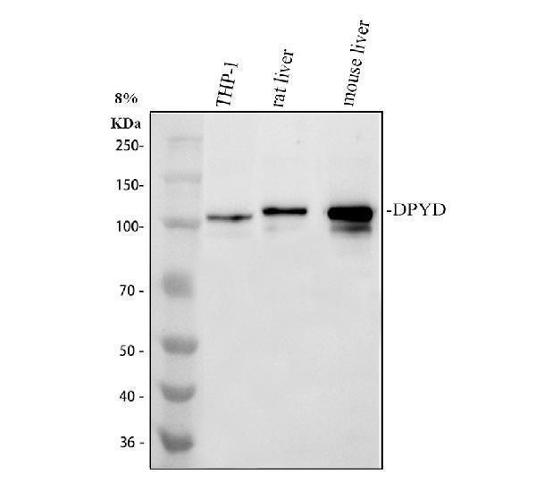
Western blot analysis of DPYD using anti-DPYD antibody (A01749).
Electrophoresis was performed on a 8% SDS-PAGE gel at 80V (Stacking gel) / 120V (Resolving gel) for 2 hours. The sample well of each lane was loaded with 30 ug of sample under reducing conditions.
Lane 1: human THP-1 whole cell lysates,
Lane 2: rat liver tissue lysates,
Lane 3: mouse liver tissue lysates.
After electrophoresis, proteins were transferred to a nitrocellulose membrane at 150 mA for 50-90 minutes. Blocked the membrane with 5% non-fat milk/TBS for 1.5 hour at RT. The membrane was incubated with rabbit anti-DPYD antigen affinity purified polyclonal antibody (A01749) at 0.5 μg/mL overnight at 4°C, then washed with TBS-0.1%Tween 3 times with 5 minutes each and probed with a goat anti-rabbit IgG-HRP secondary antibody (Catalog # BA1054) at a dilution of 1:5000 for 1.5 hour at RT. The signal is developed using an ECL Plus Western Blotting Substrate (Catalog # AR1196-200) with Tanon 5200 system. A specific band was detected for DPYD at approximately 111 kDa. The expected band size for DPYD is at 111 kDa.
Specific Publications For Anti-DPYD Antibody Picoband® (A01749)
Loading publications
Recommended Resources
Here are featured tools and databases that you might find useful.
- Boster's Pathways Library
- Protein Databases
- Bioscience Research Protocol Resources
- Data Processing & Analysis Software
- Photo Editing Software
- Scientific Literature Resources
- Research Paper Management Tools
- Molecular Biology Software
- Primer Design Tools
- Bioinformatics Tools
- Phylogenetic Tree Analysis
Customer Reviews
Have you used Anti-DPYD Antibody Picoband®?
Share your experimental results or join a short interview to earn up to $1,000 in product credits or other rewards.
0 Reviews For Anti-DPYD Antibody Picoband®
Customer Q&As
Have a question?
Find answers in Q&As, reviews.
Can't find your answer?
Submit your question
