Click image to see more details
Product Info Summary
| SKU: | PA1761 |
|---|---|
| Size: | 100 μg/vial |
| Reactive Species: | Human, Mouse, Rat |
| Host: | Rabbit |
| Application: | Flow Cytometry, WB |
Customers Who Bought This Also Bought

Product info
Product Name
Anti-DPYD Antibody Picoband®
SKU/Catalog Number
PA1761
BA3774-2 is an alternative SKU for this antibody, used in previous lots.
Size
100 μg/vial
Form
Lyophilized
Description
Boster Bio Anti-DPYD Antibody catalog # PA1761. Tested in Flow Cytometry, WB applications. This antibody reacts with Human, Mouse, Rat. The brand Picoband indicates this is a premium antibody that guarantees superior quality, high affinity, and strong signals with minimal background in Western blot applications. Only our best-performing antibodies are designated as Picoband, ensuring unmatched performance.
Storage & Handling
Store at -20˚C for one year from date of receipt. After reconstitution, at 4˚C for one month. It can also be aliquotted and stored frozen at -20˚C for six months. Avoid repeated freeze-thaw cycles.
Cite This Product
Anti-DPYD Antibody Picoband® (Boster Biological Technology, Pleasanton CA, USA, Catalog # PA1761)
Host
Rabbit
Contents
Each vial contains 4 mg Trehalose, 0.9 mg NaCl and 0.2 mg Na2HPO4.
Clonality
Polyclonal
Isotype
Rabbit IgG
Immunogen
A synthetic peptide corresponding to a sequence at the N-terminus of human DPYD, different from the related rat and mouse sequences by one amino acid.
Cross-reactivity
No cross-reactivity with other proteins
Reactive Species
PA1761 is reactive to DPYD in Human, Mouse, Rat
Observed Molecular Weight
111 kDa
Calculated molecular weight
111.4 kDa
Background of DPYD
DPYD (Dihydropyrimidine Dehydrogenase), also called DPD, is an enzyme that in humans is encoded by the DPYD gene. The protein encoded by this gene is a pyrimidine catabolic enzyme and the initial and rate-limiting factor in the pathway of uracil and thymidine catabolism. The structure of the DPYD gene contains 23 exons spanning about 950 kb. Using somatic cell hybrid strategies, the DPYD gene is mapped to the centromeric region of chromosome 1 between 1p22 and 1q21. By fluorescence in situ hybridization, the DPYD gene is mapped to 1p22. The highest level of DPD was found in monocytes followed by that in lymphocytes, granulocytes, and platelets, whereas no significant activity of DPD could be detected in erythrocytes. The activity of DPD in peripheral blood mononuclear cells was intermediate between that observed in monocytes and lymphocytes. By cDNA microarray, Western blot analysis, and luciferase reporter assay, the transcription factor LSF was identified as a positive regulator of DPYD.
Antibody Validation
Boster validates all antibodies on WB, IHC, ICC, Immunofluorescence, and ELISA with known positive control and negative samples to ensure specificity and high affinity, including thorough antibody incubations.
Application & Images
Applications
PA1761 is guaranteed for Flow Cytometry, WB Boster Guarantee
Recommend Dilution
| Application | Dilution | Species |
|---|---|---|
| Western blot | 0.1-0.5μg/ml | Human, Mouse, Rat |
| Flow Cytometry(Fixed) | 1-3 μg/1x106 cells | Human |
Tested application
Suggested blocking solution with 5% non-fat milk or BSA; (*)Recommended protein loading: 20-40 µg per lane
Validation Images & Assay Conditions
Click image to see more details
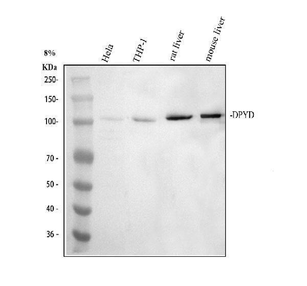
Western blot analysis of DPYD using anti-DPYD antibody (PA1761).
Electrophoresis was performed on a 8% SDS-PAGE gel at 80V (Stacking gel) / 120V (Resolving gel) for 2 hours. The sample well of each lane was loaded with 30 ug of sample under reducing conditions.
Lane 1: human Hela whole cell lysates,
Lane 2: human THP-1 whole cell lysates,
Lane 3: rat liver tissue lysates,
Lane 4: mouse liver tissue lysates.
After electrophoresis, proteins were transferred to a nitrocellulose membrane at 150 mA for 50-90 minutes. Blocked the membrane with 5% non-fat milk/TBS for 1.5 hour at RT. The membrane was incubated with rabbit anti-DPYD antigen affinity purified polyclonal antibody (PA1761) at 0.5 μg/mL overnight at 4°C, then washed with TBS-0.1%Tween 3 times with 5 minutes each and probed with a goat anti-rabbit IgG-HRP secondary antibody (Catalog # BA1054) at a dilution of 1:5000 for 1.5 hour at RT. The signal is developed using an ECL Plus Western Blotting Substrate (Catalog # AR1196-200) with Tanon 5200 system. A specific band was detected for DPYD at approximately 111 kDa. The expected band size for DPYD is at 111 kDa.

Click image to see more details
Flow Cytometry analysis of HepG2 cells using anti-DPYD antibody (PA1761).
Overlay histogram showing HepG2 cells stained with PA1761 (Blue line). To facilitate intracellular staining, cells were fixed with 4% paraformaldehyde and permeabilized with permeabilization buffer. The cells were blocked with 10% normal goat serum. And then incubated with rabbit anti-DPYD Antibody (PA1761, 1 μg/1x106 cells) for 30 min at 20°C. DyLight®488 conjugated goat anti-rabbit IgG (BA1127, 5-10 μg/1x106 cells) was used as secondary antibody for 30 minutes at 20°C. Isotype control antibody (Green line) was rabbit IgG (1 μg/1x106) used under the same conditions. Unlabelled sample without incubation with primary antibody and secondary antibody (Red line) was used as a blank control.
Specific Publications For Anti-DPYD Antibody Picoband® (PA1761)
Loading publications
Recommended Resources
Here are featured tools and databases that you might find useful.
- Boster's Pathways Library
- Protein Databases
- Bioscience Research Protocol Resources
- Data Processing & Analysis Software
- Photo Editing Software
- Scientific Literature Resources
- Research Paper Management Tools
- Molecular Biology Software
- Primer Design Tools
- Bioinformatics Tools
- Phylogenetic Tree Analysis
Customer Reviews
Have you used Anti-DPYD Antibody Picoband®?
Share your experimental results or join a short interview to earn up to $1,000 in product credits or other rewards.
0 Reviews For Anti-DPYD Antibody Picoband®
Customer Q&As
Have a question?
Find answers in Q&As, reviews.
Can't find your answer?
Submit your question
7 Customer Q&As for Anti-DPYD Antibody Picoband®
Question
Please see the WB image, lot number and protocol we used for cervix carcinoma using anti-DPYD antibody PA1761. Please let me know if you require anything else.
Verified Customer
Verified customer
Asked: 2019-12-27
Answer
Thank you very much for the data. Our lab team are working to resolve this as quickly as possible, and we appreciate your patience and understanding! You have provided everything we needed. Please let me know if there is anything you need in the meantime.
Boster Scientific Support
Answered: 2019-12-27
Question
Can you help my question with product PA1761, anti-DPYD antibody. I was wondering if it would be possible to conjugate this antibody with biotin. I would need it to be without BSA or sodium azide. I am planning on using a buffer exchange of sodium azide with PBS only. Would there be problems for me to conjugate the antibody and store it in -20 degrees in small aliquots?
Verified Customer
Verified customer
Asked: 2019-01-09
Answer
We do not advise storing this antibody with PBS buffer only in -20 degrees. If you want to store it in -20 degrees it is best to add some cryoprotectant like glycerol. If you want carrier free PA1761 anti-DPYD antibody, we can provide it to you in a special formula with trehalose and/or glycerol. These molecules will not interfere with conjugation chemistry and provide a good level of protection for the antibody from degradation. Please be sure to specify this in your purchase order.
Boster Scientific Support
Answered: 2019-01-09
Question
Will anti-DPYD antibody PA1761 work on primate WB with cervix carcinoma?
H. Dhar
Verified customer
Asked: 2018-06-27
Answer
Our lab technicians have not validated anti-DPYD antibody PA1761 on primate. You can run a BLAST between primate and the immunogen sequence of anti-DPYD antibody PA1761 to see if they may cross-react. If the sequence homology is close, then you can perform a pilot test. Keep in mind that since we have not validated primate samples, this use of the antibody is not covered by our guarantee. However we have an innovator award program that if you test this antibody and show it works in primate cervix carcinoma in WB, you can get your next antibody for free.
Boster Scientific Support
Answered: 2018-06-27
Question
Would PA1761 anti-DPYD antibody work on parafin embedded sections? If so, which fixation method do you recommend we use (PFA, paraformaldehyde, other)?
M. Huang
Verified customer
Asked: 2017-11-28
Answer
You can see on the product datasheet, PA1761 anti-DPYD antibody as been validated on WB. It is best to use PFA for fixation because it has better tissue penetration ability. PFA needs to be prepared fresh before use. Long term stored PFA turns into formalin, as the PFA molecules congregate and become formalin.
Boster Scientific Support
Answered: 2017-11-28
Question
We are currently using anti-DPYD antibody PA1761 for human tissue, and we are happy with the WB results. The species of reactivity given in the datasheet says human, mouse, rat. Is it true that the antibody can work on bovine tissues as well?
Verified Customer
Verified customer
Asked: 2017-09-19
Answer
The anti-DPYD antibody (PA1761) has not been validated for cross reactivity specifically with bovine tissues, but there is a good chance of cross reactivity. We have an innovator award program that if you test this antibody and show it works in bovine you can get your next antibody for free. Please contact me if I can help you with anything.
Boster Scientific Support
Answered: 2017-09-19
Question
Is this PA1761 anti-DPYD antibody reactive to the isotypes of DPYD?
P. Baker
Verified customer
Asked: 2017-04-07
Answer
The immunogen of PA1761 anti-DPYD antibody is A synthetic peptide corresponding to a sequence at the N-terminus of human DPYD(33-52aa AKKLDKKHWKRNPDKNCFNC), different from the related rat and mouse sequences by one amino acid. Could you tell me which isotype you are interested in so I can help see if the immunogen is part of this isotype?
Boster Scientific Support
Answered: 2017-04-07
Question
I appreciate helping with my inquiry over the phone. Here are the WB image, lot number and protocol we used for cervix carcinoma using anti-DPYD antibody PA1761. Let me know if you need anything else.
B. Mangal
Verified customer
Asked: 2016-05-09
Answer
We appreciate the data. You have provided everything we needed. Our lab team are working to resolve your inquiry as quickly as possible, and we appreciate your patience and understanding! Please let me know if there is anything you need in the meantime.
Boster Scientific Support
Answered: 2016-05-09
