Click image to see more details
-

-
-
-
-
+5
Product Info Summary
| SKU: | A00410 |
|---|---|
| Size: | 100 μg/vial |
| Reactive Species: | Human, Mouse, Rat |
| Host: | Rabbit |
| Application: | Flow Cytometry, IHC, WB |
Customers Who Bought This Also Bought

Product info
Product Name
Anti-DR5/TNFRSF10B Antibody Picoband®
SKU/Catalog Number
A00410
Size
100 μg/vial
Form
Lyophilized
Description
Boster Bio Anti-DR5/TNFRSF10B Antibody Picoband® catalog # A00410. Tested in Flow Cytometry, IHC, WB applications. This antibody reacts with Human, Mouse, Rat. The brand Picoband indicates this is a premium antibody that guarantees superior quality, high affinity, and strong signals with minimal background in Western blot applications. Only our best-performing antibodies are designated as Picoband, ensuring unmatched performance.
Storage & Handling
Store at -20˚C for one year from date of receipt. After reconstitution, at 4˚C for one month. It can also be aliquotted and stored frozen at -20˚C for six months. Avoid repeated freeze-thaw cycles.
Cite This Product
Anti-DR5/TNFRSF10B Antibody Picoband® (Boster Biological Technology, Pleasanton CA, USA, Catalog # A00410)
Host
Rabbit
Contents
Each vial contains antibody formulated with stabilizing components, 0.9 mg NaCl, 0.2 mg Na2HPO4, and 0.05 mg NaN3.
*This antibody is supplied in a stabilized formulation.
Compatibility with conjugation reactions depends on the chemistry of the conjugation method used.
For conjugation methods that are not compatible with the stabilizing components present in this formulation, a carrier-free antibody format is required.
Clonality
Polyclonal
Isotype
Rabbit IgG
Immunogen
E.coli-derived human DR5 recombinant protein (Position: K233-S440). Human DR5 shares 48.4% amino acid (aa) sequence identity with mouse DR5.
Cross-reactivity
No cross-reactivity with other proteins
Reactive Species
A00410 is reactive to TNFRSF10B in Human, Mouse, Rat
Observed Molecular Weight
38, 48 kDa
Calculated molecular weight
47.9 kDa
Background of TNFRSF10B
TNFRSF10B (Tumor necrosis factor receptor superfamily, member 10b) is a human gene. It is also known as DR5, CD262, KILLER, TRICK2, TRICKB, ZTNFR9, TRAILR2, TRICK2A, TRICK2B, TRAIL-R2, KILLER/DR5. The protein encoded by this gene is a member of the TNF-receptor superfamily, and contains an intracellular death domain. This receptor can be activated by tumor necrosis factor-related apoptosis inducing ligand (TNFSF10/TRAIL/APO-2L), and transduces apoptosis signal. Mice have a homologous gene, tnfrsf10b that has been essential in the elucidation of the function of this gene in humans. Studies with FADD-deficient mice suggested that FADD, a death domain containing adaptor protein, is required for the apoptosis mediated by this protein.By analysis of radiation hybrid panels, this gene is mapped to chromosome 8p22-p21. Northern blot analysis indicated that TRAILR2 was expressed as a 4.4-kb mRNA in all tissues tested, with the highest levels of expression in peripheral blood lymphocytes, spleen, and ovary.
Antibody Validation
Boster validates all antibodies on WB, IHC, ICC, Immunofluorescence, and ELISA with known positive control and negative samples to ensure specificity and high affinity, including thorough antibody incubations.
Application & Images
Applications
A00410 is guaranteed for Flow Cytometry, IHC, WB Boster Guarantee
Recommend Dilution
| Application | Dilution | Species |
|---|---|---|
| Western blot | 0.1-0.5μg/ml | Human, Mouse, Rat |
| Immunohistochemistry (Paraffin-embedded Section) | 0.5-1μg/ml | Human, Mouse, Rat |
| Flow Cytometry (Fixed) | 1-3μg/1x106 cells | Human |
Tested application
Suggested blocking solution with 5% non-fat milk or BSA; (*)Recommended protein loading: 20-40 µg per lane
Use TE buffer pH 9.0 for antigen retrieval; (*) citrate buffer pH 6.0 is an alternative.
Validation Images & Assay Conditions
Click image to see more details
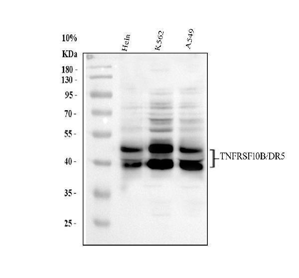
Western blot analysis of DR5 using anti-DR5 antibody (A00410).
Electrophoresis was performed on a 10% SDS-PAGE gel at 80V (Stacking gel) / 120V (Resolving gel) for 2 hours. The sample well of each lane was loaded with 30 ug of sample under reducing conditions.
Lane 1: human Hela whole cell lysates,
Lane 2: human K562 whole cell lysates,
Lane 3: human A549 whole cell lysates.
After electrophoresis, proteins were transferred to a nitrocellulose membrane at 150 mA for 50-90 minutes. Blocked the membrane with 5% non-fat milk/TBS for 1.5 hour at RT. The membrane was incubated with rabbit anti-DR5 antigen affinity purified polyclonal antibody (A00410) at 0.5 μg/mL overnight at 4°C, then washed with TBS-0.1%Tween 3 times with 5 minutes each and probed with a goat anti-rabbit IgG-HRP secondary antibody (Catalog # BA1054) at a dilution of 1:5000 for 1.5 hour at RT. The signal is developed using an ECL Plus Western Blotting Substrate (Catalog # AR1196-200) with Tanon 5200 system. A specific band was detected for DR5 at approximately 38, 48 kDa. The expected band size for DR5 is at 48 kDa.

Click image to see more details
IHC analysis of DR5 using anti-DR5 antibody (A00410).
DR5 was detected in a paraffin-embedded section of mouse intestine tissue. Heat mediated antigen retrieval was performed in EDTA buffer (pH 8.0, epitope retrieval solution). The tissue section was blocked with 10% goat serum. The tissue section was then incubated with 1 μg/ml rabbit anti-DR5 Antibody (A00410) overnight at 4°C. Biotinylated goat anti-rabbit IgG was used as secondary antibody and incubated for 30 minutes at 37°C. The tissue section was developed using Strepavidin-Biotin-Complex (SABC) (Catalog # SA1022) with DAB as the chromogen.

Click image to see more details
The cytotoxic effect of TRAIL on human HCC and GC cell lines. (A) Detection of DNA fragmentation by DNA ladder assay. LH86, Huh7, HLCZ01 and HLCZ02 cells were exposed to TRAIL for 4 hours. 2 μg of cellular DNA was separated on 1% agarose gel at 50V for one hour. The data are one representative of three independent experiments. (B) Detection of apoptosis by flow cytometry and western blot. LH86, Huh7, HLCZ01, HLCZ02, HGC-27 and BGC-823 cells were exposed to TRAIL for 4 hours. Samples were analyzed on a FACS Caliber Cytometer. A minimum of 30000 events per samples were acquired, and subsequently analyzed with CellQuest software. The results are the average of at least three independent experiments. (C) Cleaved PARP was detected by western blot. β–actin was used as control. The data are one representative of three independent experiments. (D) Detection of DR4 and DR5 in HCC and GC cell lines by real-time PCR and western blot analysis. DR4 or DR5 mRNA were detected by real-time RT-PCR and normalized with GAPDH respectively. The results are the average of three independent experiments performed in triplicate. DR4 and DR5 protein was detected by western blot. The data are one representative of three independent experiments.
Index in PubMed under a CC BY license. PMID: 24970806

Click image to see more details
IHC analysis of DR5 using anti-DR5 antibody (A00410).
DR5 was detected in a paraffin-embedded section of rat kidney tissue. Heat mediated antigen retrieval was performed in EDTA buffer (pH 8.0, epitope retrieval solution). The tissue section was blocked with 10% goat serum. The tissue section was then incubated with 1 μg/ml rabbit anti-DR5 Antibody (A00410) overnight at 4°C. Biotinylated goat anti-rabbit IgG was used as secondary antibody and incubated for 30 minutes at 37°C. The tissue section was developed using Strepavidin-Biotin-Complex (SABC) (Catalog # SA1022) with DAB as the chromogen.

Click image to see more details
IHC analysis of DR5 using anti-DR5 antibody (A00410).
DR5 was detected in a paraffin-embedded section of human intetsinal cancer tissue. Heat mediated antigen retrieval was performed in EDTA buffer (pH 8.0, epitope retrieval solution). The tissue section was blocked with 10% goat serum. The tissue section was then incubated with 1 μg/ml rabbit anti-DR5 Antibody (A00410) overnight at 4°C. Biotinylated goat anti-rabbit IgG was used as secondary antibody and incubated for 30 minutes at 37°C. The tissue section was developed using Strepavidin-Biotin-Complex (SABC) (Catalog # SA1022) with DAB as the chromogen.

Click image to see more details
Flow Cytometry analysis of A549 cells using anti-DR5 antibody (A00410).
Overlay histogram showing A549 cells stained with A00410 (Blue line). To facilitate intracellular staining, cells were fixed with 4% paraformaldehyde and permeabilized with permeabilization buffer. The cells were blocked with 10% normal goat serum. And then incubated with rabbit anti-DR5 Antibody (A00410,1μg/1x106 cells) for 30 min at 20°C. DyLight®488 conjugated goat anti-rabbit IgG (BA1127, 5-10μg/1x106 cells) was used as secondary antibody for 30 minutes at 20°C. Isotype control antibody (Green line) was rabbit IgG (1μg/1x106) used under the same conditions. Unlabelled sample (Red line) was also used as a control.

Click image to see more details
Flow Cytometry analysis of U87 cells using anti-DR5 antibody (A00410).
Overlay histogram showing U87 cells stained with A00410 (Blue line). To facilitate intracellular staining, cells were fixed with 4% paraformaldehyde and permeabilized with permeabilization buffer. The cells were blocked with 10% normal goat serum. And then incubated with rabbit anti-DR5 Antibody (A00410,1μg/1x106 cells) for 30 min at 20°C. DyLight®488 conjugated goat anti-rabbit IgG (BA1127, 5-10μg/1x106 cells) was used as secondary antibody for 30 minutes at 20°C. Isotype control antibody (Green line) was rabbit IgG (1μg/1x106) used under the same conditions. Unlabelled sample (Red line) was also used as a control.

Click image to see more details
Flow Cytometry analysis of THP-1 cells using anti-DR5 antibody (A00410). Overlay histogram showing THP-1 cells stained with A00410 (Blue line). To facilitate intracellular staining, cells were fixed with 4% paraformaldehyde and permeabilized with permeabilization buffer. The cells were blocked with 10% normal goat serum. And then incubated with rabbit anti-DR5 Antibody (A00410,1μg/1x106 cells) for 30 min at 20°C. FITC conjugated goat anti-rabbit IgG (BA1105, 5-10μg/1x106 cells) was used as secondary antibody for 30 minutes at 20°C. Isotype control antibody (Green line) was rabbit IgG (1μg/1x106) used under the same conditions. Unlabelled sample without incubation with primary antibody and secondary antibody (Red line) was used as a blank control.

Click image to see more details
Flow Cytometry analysis of PC-3 cells using anti-DR5 antibody (A00410). Overlay histogram showing PC-3 cells stained with A00410 (Blue line). To facilitate intracellular staining, cells were fixed with 4% paraformaldehyde and permeabilized with permeabilization buffer. The cells were blocked with 10% normal goat serum. And then incubated with rabbit anti-DR5 Antibody (A00410,1μg/1x106 cells) for 30 min at 20°C. FITC conjugated goat anti-rabbit IgG (BA1105, 5-10μg/1x106 cells) was used as secondary antibody for 30 minutes at 20°C. Isotype control antibody (Green line) was rabbit IgG (1μg/1x106) used under the same conditions. Unlabelled sample without incubation with primary antibody and secondary antibody (Red line) was used as a blank control.
Specific Publications For Anti-DR5/TNFRSF10B Antibody Picoband® (A00410)
Loading publications
Recommended Resources
Here are featured tools and databases that you might find useful.
- Boster's Pathways Library
- Protein Databases
- Bioscience Research Protocol Resources
- Data Processing & Analysis Software
- Photo Editing Software
- Scientific Literature Resources
- Research Paper Management Tools
- Molecular Biology Software
- Primer Design Tools
- Bioinformatics Tools
- Phylogenetic Tree Analysis
Customer Reviews
Have you used Anti-DR5/TNFRSF10B Antibody Picoband®?
Share your experimental results or join a short interview to earn up to $1,000 in product credits or other rewards.
0 Reviews For Anti-DR5/TNFRSF10B Antibody Picoband®
Customer Q&As
Have a question?
Find answers in Q&As, reviews.
Can't find your answer?
Submit your question
