Click image to see more details
-

-
-
-
-
+11
Product Info Summary
| SKU: | A00410-2 |
|---|---|
| Size: | 0.1 mg |
| Reactive Species: | Human, Mouse, Rat |
| Host: | Rabbit |
| Application: | ELISA, IF, IHC-P, WB |
Customers Who Bought This Also Bought

Product info
Product Name
Anti-DR5 TNFRSF10B Antibody
SKU/Catalog Number
A00410-2
Size
0.1 mg
Form
Liquid
Description
Boster Bio Anti-DR5 TNFRSF10B Antibody (Catalog # A00410-2). Tested in ELISA, WB, IHC-P, IF applications. This antibody reacts with Human, Mouse, Rat.
Storage & Handling
DR5 antibody can be stored at 4°C up to one year. Antibodies should not be exposed to prolonged high temperatures.
Cite This Product
Anti-DR5 TNFRSF10B Antibody (Boster Biological Technology, Pleasanton CA, USA, Catalog # A00410-2)
Host
Rabbit
Contents
DR5 Antibody is supplied in PBS containing 0.02% sodium azide.
Clonality
Polyclonal
Isotype
IgG
Immunogen
DR5 antibody was raised against a peptide corresponding to 20 amino acids near the carboxy terminus of human DR5 precursor. The immunogen is located within the last 50 amino acids of DR5.
Cross-reactivity
Antibody has no cross reaction to DR4.
Reactive Species
A00410-2 is reactive to TNFRSF10B in Human, Mouse, Rat
Observed Molecular Weight
68 kDa
Calculated molecular weight
47.9 kDa
Background of TNFRSF10B
Apoptosis is induced by certain cytokines including TNF and Fas ligand in the TNF family through their death domain containing receptors. TRAIL/Apo2L is a new member of the TNF family. DR4 was recently identified as the receptor for TRAIL. A novel death domain containing receptor for TRAIL was more recently identified and designated DR5, Apo2, TRAIL-R2, TRICK2, or KILLER by several groups independently. Like DR4, DR5 transcript is widely expressed in normal tissues and in many types of tumor cells. DR5 binds to TRAIL and mediates TRAIL induced cell death. Overexpression of DR5 induces apoptosis and activates NF-κB.
Antibody Validation
Boster validates all antibodies on WB, IHC, ICC, Immunofluorescence, and ELISA with known positive control and negative samples to ensure specificity and high affinity, including thorough antibody incubations.
Application & Images
Applications
A00410-2 is guaranteed for ELISA, IF, IHC-P, WB Boster Guarantee
Recommend Dilution
| Application | Dilution | Species |
|---|---|---|
| Antibody validated: Western Blot in human | mouse and rat samples; Immunohistochemistry in mouse samples; Immunocytochemistry in human samples and Immunofluorescence in human | mouse and rat samples. All other applications and species not yet tested. Optimal dilutions for each application should be determined by the researcher. |
Validation Images & Assay Conditions
Click image to see more details
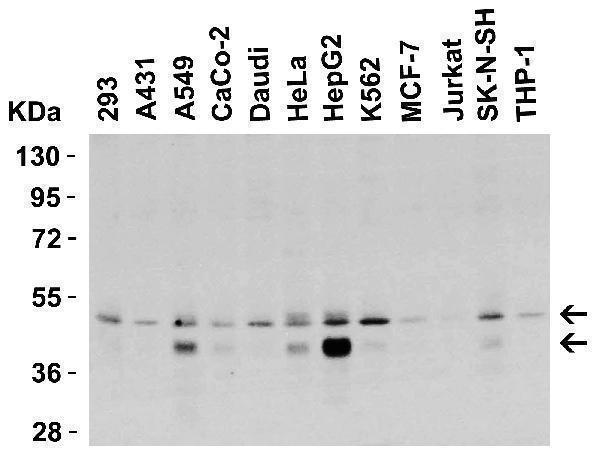
Western Blot Validation in Human Cell Lines
Loading: 15 μg of lysates per lane.
Antibodies: DR5 A00410-2, (0.5 μg/mL), 1h incubation at RT in 5% NFDM/TBST.
Secondary: Goat anti-rabbit IgG HRP conjugate at 1:10000 dilution.

Click image to see more details
Western Blot Validation in Human HepG2 Cells
Loading: 15 μg of lysates per lane.
Antibodies: DR5 A00410-2, 1h incubation at RT in 5% NFDM/TBST.
Secondary: Goat anti-rabbit IgG HRP conjugate at 1:10000 dilution.
Lane 1: 1 μg/mL
Lane 2: 2 μg/mL
Lane 3: 4 μg/mL

Click image to see more details
Western Blot Validation in Mouse and Rat Cell Lines
Loading: 15 μg of lysates per lane.
Antibodies: DR5 A00410-2, (2 μg/mL), 1h incubation at RT in 5% NFDM/TBST.
Secondary: Goat anti-rabbit IgG HRP

Click image to see more details
Western Blot Validation in Mouse Cell Lines
Loading: 15 μg of lysates per lane.
Antibodies: DR5 A00410-2, (1 μg/mL), 1h incubation at RT in 5% NFDM/TBST.
Secondary: Goat anti-rabbit IgG HRP conjugate at 1:10000 dilution.

Click image to see more details
Western Blot Validation in Mouse Heart
Loading: 15 μg of lysatesper lane.
Antibodies: DR5 A00410-2, (1 μg/mL), 1h incubation at RT in 5% NFDM/TBST.
Secondary: Goat anti-rabbit IgG HRP conjugate at 1:10000 dilution.

Click image to see more details
Western Blot Validation in Rat Skeletal Muscle
Loading: 15 μg of lysate per lane.
Antibodies: DR5 A00410-2, (1 μg/mL), 1h incubation at RT in 5% NFDM/TBST.
Secondary: Goat anti-rabbit IgG HRP conjugate at 1:10000 dilution.

Click image to see more details
Immunofluorescence Validation of DR5 in Human HepG2 Cells
Immunofluorescent analysis of 4% paraformaldehyde-fixed human HepG2 cells labeling DR5 with A00410-2 at 5 μg/mL, followed by goat anti-rabbit IgG secondary antibody at 1/500 dilution (green) and DAPI (blue).

Click image to see more details
Immunofluorescence Validation of DR5 in Human Testis
Immunofluorescent analysis of 4% paraformaldehyde-fixed human testis tissue labeling DR5 with A00410-2 at 10 μg/mL, followed by goat anti-rabbit IgG secondary antibody at 1/500 dilution (green) and DAPI (blue).

Click image to see more details
Immunofluorescence Validation of DR5 in Mouse Pancreas
Immunofluorescent analysis of 4% paraformaldehyde-fixed mouse pancreas tissue labeling DR5 with A00410-2 at 10 μg/mL, followed by goat anti-rabbit IgG secondary antibody at 1/500 dilution (green) and DAPI (blue).

Click image to see more details
Immunofluorescence Validation of DR5 in Rat Brain
Immunofluorescent analysis of 4% paraformaldehyde-fixed rat brain tissue labeling DR5 with A00410-2 at 5 μg/mL, followed by goat anti-rabbit IgG secondary antibody at 1/500 dilution (green) and DAPI (blue).

Click image to see more details
KO Validation of DR5 in HCT116 Cells (Han et al., 2015)
Anti-cancer drug, Carfilzomib (CFZ), induced up-regulation of DR5 and the expression of DR5 was not detected in DR5-KO HCT 116 cell line with anti-DR5 antibodies (A00410-2).

Click image to see more details
KD Validation of DR5 in MB231 Cells (Rahman et al., 2009)
Western blot analysis with anti-DR5 antibodies was performed for DR5 in MB231 cells transfected with control siRNA or DR5 siRNA. DR5 expression was disrupted after DR5 siRNA knockdown.

Click image to see more details
Immunohistochemistry Validation of DR5 in Mouse kidney tissue
Immunohistochemical analysis of paraffin-embedded mouse kidney tissue using anti-DR5 antibody (A00410-2) at 5μg/ml. Tissue was fixed with formaldehyde and blocked with 10% serum for 1 h at RT; antigen retrieval was by heat mediation with a citrate buffer (pH6). Samples were incubated with primary antibody overnight at 4˚C. A goat anti-rabbit IgG H&L (HRP) at 1/250 was used as secondary. Counter stained with Hematoxylin.

Click image to see more details
Immunohistochemistry Validation of BIM in Human Colon Tumors (Devetzi et al., 2016)
Protein analysis for DR5 by immunohistochemistry with anti-DR5 antibodies in human colon tumors. Strong immunoreactivity is shown for DR5 in T167 patient with colorectal cancer.

Click image to see more details
Regulated Expression Validation of DR5 in Thyroid Epithelial Cells (Bretz et al., 2002)
Immunostaining with anti-DR5 antibodies shows high levels of DR5 expression in untreated cells and cells treated with each of the three cytokines alone or TNFalpha combined with IL-1b. In contrast, treatment with both IFNg and TNFalpha or all three cytokines greatly reduces DR5 staining. The reduction in staining appears most significant in cytoplasmic regions while some staining is maintained in or around the nucleus.
Specific Publications For Anti-DR5 TNFRSF10B Antibody (A00410-2)
Loading publications
Recommended Resources
Here are featured tools and databases that you might find useful.
- Boster's Pathways Library
- Protein Databases
- Bioscience Research Protocol Resources
- Data Processing & Analysis Software
- Photo Editing Software
- Scientific Literature Resources
- Research Paper Management Tools
- Molecular Biology Software
- Primer Design Tools
- Bioinformatics Tools
- Phylogenetic Tree Analysis
Customer Reviews
Have you used Anti-DR5 TNFRSF10B Antibody?
Share your experimental results or join a short interview to earn up to $1,000 in product credits or other rewards.
0 Reviews For Anti-DR5 TNFRSF10B Antibody
Customer Q&As
Have a question?
Find answers in Q&As, reviews.
Can't find your answer?
Submit your question
