Click image to see more details
Product Info Summary
| SKU: | A00410-3 |
|---|---|
| Size: | 100 μg/vial |
| Reactive Species: | Human |
| Host: | Rabbit |
| Application: | ELISA, WB |
Customers Who Bought This Also Bought

Product info
Product Name
Anti-DR5/TNFRSF10B Antibody Picoband®
SKU/Catalog Number
A00410-3
Size
100 μg/vial
Form
Lyophilized
Description
Boster Bio Anti-DR5/TNFRSF10B Antibody Picoband® catalog # A00410-3. Tested in ELISA, WB applications. This antibody reacts with Human. The brand Picoband indicates this is a premium antibody that guarantees superior quality, high affinity, and strong signals with minimal background in Western blot applications. Only our best-performing antibodies are designated as Picoband, ensuring unmatched performance.
Storage & Handling
Store at -20˚C for one year from date of receipt. After reconstitution, at 4˚C for one month. It can also be aliquotted and stored frozen at -20˚C for six months. Avoid repeated freeze-thaw cycles.
Cite This Product
Anti-DR5/TNFRSF10B Antibody Picoband® (Boster Biological Technology, Pleasanton CA, USA, Catalog # A00410-3)
Host
Rabbit
Contents
Each vial contains 4mg Trehalose, 0.9mg NaCl, 0.2mg Na2HPO4, 0.01mg NaN3.
Clonality
Polyclonal
Isotype
Rabbit IgG
Immunogen
E.coli-derived human DR5/TNFRSF10B recombinant protein (Position: Q70-K427).
Cross-reactivity
No cross-reactivity with other proteins
Reactive Species
A00410-3 is reactive to TNFRSF10B in Human
Observed Molecular Weight
43 kDa, 48 kDa
Calculated molecular weight
47.9 kDa
Background of TNFRSF10B
TNFRSF10B (Tumor necrosis factor receptor superfamily, member 10b) is a human gene. It is also known as DR5, CD262, KILLER, TRICK2, TRICKB, ZTNFR9, TRAILR2, TRICK2A, TRICK2B, TRAIL-R2, KILLER/DR5. The protein encoded by this gene is a member of the TNF-receptor superfamily, and contains an intracellular death domain. This receptor can be activated by tumor necrosis factor-related apoptosis inducing ligand (TNFSF10/TRAIL/APO-2L), and transduces apoptosis signal. Mice have a homologous gene, tnfrsf10b that has been essential in the elucidation of the function of this gene in humans. Studies with FADD-deficient mice suggested that FADD, a death domain containing adaptor protein, is required for the apoptosis mediated by this protein.By analysis of radiation hybrid panels, this gene is mapped to chromosome 8p22-p21. Northern blot analysis indicated that TRAILR2 was expressed as a 4.4-kb mRNA in all tissues tested, with the highest levels of expression in peripheral blood lymphocytes, spleen, and ovary.
Antibody Validation
Boster validates all antibodies on WB, IHC, ICC, Immunofluorescence, and ELISA with known positive control and negative samples to ensure specificity and high affinity, including thorough antibody incubations.
Application & Images
Applications
A00410-3 is guaranteed for ELISA, WB Boster Guarantee
Recommend Dilution
| Application | Dilution | Species |
|---|---|---|
| Western blot | 0.25-0.5μg/ml | Human |
| ELISA | 0.1-0.5μg/ml | - |
Tested application
Suggested blocking solution with 5% non-fat milk or BSA; (*)Recommended protein loading: 20-40 µg per lane
Validation Images & Assay Conditions
Click image to see more details
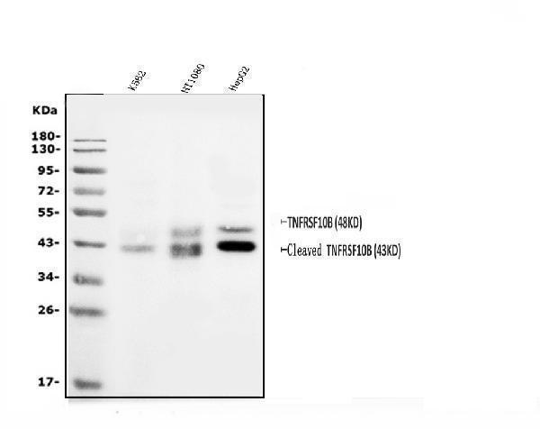
Western blot analysis of DR5/TNFRSF10B using anti-DR5/TNFRSF10B antibody (A00410-3).
Electrophoresis was performed on a 5-20% SDS-PAGE gel at 70V (Stacking gel) / 90V (Resolving gel) for 2-3 hours. The sample well of each lane was loaded with 50ug of sample under reducing conditions.
Lane 1: human K562 whole cell lysates,
Lane 2: human HT1080 whole cell lysates,
Lane 3: human HepG2 whole cell lysates.
After Electrophoresis, proteins were transferred to a Nitrocellulose membrane at 150mA for 50-90 minutes. Blocked the membrane with 5% Non-fat Milk/ TBS for 1.5 hour at RT. The membrane was incubated with rabbit anti-DR5/TNFRSF10B antigen affinity purified polyclonal antibody (Catalog # A00410-3) at 0.5 μg/mL overnight at 4°C, then washed with TBS-0.1%Tween 3 times with 5 minutes each and probed with a goat anti-rabbit IgG-HRP secondary antibody at a dilution of 1:5000 for 1.5 hour at RT. The signal is developed using an Enhanced Chemiluminescent detection (ECL) kit (Catalog # EK1002) with Tanon 5200 system. A specific band was detected for DR5/TNFRSF10B at approximately 43KD,48KD. The expected band size for DR5/TNFRSF10B is at 48KD.
Specific Publications For Anti-DR5/TNFRSF10B Antibody Picoband® (A00410-3)
Loading publications
Recommended Resources
Here are featured tools and databases that you might find useful.
- Boster's Pathways Library
- Protein Databases
- Bioscience Research Protocol Resources
- Data Processing & Analysis Software
- Photo Editing Software
- Scientific Literature Resources
- Research Paper Management Tools
- Molecular Biology Software
- Primer Design Tools
- Bioinformatics Tools
- Phylogenetic Tree Analysis
Customer Reviews
Have you used Anti-DR5/TNFRSF10B Antibody Picoband®?
Share your experimental results or join a short interview to earn up to $1,000 in product credits or other rewards.
0 Reviews For Anti-DR5/TNFRSF10B Antibody Picoband®
Customer Q&As
Have a question?
Find answers in Q&As, reviews.
Can't find your answer?
Submit your question
