Click image to see more details
Product Info Summary
| SKU: | A04265-2 |
|---|---|
| Size: | 100 μg/vial |
| Reactive Species: | Human |
| Host: | Rabbit |
| Application: | IF, ICC, WB |
Customers Who Bought This Also Bought

Product info
Product Name
Anti-EHD2 Antibody Picoband®
SKU/Catalog Number
A04265-2
Size
100 μg/vial
Form
Lyophilized
Description
Boster Bio Anti-EHD2 Antibody Picoband® catalog # A04265-2. Tested in WB applications. This antibody reacts with Human. The brand Picoband indicates this is a premium antibody that guarantees superior quality, high affinity, and strong signals with minimal background in Western blot applications. Only our best-performing antibodies are designated as Picoband, ensuring unmatched performance.
Storage & Handling
Store at -20˚C for one year from date of receipt. After reconstitution, at 4˚C for one month. It can also be aliquotted and stored frozen at -20˚C for six months. Avoid repeated freeze-thaw cycles.
Cite This Product
Anti-EHD2 Antibody Picoband® (Boster Biological Technology, Pleasanton CA, USA, Catalog # A04265-2)
Host
Rabbit
Contents
Each vial contains 4mg Trehalose, 0.9mg NaCl, 0.2mg Na2HPO4.
Clonality
Polyclonal
Isotype
Rabbit IgG
Immunogen
A synthetic peptide corresponding to a sequence in the middle region of human EHD2, which shares 100% and 95% amino acid (aa) sequence identity with mouse and rat EHD2, respectively.
Cross-reactivity
No cross-reactivity with other proteins.
Reactive Species
A04265-2 is reactive to EHD2 in Human
Observed Molecular Weight
61 kDa
Calculated molecular weight
61.2 kDa
Background of EHD2
EH-domain containing 2, also known as EHD2, is a human gene[5] belonging to the EHD protein family. This gene encodes a member of the EH domain-containing protein family. These proteins are characterized by a C-terminal EF-hand domain, a nucleotide-binding consensus site at the N terminus and a bipartite nuclear localization signal. The encoded protein interacts with the actin cytoskeleton through an N-terminal domain and also binds to an EH domain-binding protein through the C-terminal EH domain. This interaction appears to connect clathrin-dependent endocytosis to actin, suggesting that this gene product participates in the endocytic pathway.
Antibody Validation
Boster validates all antibodies on WB, IHC, ICC, Immunofluorescence, and ELISA with known positive control and negative samples to ensure specificity and high affinity, including thorough antibody incubations.
Application & Images
Applications
A04265-2 is guaranteed for IF, ICC, WB Boster Guarantee
Recommend Dilution
| Application | Dilution | Species |
|---|---|---|
| Western blot | 0.25-0.5μg/ml | Human |
| Immunocytochemistry/Immunofluorescence | 5μg/ml | Human |
Tested application
Suggested blocking solution with 5% non-fat milk or BSA; (*)Recommended protein loading: 20-40 µg per lane
Validation Images & Assay Conditions
Click image to see more details
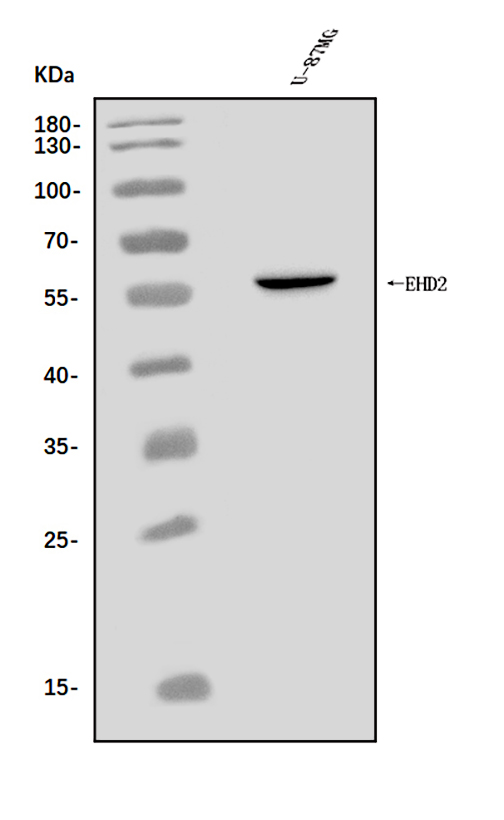
Western blot analysis of EHD2 using anti-EHD2 antibody (A04265-2).
Electrophoresis was performed on a 5-20% SDS-PAGE gel at 70V (Stacking gel) / 90V (Resolving gel) for 2-3 hours. The sample well of each lane was loaded with 30 ug of sample under reducing conditions.
Lane 1: human U-87MG whole cell lysates.
After electrophoresis, proteins were transferred to a nitrocellulose membrane at 150 mA for 50-90 minutes. Blocked the membrane with 5% non-fat milk/TBS for 1.5 hour at RT. The membrane was incubated with rabbit anti-EHD2 antigen affinity purified polyclonal antibody (Catalog # A04265-2) at 0.5 μg/mL overnight at 4°C, then washed with TBS-0.1%Tween 3 times with 5 minutes each and probed with a goat anti-rabbit IgG-HRP secondary antibody at a dilution of 1:5000 for 1.5 hour at RT. The signal is developed using an Enhanced Chemiluminescent detection (ECL) kit (Catalog # EK1002) with Tanon 5200 system. A specific band was detected for EHD2 at approximately 61 kDa. The expected band size for EHD2 is at 51 kDa.

Click image to see more details
IF analysis of EHD2 using anti-EHD2 antibody (A04265-2).
EHD2 was detected in an immunocytochemical section of A549 cells. Enzyme antigen retrieval was performed using IHC enzyme antigen retrieval reagent (AR0022) for 15 mins. The cells were blocked with 10% goat serum. And then incubated with 5 μg/mL rabbit anti-EHD2 Antibody (A04265-2) overnight at 4°C. DyLight®594 Conjugated Goat Anti-Rabbit IgG (BA1142) was used as secondary antibody at 1:100 dilution and incubated for 30 minutes at 37°C. The section was counterstained with DAPI. Visualize using a fluorescence microscope and filter sets appropriate for the label used.
Specific Publications For Anti-EHD2 Antibody Picoband® (A04265-2)
Loading publications
Recommended Resources
Here are featured tools and databases that you might find useful.
- Boster's Pathways Library
- Protein Databases
- Bioscience Research Protocol Resources
- Data Processing & Analysis Software
- Photo Editing Software
- Scientific Literature Resources
- Research Paper Management Tools
- Molecular Biology Software
- Primer Design Tools
- Bioinformatics Tools
- Phylogenetic Tree Analysis
Customer Reviews
Have you used Anti-EHD2 Antibody Picoband®?
Share your experimental results or join a short interview to earn up to $1,000 in product credits or other rewards.
0 Reviews For Anti-EHD2 Antibody Picoband®
Customer Q&As
Have a question?
Find answers in Q&As, reviews.
Can't find your answer?
Submit your question
