Click image to see more details
-

-
-
-
-
+6
Product Info Summary
| SKU: | A00578 |
|---|---|
| Size: | 100 μg/vial |
| Reactive Species: | Human, Mouse, Rat |
| Host: | Rabbit |
| Application: | ELISA, Flow Cytometry, IF, ICC, WB |
Customers Who Bought This Also Bought

Product info
Product Name
Anti-Eph receptor A2/EPHA2 Antibody Picoband®
SKU/Catalog Number
A00578
Size
100 μg/vial
Form
Lyophilized
Description
Boster Bio Anti-Eph receptor A2/EPHA2 Antibody Picoband® catalog # A00578. Tested in ELISA, Flow Cytometry, IF, ICC, WB applications. This antibody reacts with Human, Mouse, Rat. The brand Picoband indicates this is a premium antibody that guarantees superior quality, high affinity, and strong signals with minimal background in Western blot applications. Only our best-performing antibodies are designated as Picoband, ensuring unmatched performance.
Storage & Handling
Store at -20˚C for one year from date of receipt. After reconstitution, at 4˚C for one month. It can also be aliquotted and stored frozen at -20˚C for six months. Avoid repeated freeze-thaw cycles.
Cite This Product
Anti-Eph receptor A2/EPHA2 Antibody Picoband® (Boster Biological Technology, Pleasanton CA, USA, Catalog # A00578)
Host
Rabbit
Contents
Each vial contains 4mg Trehalose, 0.9mg NaCl, 0.2mg Na2HPO4, 0.05mg NaN3.
Clonality
Polyclonal
Isotype
Rabbit IgG
Immunogen
E. coli-derived human Eph receptor A2 recombinant protein (Position: M851-N970).
Cross-reactivity
No cross-reactivity with other proteins.
Reactive Species
A00578 is reactive to EPHA2 in Human, Mouse, Rat
Observed Molecular Weight
125 kDa
Calculated molecular weight
108.3 kDa
Background of EPHA2
EPHA2 (ephrin type-A receptor 2) also known as ECK, is a protein that in humans is encoded by the EPHA2 gene. This gene belongs to the ephrin receptor subfamily of the protein-tyrosine kinase family. Receptors in the EPH subfamily typically have a single kinase domain and an extracellular region containing a Cys-rich domain and 2 fibronectin type III repeats. By somatic cell hybrid analysis and fluorescence in situ hybridization, the EPHA2 gene is mapped to chromosome 1p36.1. EPHA2 was readily detectable in human lens fiber cells using immunoblot and immunohistochemistry. EGFR and EPHA2 mediated HCV entry by regulating CD81 -claudin-1 (CLDN1) coreceptor associations and viral glycoprotein-dependent membrane fusion.
Antibody Validation
Boster validates all antibodies on WB, IHC, ICC, Immunofluorescence, and ELISA with known positive control and negative samples to ensure specificity and high affinity, including thorough antibody incubations.
Application & Images
Applications
A00578 is guaranteed for ELISA, Flow Cytometry, IF, ICC, WB Boster Guarantee
Recommend Dilution
| Application | Dilution | Species |
|---|---|---|
| Western blot | 0.1-0.5μg/ml | |
| Immunocytochemistry/Immunofluorescence | 5μg/ml | |
| Flow Cytometry (Fixed) | 1-3μg/1x106 cells | |
| ELISA | 0.1-0.5μg/ml |
Tested application
Suggested blocking solution with 5% non-fat milk or BSA; (*)Recommended protein loading: 20-40 µg per lane
Validation Images & Assay Conditions
Click image to see more details
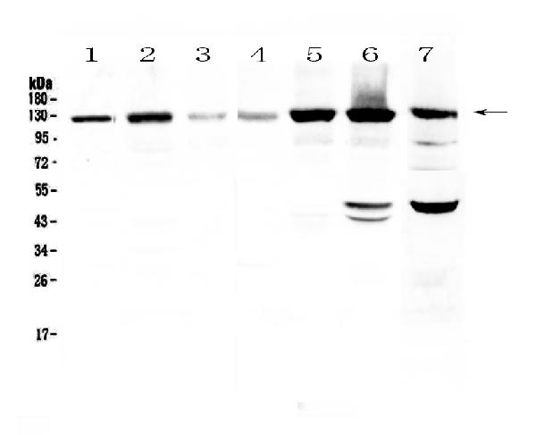
Western blot analysis of Eph receptor A2 using anti-Eph receptor A2 antibody (A00578).
Electrophoresis was performed on a 5-20% SDS-PAGE gel at 70V (Stacking gel) / 90V (Resolving gel) for 2-3 hours. The sample well of each lane was loaded with 50ug of sample under reducing conditions.
Lane 1: human Hela cell lysate,
Lane 2: human U-87MG cell lysate,
Lane 3: human SHG-44 cell lysate,
Lane 4: human COLO-320 cell lysate,
Lane 5: human SK-OV-3 cell lysate,
Lane 6: human A549 cell lysate,
Lane 7: mouse HEPA1-6 cell lysate.
After Electrophoresis, proteins were transferred to a Nitrocellulose membrane at 150mA for 50-90 minutes. Blocked the membrane with 5% Non-fat Milk/ TBS for 1.5 hour at RT. The membrane was incubated with rabbit anti-Eph receptor A2 antigen affinity purified polyclonal antibody (Catalog # A00578) at 0.5 μg/mL overnight at 4°C, then washed with TBS-0.1%Tween 3 times with 5 minutes each and probed with a goat anti-rabbit IgG-HRP secondary antibody at a dilution of 1:10000 for 1.5 hour at RT. The signal is developed using an Enhanced Chemiluminescent detection (ECL) kit (Catalog # EK1002) with Tanon 5200 system. A specific band was detected for Eph receptor A2 at approximately 125KD. The expected band size for Eph receptor A2 is at 108KD.

Click image to see more details
Analysis of EPHA2 expression (A) High EPHA2 were associated with shorter relapse-free survival (RFS) in TNBC (Logrank p = 0.037) as demonstrated by Kaplan-Meier analysis. (B) According to the results of UCSC Xena in different BRCA subtypes, the expression level of EPHA2 was higher in TNBC than that in Luminal A ( p = 0.034), Luminal B ( p = 0.019) and normal-like ( p = 0.031) (Mann–Whitney test). (C) Relative protein expression of EPHA2 in different BRCA cell lines derived from metastatic tumors and primary tumors using DepMap portal. (D) The confirmation of relative protein expression of EPHA2 in different BRCA cell lines using western blot. *p < 0.05, **p < 0.01, ****p < 0.0001.
Index in PubMed under a CC BY license. PMID: 40919148

Click image to see more details
Spatial distribution of cell types expressing EPHA2. (A) UMAP plots of stromal cells. (B) UMAP plots of epithelial cells. (C) EPHA2 expression distribution in stromal cells by subtype and celltype_subset. (D) EPHA2 expression distribution in epithelial cells by celltype_subset and subtype. (E) EPHA2 expression in different stromal cells. (F) EPHA2 expression in different epithelial cells. All these were analyzed using Single Cell Portal ( ). * p < 0.05, ns indicates no significant difference (Mann–Whitney test).
Index in PubMed under a CC BY license. PMID: 40919148

Click image to see more details
EPHA2 knockdown affected MDA-MB-231 proliferation and invasion. (A) The verification of the knockdown efficiency of three EPHA2 shRNAs. (B) The transduction efficiency of EPHA2 shRNA was confirmed using Fluorescence microscopy. (C) Relative proliferation of MDA-MB-231 cells was decreased in EPHA2 shRNA group, compared to the control shRNA group ( p = 0.0007, t -test). (D) Knockdown of EPHA2 inhibited invasion of MDA-MB-231 as assessed by crystal violet staining ( p = 0.0003, t -test). Bars represent the mean ± SD. ***p < 0.001.
Index in PubMed under a CC BY license. PMID: 40919148

Click image to see more details
EPHA2 knockdown induced MDA-MB-231 pyroptosis. (A) Volcano plot demonstrating an overview of the differential expression of all genes. (B) Enrichment ratio and enriched function of these DEGs was analyzed using KOBAS online server. C1-C5 represent different clusters. (C) STRING protein-protein interaction network. Proteins are represented as nodes while interactions appear as edges. Relative NLRP3 expression (D) and IL1β expression (E) in EPHA2 shRNA groups were all lower than in control shRNA groups ( p = 0.0006 and p = 0.0001, respectively, t -test). (F) The electron microscopy images of control shRNA and EPHA2 shRNA, red arrows indicating the apoptotic bodies for pyroptotic morphology. Bars represent the mean ± SD.
Index in PubMed under a CC BY license. PMID: 40919148

Click image to see more details
EPHA2 knockdown induced pyroptosis pathway activation and inhibits PI3K/AKT/mTOR signal pathway activation. (A) Western blot analysis of NLRP3, GSDMD, caspase-1, AKT, p-AKT, PI3K, p-PI3K, mTOR and p-mTOR expression. * p <0.05, ** p < 0.01, *** p < 0.001. Statistical analysis of the two groups were performed using two-tailed Student’s t test. (B) Analysis of pathways and co-expression data for pyroptosis and genes related to the PI3K/AKT/mTOR signaling pathway using GeneMANIA. (C) Correlation of EPHA2 and IL18 expression in epithelial cells by subtype was analyzed based on Single Cell Portal. (D) The concentration of IL18 in cell culture supernatant of shEPHA2–1 was lower than in shControl RNA group ( p =0.0174, t -test). (E) Correlation of EPHA2 and IL1β expression in epithelial cells by celltype_subset was analyzed based on Single Cell Portal. (F) The concentration of IL1β in cell culture supernatant of shEPHA2–1 was lower than in shControl RNA group ( p =0.0259, t -test). Bars represent the mean ± SD.
Index in PubMed under a CC BY license. PMID: 40919148

Click image to see more details
AKT inhibition induced MDA-MB-231 pyroptosis and decrease SLC7A11 expression. (A) Western blot analysis of NLRP3, GSDMD, caspase-1, AKT, p-AKT, PI3K, p-PI3K, mTOR and p-mTOR expression in MDA-MB-231 cells treated with AKT inhibitor or control buffer. * p <0.05, ** p < 0.01. Statistical analysis of the two groups were performed using two-tailed Student’s t test. The concentration of IL1β (B) and IL18 (C) in cell culture supernatant were all significantly elevated in the MK-2206-treated group, compared to the control group ( p = 0.0134 and p = 0.0015, respectively, t -test). (D) Analysis of pathways and co-expression data of SLC7A11, EHPA2, pyroptosis related genes and AKT/PI3K/mTOR related genes. Relative mRNA expression levels ( p = 0.0002, t -test) (E) and protein expression levels (F) of ferroptosis-associated gene SLC7A11 were all decreased in shEPHA2–1 group. Bars represent the mean ± SD.
Index in PubMed under a CC BY license. PMID: 40919148

Click image to see more details
Flow Cytometry analysis of A549 cells using anti-Eph receptor A2 antibody (A00578).
Overlay histogram showing A549 cells stained with A00578 (Blue line). To facilitate intracellular staining, cells were fixed with 4% paraformaldehyde and permeabilized with permeabilization buffer. The cells were blocked with 10% normal goat serum. And then incubated with rabbit anti-Eph receptor A2 Antibody (A00578,1μg/1x106 cells) for 30 min at 20°C. DyLight®488 conjugated goat anti-rabbit IgG (BA1127, 5-10μg/1x106 cells) was used as secondary antibody for 30 minutes at 20°C. Isotype control antibody (Green line) was rabbit IgG (1μg/1x106) used under the same conditions. Unlabelled sample without incubation with primary antibody and secondary antibody (Red line) was used as a blank control.

Click image to see more details
Flow Cytometry analysis of U20S cells using anti-Eph receptor A2 antibody (A00578).
Overlay histogram showing U20S cells stained with A00578 (Blue line). To facilitate intracellular staining, cells were fixed with 4% paraformaldehyde and permeabilized with permeabilization buffer. The cells were blocked with 10% normal goat serum. And then incubated with rabbit anti-Eph receptor A2 Antibody (A00578,1μg/1x106 cells) for 30 min at 20°C. DyLight®488 conjugated goat anti-rabbit IgG (BA1127, 5-10μg/1x106 cells) was used as secondary antibody for 30 minutes at 20°C. Isotype control antibody (Green line) was rabbit IgG (1μg/1x106) used under the same conditions. Unlabelled sample without incubation with primary antibody and secondary antibody (Red line) was used as a blank control.

Click image to see more details
IF analysis of Eph receptor A2 using anti-Eph receptor A2 antibody (A00578).
Eph receptor A2 was detected in immunocytochemical section of PC-3 cells. Enzyme antigen retrieval was performed using IHC enzyme antigen retrieval reagent (AR0022) for 15 mins. The cells were blocked with 10% goat serum. And then incubated with 5μg/mL rabbit anti-Eph receptor A2 Antibody (A00578) overnight at 4°C. DyLight®488 Conjugated Goat Anti-Rabbit IgG (BA1127) was used as secondary antibody at 1:100 dilution and incubated for 30 minutes at 37°C. The section was counterstained with DAPI. Visualize using a fluorescence microscope and filter sets appropriate for the label used.
Specific Publications For Anti-Eph receptor A2/EPHA2 Antibody Picoband® (A00578)
Loading publications
Recommended Resources
Here are featured tools and databases that you might find useful.
- Boster's Pathways Library
- Protein Databases
- Bioscience Research Protocol Resources
- Data Processing & Analysis Software
- Photo Editing Software
- Scientific Literature Resources
- Research Paper Management Tools
- Molecular Biology Software
- Primer Design Tools
- Bioinformatics Tools
- Phylogenetic Tree Analysis
Customer Reviews
Have you used Anti-Eph receptor A2/EPHA2 Antibody Picoband®?
Share your experimental results or join a short interview to earn up to $1,000 in product credits or other rewards.
0 Reviews For Anti-Eph receptor A2/EPHA2 Antibody Picoband®
Customer Q&As
Have a question?
Find answers in Q&As, reviews.
Can't find your answer?
Submit your question
16 Customer Q&As for Anti-Eph receptor A2/EPHA2 Antibody Picoband®
Question
Will anti-Eph receptor A2/EPHA2 antibody A00578 work for WB with pancreas?
Verified Customer
Verified customer
Asked: 2020-05-05
Answer
According to the expression profile of pancreas, EPHA2 is highly expressed in pancreas. So, it is likely that anti-Eph receptor A2/EPHA2 antibody A00578 will work for WB with pancreas.
Boster Scientific Support
Answered: 2020-05-05
Question
We are currently using anti-Eph receptor A2/EPHA2 antibody A00578 for human tissue, and we are well pleased with the WB results. The species of reactivity given in the datasheet says human, mouse, rat. Is it likely that the antibody can work on pig tissues as well?
Verified Customer
Verified customer
Asked: 2020-03-30
Answer
The anti-Eph receptor A2/EPHA2 antibody (A00578) has not been tested for cross reactivity specifically with pig tissues, though there is a good chance of cross reactivity. We have an innovator award program that if you test this antibody and show it works in pig you can get your next antibody for free. Please contact me if I can help you with anything.
Boster Scientific Support
Answered: 2020-03-30
Question
Is a blocking peptide available for product anti-Eph receptor A2/EPHA2 antibody (A00578)?
Verified Customer
Verified customer
Asked: 2020-01-08
Answer
We do provide the blocking peptide for product anti-Eph receptor A2/EPHA2 antibody (A00578). If you would like to place an order for it please contact support@bosterbio.com and make a special request.
Boster Scientific Support
Answered: 2020-01-08
Question
My question regarding product A00578, anti-Eph receptor A2/EPHA2 antibody. I was wondering if it would be possible to conjugate this antibody with biotin. I would need it to be without BSA or sodium azide. I am planning on using a buffer exchange of sodium azide with PBS only. Would there be problems for me to conjugate the antibody and store it in -20 degrees in small aliquots?
Verified Customer
Verified customer
Asked: 2019-12-05
Answer
It is not recommended storing this antibody with PBS buffer only in -20 degrees. If you want to store it in -20 degrees it is best to add some cryoprotectant like glycerol. If you want carrier free A00578 anti-Eph receptor A2/EPHA2 antibody, we can provide it to you in a special formula with trehalose and/or glycerol. These molecules will not interfere with conjugation chemistry and provide a good level of protection for the antibody from degradation. Please be sure to specify this in your purchase order.
Boster Scientific Support
Answered: 2019-12-05
Question
I would like using your anti-Eph receptor A2/EPHA2 antibody for regulation of blood vessel endothelial cell migration studies. Has this antibody been tested with western blotting on human hela? We would like to see some validation images before ordering.
Verified Customer
Verified customer
Asked: 2019-10-17
Answer
We appreciate your inquiry. This A00578 anti-Eph receptor A2/EPHA2 antibody is tested on human hela, hela cell lysate, a549 cell lysate, u20s cells. It is guaranteed to work for ELISA, Flow Cytometry, WB in human, mouse, rat. Our Boster guarantee will cover your intended experiment even if the sample type has not been be directly tested.
Boster Scientific Support
Answered: 2019-10-17
Question
I appreciate helping with my inquiry over the phone. Here are the WB image, lot number and protocol we used for pancreas using anti-Eph receptor A2/EPHA2 antibody A00578. Let me know if you need anything else.
Verified Customer
Verified customer
Asked: 2019-09-06
Answer
Thanks for the data. You have provided everything we needed. Our lab team are working to resolve your inquiry as quickly as possible, and we appreciate your patience and understanding! Please let me know if there is anything you need in the meantime.
Boster Scientific Support
Answered: 2019-09-06
Question
I see that the anti-Eph receptor A2/EPHA2 antibody A00578 works with WB, what is the protocol used to produce the result images on the product page?
B. Evans
Verified customer
Asked: 2019-08-27
Answer
You can find protocols for WB on the "support/technical resources" section of our navigation menu. If you have any further questions, please send an email to support@bosterbio.com
Boster Scientific Support
Answered: 2019-08-27
Question
We ordered your anti-Eph receptor A2/EPHA2 antibody for WB on ectocervix in the past. I am using human, and We intend to use the antibody for Flow Cytometry next. We need examining ectocervix as well as liver in our next experiment. Could give a recommendation on which antibody would work the best for Flow Cytometry?
Verified Customer
Verified customer
Asked: 2019-07-23
Answer
I have checked the website and datasheets of our anti-Eph receptor A2/EPHA2 antibody and it seems that A00578 has been tested on human in both WB and Flow Cytometry. Thus A00578 should work for your application. Our Boster satisfaction guarantee will cover this product for Flow Cytometry in human even if the specific tissue type has not been validated. We do have a comprehensive range of products for Flow Cytometry detection and you can check out our website bosterbio.com to find out more information about them.
Boster Scientific Support
Answered: 2019-07-23
Question
Please see the WB image, lot number and protocol we used for pancreas using anti-Eph receptor A2/EPHA2 antibody A00578. Please let me know if you require anything else.
Verified Customer
Verified customer
Asked: 2018-11-22
Answer
Thank you very much for the data. Our lab team are working to resolve this as quickly as possible, and we appreciate your patience and understanding! You have provided everything we needed. Please let me know if there is anything you need in the meantime.
Boster Scientific Support
Answered: 2018-11-22
Question
Would A00578 anti-Eph receptor A2/EPHA2 antibody work on parafin embedded sections? If so, which fixation method do you recommend we use (PFA, paraformaldehyde, other)?
B. Mitchell
Verified customer
Asked: 2018-10-05
Answer
As indicated on the product datasheet, A00578 anti-Eph receptor A2/EPHA2 antibody as been validated on WB. It is best to use PFA for fixation because it has better tissue penetration ability. PFA needs to be prepared fresh before use. Long term stored PFA turns into formalin, as the PFA molecules congregate and become formalin.
Boster Scientific Support
Answered: 2018-10-05
Question
Is there a BSA free version of anti-Eph receptor A2/EPHA2 antibody A00578 available?
Verified Customer
Verified customer
Asked: 2018-10-02
Answer
Thank you for your recent telephone inquiry. I can confirm that some lots of this anti-Eph receptor A2/EPHA2 antibody A00578 are BSA free. For now, these lots are available and we can make a BSA free formula for you free of charge. It will take 3 extra days to prepare. If you require this antibody BSA free again in future, please do not hesitate to contact me and I will be pleased to check which lots we have in stock that are BSA free.
Boster Scientific Support
Answered: 2018-10-02
Question
My lab would like to test anti-Eph receptor A2/EPHA2 antibody A00578 on rat pancreas for research purposes, then I may be interested in using anti-Eph receptor A2/EPHA2 antibody A00578 for diagnostic purposes as well. Is the antibody suitable for diagnostic purposes?
Verified Customer
Verified customer
Asked: 2018-08-31
Answer
The products we sell, including anti-Eph receptor A2/EPHA2 antibody A00578, are only intended for research use. They would not be suitable for use in diagnostic work. If you have the means to develop a product into diagnostic use, and are interested in collaborating with us and develop our product into an IVD product, please contact us for more discussions.
Boster Scientific Support
Answered: 2018-08-31
Question
Is this A00578 anti-Eph receptor A2/EPHA2 antibody reactive to the isotypes of EPHA2?
Verified Customer
Verified customer
Asked: 2018-01-10
Answer
The immunogen of A00578 anti-Eph receptor A2/EPHA2 antibody is E. coli-derived human Eph receptor A2 recombinant protein (Position: M851-N970). Could you tell me which isotype you are interested in so I can help see if the immunogen is part of this isotype?
Boster Scientific Support
Answered: 2018-01-10
Question
I was wanting to use your anti-Eph receptor A2/EPHA2 antibody for WB for rat pancreas on frozen tissues, but I want to know if it has been validated for this particular application. Has this antibody been validated and is this antibody a good choice for rat pancreas identification?
E. Krishna
Verified customer
Asked: 2016-06-21
Answer
You can see on the product datasheet, A00578 anti-Eph receptor A2/EPHA2 antibody has been tested for ELISA, Flow Cytometry, WB on human, mouse, rat tissues. We have an innovator award program that if you test this antibody and show it works in rat pancreas in IHC-frozen, you can get your next antibody for free.
Boster Scientific Support
Answered: 2016-06-21
Question
My colleagues were satisfied with the WB result of your anti-Eph receptor A2/EPHA2 antibody. However we have seen positive staining in liver cell membrane using this antibody. Is that expected? Could you tell me where is EPHA2 supposed to be expressed?
R. Yang
Verified customer
Asked: 2016-01-08
Answer
From what I have seen in literature, liver does express EPHA2. Generally EPHA2 expresses in cell membrane. Regarding which tissues have EPHA2 expression, here are a few articles citing expression in various tissues:
Cervix carcinoma, Pubmed ID: 18669648, 18691976, 20068231, 23186163
Epithelium, Pubmed ID: 2174105
Liver, Pubmed ID: 19159218
Pancreas, Pubmed ID: 15489334
Boster Scientific Support
Answered: 2016-01-08
Question
We have observed staining in human epithelium. Any tips? Is anti-Eph receptor A2/EPHA2 antibody supposed to stain epithelium positively?
G. Evans
Verified customer
Asked: 2015-01-27
Answer
From literature epithelium does express EPHA2. From Uniprot.org, EPHA2 is expressed in ectocervix, epithelium, pancreas, cervix carcinoma, liver, among other tissues. Regarding which tissues have EPHA2 expression, here are a few articles citing expression in various tissues:
Cervix carcinoma, Pubmed ID: 18669648, 18691976, 20068231, 23186163
Epithelium, Pubmed ID: 2174105
Liver, Pubmed ID: 19159218
Pancreas, Pubmed ID: 15489334
Boster Scientific Support
Answered: 2015-01-27
