Click image to see more details
-

-
-
-
-
+3
Product Info Summary
| SKU: | A00690 |
|---|---|
| Size: | 100μl |
| Reactive Species: | Human, Mouse |
| Host: | Rabbit |
| Application: | ELISA, IF, IHC, WB |
Customers Who Bought This Also Bought

Product info
Product Name
Anti-Ephrin type-B receptor 4 EphB4 Antibody
SKU/Catalog Number
A00690
Size
100μl
Form
Liquid
Description
Boster Bio Anti-Ephrin type-B receptor 4 EphB4 Antibody catalog # A00690. Tested in WB, IHC, IF, ELISA applications. This antibody reacts with Human, Mouse.
Storage & Handling
Store at -20°C for one year. For short term storage and frequent use, store at 4°C for up to one month. Avoid repeated freeze-thaw cycles.
Cite This Product
Anti-Ephrin type-B receptor 4 EphB4 Antibody (Boster Biological Technology, Pleasanton CA, USA, Catalog # A00690)
Host
Rabbit
Contents
Liquid in PBS containing 50% glycerol, 0.5% stabilizing protein and 0.02% sodium azide.
*This antibody is supplied in a stabilized formulation.
Compatibility with conjugation reactions depends on the chemistry of the conjugation method used.
For conjugation methods that are not compatible with the stabilizing components present in this formulation, a carrier-free antibody format is required.
Clonality
Polyclonal
Isotype
IgG
Immunogen
The antiserum was produced against synthesized peptide derived from human EPHB4. AA range:571-620
Cross-reactivity
No cross reactivity with other proteins.
Reactive Species
A00690 is reactive to EPHB4 in Human, Mouse
Observed Molecular Weight
39 kDa
Calculated molecular weight
108.3 kDa
Antibody Validation
Boster validates all antibodies on WB, IHC, ICC, Immunofluorescence, and ELISA with known positive control and negative samples to ensure specificity and high affinity, including thorough antibody incubations.
Application & Images
Applications
A00690 is guaranteed for ELISA, IF, IHC, WB Boster Guarantee
Recommend Dilution
WB 1:500-1:2000
IHC 1:100-1:300
IF 1:200-1:1000
ELISA 1:20000
Validation Images & Assay Conditions
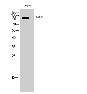
Click image to see more details
Western Blot analysis of HY926 cells using EphB4 Polyclonal Antibody diluted at 1:2000

Click image to see more details
Western blot analysis of lysates from Jurkat and 293 cells, using EPHB4 Antibody. The lane on the right is blocked with the synthesized peptide.

Click image to see more details
Immunohistochemical analysis of paraffin-embedded human tonsil. 1, Antibody was diluted at 1:200 (4° overnight). 2, Tris-EDTA, pH9.0 was used for antigen retrieval. 3, Secondary antibody was diluted at 1:200 (room temperature, 45min).

Click image to see more details
The effect of the lentivirus-mediated shRNA interference of TNFR2 on TNF-α-stimulated EphB4 expression and osteogenic differentiation. a MC3T3-E1 cells stably transduced with lentiviral particles were selected with puromycin and named as pHBLV-TNFR2siRNA1 cells, pHBLV-TNFR2siRNA2 cells, pHBLV-TNFR2siRNA3 cells and pHBLV-NC cells, respectively. The mRNA levels of Tnfr2 were determined in these cells, among which the pHBLV-TNFR2siRNA1 cells displayed the highest TNFR2 gene silencing efficiency and were selected to continue the following studies. b TNFR2 protein levels in pHBLV-TNFR2siRNA1 cells and pHBLV-NC cells. c , d mRNA levels of Ephb4 , Runx2 and Bsp in pHBLV-TNFR2siRNA1 cells and pHBLV-NC cells cultured in the osteogenic induction medium supplemented with 0.5 ng/ml TNF-α for 24 h ( c ) or 48 h ( d ). e , f Protein levels of EphB4, RUNX2 and BSP in pHBLV-TNFR2siRNA1 cells and pHBLV-NC cells cultured in the osteogenic induction medium supplemented with 0.5 ng/ml TNF-α for 24 h ( e ) or 48 h ( f ). *, p < 0.05 vs. the pHBLV-NC group; **, p < 0.01 vs. the pHBLV-NC group
Index in PubMed under a CC BY license. PMID: 32299362

Click image to see more details
The effect of the impaired binding between TNF-α and TNFR2 on TNF-α-stimulated EphB4 expression and osteogenic differentiation. MC3T3-E1 cells were treated with an anti-mouse TNFR2/ CD120b/TNFRSF1B neutralizing antibody (TNFR2 NAb) at the concentration of 0.2 μg/ml, and were cultured in the osteogenic induction medium supplemented with or without 0.5 ng/ml TNF-α. Cells treated with 0.2 μg/ml of the normal rabbit IgG negative control antibody (control Ab) served as negative controls. (a) ALP activities were determined 7d or 14d after the treatment. (b, c) mRNA levels of Ephb4 , Runx2 and Bsp were determined after 24 h (b) or 48 h (c). (d, e) Protein levels of EphB4, RUNX2 and BSP were determined after 24 h (d) or 48 h (e). a, p < 0.05 vs. the control Ab group; b, p < 0.05 vs. the TNFR2 NAb group; c, p < 0.05 vs. the TNF-α + control Ab group
Index in PubMed under a CC BY license. PMID: 32299362

Click image to see more details
The effect of inhibited EphB4 forward signaling on TNF-α-stimulated TNFR2 expression and osteogenic differentiation. (a) A potent inhibitor of EphB4 auto-phosphorylation, NVP-BHG712, was used to suppress EphB4 forward signaling. MC3T3-E1 cells were pretreated with 200 nM NVP-BHG712 in the regular culture medium for 1 h. Cells were then incubated in osteogenic induction medium supplemented with 200 nM NVP-BHG712 and/or 0.5 ng/ml TNF-α for 7d or 14d. MC3T3-E1 cells cultured in osteogenic induction medium served as controls. The ALP activities were determined. (b, c) MC3T3-E1 cells were pretreated with 200 nM NVP-BHG712 for 1 h in the regular culture medium, and then incubated in osteogenic induction medium supplemented with 200 nM NVP-BHG712 and/or 0.5 ng/ml TNF-α. Cells cultured in osteogenic induction medium served as controls. mRNA levels of Tnfr2 , Runx2 and Bsp were determined after 24 h (b) or 48 h (c) of incubation. (d, e) MC3T3-E1 cells were pretreated with 200 nM NVP-BHG712 for 1 h in the regular culture medium, and then incubated in osteogenic induction medium supplemented with 200 nM NVP-BHG712 and/or 0.5 ng/ml TNF-α. Cells cultured in osteogenic induction medium served as controls. Protein levels of TNFR2, RUNX2 and BSP were determined after 24 h (d) or 48 h (e) of incubation. a, p < 0.05 vs. the control group; b, p < 0.05 vs. the NVP-BHG712 group; c, p < 0.05 vs. the TNF-α group
Index in PubMed under a CC BY license. PMID: 32299362

Click image to see more details
EphB4, TNFR2 and MAPK signaling pathways comprise a signaling axis to mediate the positive effect of TNF-α on osteogenic differentiation. a Levels of p38, p -p38, ERK1/2, p -ERK1/2, JNK1 + 2 + 3 and p -JNK1 + 2 + 3 in MC3T3-E1 cells treated with TNF-α for 0 min, 5 min, 15 min, 30 min and 60 min. b Levels of p38, p -p38, ERK1/2, p -ERK1/2, JNK1 + 2 + 3 and p -JNK1 + 2 + 3 in the pHBLV-TNFR2siRNA1 cells and the pHBLV-NC cells treated with or without 0.5 ng/ml TNF-α in regular culture medium for 15 min. c MC3T3-E1 cells were pretreated with or without 200 nM NVP-BHG712 in the regular culture medium for 1 h, and then 0.5 ng/ml TNF-α was added into the medium. The cells were incubated for another 15 min. Levels of ERK1/2 and p -ERK1/2 were determined. d-f MC3T3-E1 cells were cultured in the regular culture medium and pretreated with the ERK inhibitor U0126 (10 μM) for 1 h. The culture medium was then switched to the osteogenic induction medium supplemented with 0.5 ng/ml TNF-α and U0126 (10 μM). Cells treated without U0126 (10 μM) served as controls. ALP activities were determined 7d or 14d after the treatment ( d ). mRNA levels ( e ) and protein levels ( f ) of BSP and RUNX2 were determined 3 days after the treatment. *, p < 0.05 vs. the control group; **, p < 0.01 vs. the control group
Index in PubMed under a CC BY license. PMID: 32299362
Specific Publications For Anti-Ephrin type-B receptor 4 EphB4 Antibody (A00690)
Loading publications
Recommended Resources
Here are featured tools and databases that you might find useful.
- Boster's Pathways Library
- Protein Databases
- Bioscience Research Protocol Resources
- Data Processing & Analysis Software
- Photo Editing Software
- Scientific Literature Resources
- Research Paper Management Tools
- Molecular Biology Software
- Primer Design Tools
- Bioinformatics Tools
- Phylogenetic Tree Analysis
Customer Reviews
Have you used Anti-Ephrin type-B receptor 4 EphB4 Antibody?
Share your experimental results or join a short interview to earn up to $1,000 in product credits or other rewards.
0 Reviews For Anti-Ephrin type-B receptor 4 EphB4 Antibody
Customer Q&As
Have a question?
Find answers in Q&As, reviews.
Can't find your answer?
Submit your question
6 Customer Q&As for Anti-Ephrin type-B receptor 4 EphB4 Antibody
Question
My lab would like to test anti-EphB4 antibody A00690 on human cervix carcinoma erythroleukemia for research purposes, then I may be interested in using anti-EphB4 antibody A00690 for diagnostic purposes as well. Is the antibody suitable for diagnostic purposes?
Verified Customer
Verified customer
Asked: 2018-12-04
Answer
The products we sell, including anti-EphB4 antibody A00690, are only intended for research use. They would not be suitable for use in diagnostic work. If you have the means to develop a product into diagnostic use, and are interested in collaborating with us and develop our product into an IVD product, please contact us for more discussions.
Boster Scientific Support
Answered: 2018-12-04
Question
Would anti-EphB4 antibody A00690 work for WB with cervix carcinoma erythroleukemia?
Verified Customer
Verified customer
Asked: 2018-11-15
Answer
According to the expression profile of cervix carcinoma erythroleukemia, EPHB4 is highly expressed in cervix carcinoma erythroleukemia. So, it is likely that anti-EphB4 antibody A00690 will work for WB with cervix carcinoma erythroleukemia.
Boster Scientific Support
Answered: 2018-11-15
Question
I was wanting to use your anti-EphB4 antibody for WB for human cervix carcinoma erythroleukemia on frozen tissues, but I want to know if it has been validated for this particular application. Has this antibody been validated and is this antibody a good choice for human cervix carcinoma erythroleukemia identification?
K. Rodriguez
Verified customer
Asked: 2018-05-30
Answer
As indicated on the product datasheet, A00690 anti-EphB4 antibody has been validated for WB on human, mouse tissues. We have an innovator award program that if you test this antibody and show it works in human cervix carcinoma erythroleukemia in IHC-frozen, you can get your next antibody for free.
Boster Scientific Support
Answered: 2018-05-30
Question
Is this A00690 anti-EphB4 antibody reactive to the isotypes of EPHB4?
Verified Customer
Verified customer
Asked: 2018-05-24
Answer
The immunogen of A00690 anti-EphB4 antibody is Synthesized peptide derived from the Internal region of human EphB4. at AA rangle: 540-620. Could you tell me which isotype you are interested in so I can help see if the immunogen is part of this isotype?
Boster Scientific Support
Answered: 2018-05-24
Question
I have attached the WB image, lot number and protocol we used for cervix carcinoma erythroleukemia using anti-EphB4 antibody A00690. Please let me know if you require anything else.
Verified Customer
Verified customer
Asked: 2017-11-27
Answer
Thank you very much for the data. Our lab team are working to resolve this as quickly as possible, and we appreciate your patience and understanding! You have provided everything we needed. Please let me know if there is anything you need in the meantime.
Boster Scientific Support
Answered: 2017-11-27
Question
Thank you for helping with my inquiry over the phone. Here are the WB image, lot number and protocol we used for cervix carcinoma erythroleukemia using anti-EphB4 antibody A00690. Let me know if you need anything else.
C. Krishna
Verified customer
Asked: 2015-09-01
Answer
Thanks for the data. You have provided everything we needed. Our lab team are working to resolve your inquiry as quickly as possible, and we appreciate your patience and understanding! Please let me know if there is anything you need in the meantime.
Boster Scientific Support
Answered: 2015-09-01
