Click image to see more details
-

-
-
-
-
+22
Product Info Summary
| SKU: | M00104-1 |
|---|---|
| Size: | 100 μl |
| Reactive Species: | Human, Mouse, Rat |
| Host: | Rabbit |
| Application: | Flow Cytometry, IP, IF, ICC, WB |
Customers Who Bought This Also Bought

Product info
Product Name
Anti-ERK1/2 Rabbit Monoclonal Antibody
View all MAPK3/MAPK1 Antibodies
SKU/Catalog Number
M00104-1
BM4326 is an alternative SKU for this antibody, used in previous lots.
Size
100 μl
Form
Liquid
Description
Boster Bio Anti-ERK1/2 Rabbit Monoclonal Antibody catalog # M00104-1. Tested in WB, ICC/IF, IP, Flow Cytometry applications. This antibody reacts with Human, Mouse, Rat.
Storage & Handling
Store at -20°C for one year. For short term storage and frequent use, store at 4°C for up to one month. Avoid repeated freeze-thaw cycles.
Cite This Product
Anti-ERK1/2 Rabbit Monoclonal Antibody (Boster Biological Technology, Pleasanton CA, USA, Catalog # M00104-1)
Host
Rabbit
Contents
Rabbit IgG in stabilizing components, phosphate buffered saline, pH 7.4, 150mM NaCl, 0.02% sodium azide and 50% glycerol.
*This antibody is supplied in a stabilized formulation.
Compatibility with conjugation reactions depends on the chemistry of the conjugation method used.
For conjugation methods that are not compatible with the stabilizing components present in this formulation, a carrier-free antibody format is required.
Clonality
Monoclonal
Clone Number
DFF-13
Isotype
Rabbit IgG
Immunogen
A synthesized peptide derived from human ERK1/2
Reactive Species
M00104-1 is reactive to MAPK3/MAPK1 in Human, Mouse, Rat
Observed Molecular Weight
42, 44 kDa
Antibody Validation
Boster validates all antibodies on WB, IHC, ICC, Immunofluorescence, and ELISA with known positive control and negative samples to ensure specificity and high affinity, including thorough antibody incubations.
Application & Images
Applications
M00104-1 is guaranteed for Flow Cytometry, IP, IF, ICC, WB Boster Guarantee
Recommend Dilution
WB 1:500-2000
ICC/IF 1:50-200
IP 1:20
FC 1:20
Tested application
Use TE buffer pH 9.0 for antigen retrieval; (*) citrate buffer pH 6.0 is an alternative.
Validation Images & Assay Conditions
Click image to see more details
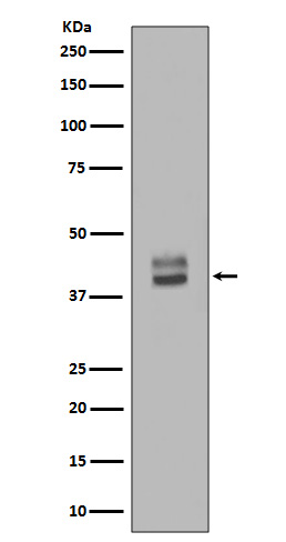
Western blot analysis of ERK1/2 Antibody expression in HepG2 whole cell lysates.

Click image to see more details
All lanes use the Antibody at 1:1W dilution for 1 hour at room temperature.

Click image to see more details
ERK/NF-κB signaling is involved in Kv1.3-related M1 macrophages. A Respective western blotting image and densitometry analysis of ERK/NF-κB signaling after transfection with LV-Kv1.3 in THP-1 cells. Data were expressed as mean ± SD (n = 4). B The phosphorylation signal data are depicted in the form of stacked histogram overlaid plots. The mean fluorescence intensity (MFI) of the ERK1/2 and NF-κB p65 phosphorylation signal was expressed as the mean ± SD (n = 3). C Effects of MgTx, PD98059, and Licochalcone B on Kv1.3 and ERK/NF-κB signaling in LV-Kv1.3-transduced THP-1 cells were analyzed by western blotting. Data were expressed as mean ± SD (n = 3). D Respective western blotting image and densitometry analysis of ERK/NF-κB signaling in HK-2 cells co-cultured with the supernatant from LV-Kv1.3-transduced THP-1 cells for 48 h. Data were expressed as mean ± SD (n = 4). E Respective western blotting image and densitometry analysis of ERK/NF-κB signaling in mice subjected to UUO (7 days post-surgery) and IRI. Data were expressed as mean ± SD (n = 4).
Index in PubMed under a CC BY license. PMID: 40324999

Click image to see more details
Effects of knocking-down ADRB1 CaMKII on mPFC activity and subsequent signals in METH-sired male F1. a Levels of c-Fos protein. b The c-Fos immunostaining. Scale bar, 500 μm /100 μm. c Density of dendritic spine. Scale bar, 50 μm /10 μm. d Levels of β-arestin2 and PKA protein. e Levels of ERK1/2, p-ERK1/2, ERK1/2/p-ERK1/2, CREB, p-CREB, p-CREB/ CREB and ΔFosB protein. F1-METH-Ctrl, METH-sired male F1 mice injected with Ctrl virus. F1-METH-KD, METH-sired male F1 mice injected with KD virus. The data are presented as the Mean ± SD. N.S., P > 0.05. * P < 0.05, ** P < 0.01 vs F1-METH-Ctrl.
Index in PubMed under a CC BY license. PMID: 37857642

Click image to see more details
Effect of MYH7 R453C mutation on TGF-β/Smad2/3, ERK1/2, NF-κB and PI3K/AKT pathways. (a) The protein expression of TGF-β/Smad2/3 and ERK1/2 cascades were detected by western blotting. (b–d) The protein expression of TGF-β1/2, p-Smad2/3/Smad2/3 and p-ERK1/2/ERK1/2 were quantitated using densitometry. (e) The protein expression of NF-κB signalling was detected by western blotting. (f) Quantitative analysis of the protein expression of p-NF-κB p65 and NF-κB p65. **p < 0.01, ****p < 0.0001. n = 3 biologically independent samples.
Index in PubMed under a CC BY license. PMID: 38862020

Click image to see more details
Effects of OPN and OVE on activation of MAPK signaling pathways in LPS-stimulated RAW264.7 cells. Expression levels of p-ERK, ERK, p-p38, p-38, p-JNK, and JNK were detected in the same samples for COX-2 detection after 24 h of LPS stimulation. (A) OVE treatment. (B) OPN treatment. All experiments were carried out in triplicates and data are presented as means ± SDs; one-way ANOVA analysis was adopted for multiple comparisons; ###P < 0.001, ####P < 0.0001, compared to the untreated control group; ***P < 0.001 and ****P < 0.0001, compared to the LPS control group.
Index in PubMed under a CC BY license. PMID: 39455284

Click image to see more details
Immunofluorescent analysis of Hela cells, using ERK1/2 Antibody.

Click image to see more details
All lanes use the Antibody at 1:2W dilution for 1 hour at room temperature.

Click image to see more details
All lanes use the Antibody at 1:2W dilution for 1 hour at room temperature.

Click image to see more details
IHC analysis of ERK1/2 using anti-ERK1/2 antibody (M00104-1).
ERK1/2 was detected in a paraffin-embedded section of mouse pancreas tissue. Heat mediated antigen retrieval was performed in EDTA buffer (pH 8.0, epitope retrieval solution). The tissue section was blocked with 10% goat serum. The tissue section was then incubated with 5 μg/ml rabbit anti-ERK1/2 Antibody (M00104-1) overnight at 4°C. HRP Conjugated Goat Anti-rabbit IgG was used as secondary antibody and incubated for 30 minutes at 37°C. The tissue section was developed using HRP Conjugated Rabbit IgG Super Vision Assay Kit (Catalog # SV0002) with DAB as the chromogen.

Click image to see more details
IHC analysis of ERK1/2 using anti-ERK1/2 antibody (M00104-1).
ERK1/2 was detected in a paraffin-embedded section of rat kidney tissue. Heat mediated antigen retrieval was performed in EDTA buffer (pH 8.0, epitope retrieval solution). The tissue section was blocked with 10% goat serum. The tissue section was then incubated with 5 μg/ml rabbit anti-ERK1/2 Antibody (M00104-1) overnight at 4°C. HRP Conjugated Goat Anti-rabbit IgG was used as secondary antibody and incubated for 30 minutes at 37°C. The tissue section was developed using HRP Conjugated Rabbit IgG Super Vision Assay Kit (Catalog # SV0002) with DAB as the chromogen.

Click image to see more details
IHC analysis of ERK1/2 using anti-ERK1/2 antibody (M00104-1).
ERK1/2 was detected in a paraffin-embedded section of human breast cancer tissue. Heat mediated antigen retrieval was performed in EDTA buffer (pH 8.0, epitope retrieval solution). The tissue section was blocked with 10% goat serum. The tissue section was then incubated with 5 μg/ml rabbit anti-ERK1/2 Antibody (M00104-1) overnight at 4°C. HRP Conjugated Goat Anti-rabbit IgG was used as secondary antibody and incubated for 30 minutes at 37°C. The tissue section was developed using HRP Conjugated Rabbit IgG Super Vision Assay Kit (Catalog # SV0002) with DAB as the chromogen.

Click image to see more details
IHC analysis of ERK1/2 using anti-ERK1/2 antibody (M00104-1).
ERK1/2 was detected in a paraffin-embedded section of human colon cancer tissue. Heat mediated antigen retrieval was performed in EDTA buffer (pH 8.0, epitope retrieval solution). The tissue section was blocked with 10% goat serum. The tissue section was then incubated with 5 μg/ml rabbit anti-ERK1/2 Antibody (M00104-1) overnight at 4°C. HRP Conjugated Goat Anti-rabbit IgG was used as secondary antibody and incubated for 30 minutes at 37°C. The tissue section was developed using HRP Conjugated Rabbit IgG Super Vision Assay Kit (Catalog # SV0002) with DAB as the chromogen.

Click image to see more details
IHC analysis of ERK1/2 using anti-ERK1/2 antibody (M00104-1).
ERK1/2 was detected in a paraffin-embedded section of human liver cancer tissue. Heat mediated antigen retrieval was performed in EDTA buffer (pH 8.0, epitope retrieval solution). The tissue section was blocked with 10% goat serum. The tissue section was then incubated with 5 μg/ml rabbit anti-ERK1/2 Antibody (M00104-1) overnight at 4°C. HRP Conjugated Goat Anti-rabbit IgG was used as secondary antibody and incubated for 30 minutes at 37°C. The tissue section was developed using HRP Conjugated Rabbit IgG Super Vision Assay Kit (Catalog # SV0002) with DAB as the chromogen.

Click image to see more details
IHC analysis of ERK1/2 using anti-ERK1/2 antibody (M00104-1).
ERK1/2 was detected in a paraffin-embedded section of human lung cancer tissue. Heat mediated antigen retrieval was performed in EDTA buffer (pH 8.0, epitope retrieval solution). The tissue section was blocked with 10% goat serum. The tissue section was then incubated with 5 μg/ml rabbit anti-ERK1/2 Antibody (M00104-1) overnight at 4°C. HRP Conjugated Goat Anti-rabbit IgG was used as secondary antibody and incubated for 30 minutes at 37°C. The tissue section was developed using HRP Conjugated Rabbit IgG Super Vision Assay Kit (Catalog # SV0002) with DAB as the chromogen.

Click image to see more details
IF analysis of ERK1/2 using anti-ERK1/2 antibody (M00104-1).
ERK1/2 was detected in a paraffin-embedded section of mouse pancreas tissue. Heat mediated antigen retrieval was performed in EDTA buffer (pH 8.0, epitope retrieval solution). The tissue section was blocked with 10% goat serum. The tissue section was then incubated with 25 μg/mL rabbit anti-ERK1/2 Antibody (M00104-1) overnight at 4°C. DyLight®594 Conjugated Goat Anti-Rabbit IgG (BA1142) was used as secondary antibody at 1:100 dilution and incubated for 30 minutes at 37°C. The section was counterstained with DAPI. Visualize using a fluorescence microscope and filter sets appropriate for the label used.

Click image to see more details
IF analysis of ERK1/2 using anti-ERK1/2 antibody (M00104-1).
ERK1/2 was detected in a paraffin-embedded section of mouse brain tissue. Heat mediated antigen retrieval was performed in EDTA buffer (pH 8.0, epitope retrieval solution). The tissue section was blocked with 10% goat serum. The tissue section was then incubated with 25 μg/mL rabbit anti-ERK1/2 Antibody (M00104-1) overnight at 4°C. DyLight®594 Conjugated Goat Anti-Rabbit IgG (BA1142) was used as secondary antibody at 1:100 dilution and incubated for 30 minutes at 37°C. The section was counterstained with DAPI. Visualize using a fluorescence microscope and filter sets appropriate for the label used.

Click image to see more details
IF analysis of ERK1/2 using anti-ERK1/2 antibody (M00104-1).
ERK1/2 was detected in a paraffin-embedded section of human prostate cancer tissue. Heat mediated antigen retrieval was performed in EDTA buffer (pH 8.0, epitope retrieval solution). The tissue section was blocked with 10% goat serum. The tissue section was then incubated with 25 μg/mL rabbit anti-ERK1/2 Antibody (M00104-1) overnight at 4°C. DyLight®594 Conjugated Goat Anti-Rabbit IgG (BA1142) was used as secondary antibody at 1:100 dilution and incubated for 30 minutes at 37°C. The section was counterstained with DAPI. Visualize using a fluorescence microscope and filter sets appropriate for the label used.

Click image to see more details
IF analysis of ERK1/2 using anti-ERK1/2 antibody (M00104-1).
ERK1/2 was detected in a paraffin-embedded section of rat brain tissue. Heat mediated antigen retrieval was performed in EDTA buffer (pH 8.0, epitope retrieval solution). The tissue section was blocked with 10% goat serum. The tissue section was then incubated with 25 μg/mL rabbit anti-ERK1/2 Antibody (M00104-1) overnight at 4°C. DyLight®594 Conjugated Goat Anti-Rabbit IgG (BA1142) was used as secondary antibody at 1:100 dilution and incubated for 30 minutes at 37°C. The section was counterstained with DAPI. Visualize using a fluorescence microscope and filter sets appropriate for the label used.

Click image to see more details
IF analysis of ERK1/2 using anti-ERK1/2 antibody (M00104-1).
ERK1/2 was detected in a paraffin-embedded section of rat colon tissue. Heat mediated antigen retrieval was performed in EDTA buffer (pH 8.0, epitope retrieval solution). The tissue section was blocked with 10% goat serum. The tissue section was then incubated with 25 μg/mL rabbit anti-ERK1/2 Antibody (M00104-1) overnight at 4°C. DyLight®594 Conjugated Goat Anti-Rabbit IgG (BA1142) was used as secondary antibody at 1:100 dilution and incubated for 30 minutes at 37°C. The section was counterstained with DAPI. Visualize using a fluorescence microscope and filter sets appropriate for the label used.

Click image to see more details
IF analysis of ERK1/2 using anti-ERK1/2 antibody (M00104-1).
ERK1/2 was detected in a paraffin-embedded section of rat kidney tissue. Heat mediated antigen retrieval was performed in EDTA buffer (pH 8.0, epitope retrieval solution). The tissue section was blocked with 10% goat serum. The tissue section was then incubated with 25 μg/mL rabbit anti-ERK1/2 Antibody (M00104-1) overnight at 4°C. DyLight®594 Conjugated Goat Anti-Rabbit IgG (BA1142) was used as secondary antibody at 1:100 dilution and incubated for 30 minutes at 37°C. The section was counterstained with DAPI. Visualize using a fluorescence microscope and filter sets appropriate for the label used.

Click image to see more details
IF analysis of ERK1/2 using anti-ERK1/2 antibody (M00104-1).
ERK1/2 was detected in a paraffin-embedded section of human breast cancer tissue. Heat mediated antigen retrieval was performed in EDTA buffer (pH 8.0, epitope retrieval solution). The tissue section was blocked with 10% goat serum. The tissue section was then incubated with 25 μg/mL rabbit anti-ERK1/2 Antibody (M00104-1) overnight at 4°C. DyLight®594 Conjugated Goat Anti-Rabbit IgG (BA1142) was used as secondary antibody at 1:100 dilution and incubated for 30 minutes at 37°C. The section was counterstained with DAPI. Visualize using a fluorescence microscope and filter sets appropriate for the label used.

Click image to see more details
IF analysis of ERK1/2 using anti-ERK1/2 antibody (M00104-1).
ERK1/2 was detected in a paraffin-embedded section of human colon cancer tissue. Heat mediated antigen retrieval was performed in EDTA buffer (pH 8.0, epitope retrieval solution). The tissue section was blocked with 10% goat serum. The tissue section was then incubated with 25 μg/mL rabbit anti-ERK1/2 Antibody (M00104-1) overnight at 4°C. DyLight®594 Conjugated Goat Anti-Rabbit IgG (BA1142) was used as secondary antibody at 1:100 dilution and incubated for 30 minutes at 37°C. The section was counterstained with DAPI. Visualize using a fluorescence microscope and filter sets appropriate for the label used.

Click image to see more details
IF analysis of ERK1/2 using anti-ERK1/2 antibody (M00104-1).
ERK1/2 was detected in a paraffin-embedded section of human liver cancer tissue. Heat mediated antigen retrieval was performed in EDTA buffer (pH 8.0, epitope retrieval solution). The tissue section was blocked with 10% goat serum. The tissue section was then incubated with 25 μg/mL rabbit anti-ERK1/2 Antibody (M00104-1) overnight at 4°C. DyLight®594 Conjugated Goat Anti-Rabbit IgG (BA1142) was used as secondary antibody at 1:100 dilution and incubated for 30 minutes at 37°C. The section was counterstained with DAPI. Visualize using a fluorescence microscope and filter sets appropriate for the label used.

Click image to see more details
IF analysis of ERK1/2 using anti-ERK1/2 antibody (M00104-1).
ERK1/2 was detected in a paraffin-embedded section of human pancreatic cancer tissue. Heat mediated antigen retrieval was performed in EDTA buffer (pH 8.0, epitope retrieval solution). The tissue section was blocked with 10% goat serum. The tissue section was then incubated with 25 μg/mL rabbit anti-ERK1/2 Antibody (M00104-1) overnight at 4°C. DyLight®594 Conjugated Goat Anti-Rabbit IgG (BA1142) was used as secondary antibody at 1:100 dilution and incubated for 30 minutes at 37°C. The section was counterstained with DAPI. Visualize using a fluorescence microscope and filter sets appropriate for the label used.

Click image to see more details
Specific Publications For Anti-ERK1/2 Rabbit Monoclonal Antibody (M00104-1)
Loading publications
Recommended Resources
Here are featured tools and databases that you might find useful.
- Boster's Pathways Library
- Protein Databases
- Bioscience Research Protocol Resources
- Data Processing & Analysis Software
- Photo Editing Software
- Scientific Literature Resources
- Research Paper Management Tools
- Molecular Biology Software
- Primer Design Tools
- Bioinformatics Tools
- Phylogenetic Tree Analysis
Customer Reviews
Have you used Anti-ERK1/2 Rabbit Monoclonal Antibody?
Share your experimental results or join a short interview to earn up to $1,000 in product credits or other rewards.
0 Reviews For Anti-ERK1/2 Rabbit Monoclonal Antibody
Customer Q&As
Have a question?
Find answers in Q&As, reviews.
Can't find your answer?
Submit your question
16 Customer Q&As for Anti-ERK1/2 Rabbit Monoclonal Antibody
Question
Would M00104-1 anti-ERK1/2 Rabbit Monoclonal antibody work on parafin embedded sections? If so, which fixation method do you recommend we use (PFA, paraformaldehyde, other)?
Verified Customer
Verified customer
Asked: 2020-03-06
Answer
You can see on the product datasheet, M00104-1 anti-ERK1/2 Rabbit Monoclonal antibody as been validated on IP. It is best to use PFA for fixation because it has better tissue penetration ability. PFA needs to be prepared fresh before use. Long term stored PFA turns into formalin, as the PFA molecules congregate and become formalin.
Boster Scientific Support
Answered: 2020-03-06
Question
We ordered your anti-ERK1/2 Rabbit Monoclonal antibody for Flow Cytometry on right frontal lobe in the past. I am using human, and We want to use the antibody for IP next. I am interested in examining right frontal lobe as well as cervix carcinoma in our next experiment. Do you have any suggestion on which antibody would work the best for IP?
Verified Customer
Verified customer
Asked: 2020-02-14
Answer
I viewed the website and datasheets of our anti-ERK1/2 Rabbit Monoclonal antibody and it seems that M00104-1 has been validated on human in both Flow Cytometry and IP. Thus M00104-1 should work for your application. Our Boster satisfaction guarantee will cover this product for IP in human even if the specific tissue type has not been validated. We do have a comprehensive range of products for IP detection and you can check out our website bosterbio.com to find out more information about them.
Boster Scientific Support
Answered: 2020-02-14
Question
I am looking for using your anti-ERK1/2 Rabbit Monoclonal antibody for rsk activation studies. Has this antibody been tested with western blotting on hepg2 whole cell lysates? We would like to see some validation images before ordering.
G. Jackson
Verified customer
Asked: 2019-08-01
Answer
We appreciate your inquiry. This M00104-1 anti-ERK1/2 Rabbit Monoclonal antibody is validated on hepg2 whole cell lysates, hela cells. It is guaranteed to work for Flow Cytometry, IP, IF, ICC, WB in human, mouse, rat. Our Boster guarantee will cover your intended experiment even if the sample type has not been be directly tested.
Boster Scientific Support
Answered: 2019-08-01
Question
I was wanting to use your anti-ERK1/2 Rabbit Monoclonal antibody for IP for mouse lymph on frozen tissues, but I want to know if it has been validated for this particular application. Has this antibody been validated and is this antibody a good choice for mouse lymph identification?
Verified Customer
Verified customer
Asked: 2019-07-12
Answer
You can see on the product datasheet, M00104-1 anti-ERK1/2 Rabbit Monoclonal antibody has been validated for Flow Cytometry, IP, IF, ICC, WB on human, mouse, rat tissues. We have an innovator award program that if you test this antibody and show it works in mouse lymph in IHC-frozen, you can get your next antibody for free.
Boster Scientific Support
Answered: 2019-07-12
Question
See attached the WB image, lot number and protocol we used for lymph using anti-ERK1/2 Rabbit Monoclonal antibody M00104-1. Please let me know if you require anything else.
Verified Customer
Verified customer
Asked: 2019-07-08
Answer
Thank you very much for the data. Our lab team are working to resolve this as quickly as possible, and we appreciate your patience and understanding! You have provided everything we needed. Please let me know if there is anything you need in the meantime.
Boster Scientific Support
Answered: 2019-07-08
Question
We have observed staining in human right frontal lobe. Any tips? Is anti-ERK1/2 Rabbit Monoclonal antibody supposed to stain right frontal lobe positively?
Verified Customer
Verified customer
Asked: 2018-12-04
Answer
According to literature right frontal lobe does express MAPK3. According to Uniprot.org, MAPK3 is expressed in right frontal lobe, hepatoma, lymph, cervix carcinoma, leukemic t-cell, cervix carcinoma erythroleukemia, among other tissues. Regarding which tissues have MAPK3 expression, here are a few articles citing expression in various tissues:
Cervix carcinoma, Pubmed ID: 17081983, 18669648, 18691976, 20068231
Cervix carcinoma, and Erythroleukemia, Pubmed ID: 23186163
Hepatoma, Pubmed ID: 1540184, 7687743
Leukemic T-cell, Pubmed ID: 19690332
Lymph, Pubmed ID: 15489334
Boster Scientific Support
Answered: 2018-12-04
Question
We need to test anti-ERK1/2 Rabbit Monoclonal antibody M00104-1 on mouse lymph for research purposes, then I may be interested in using anti-ERK1/2 Rabbit Monoclonal antibody M00104-1 for diagnostic purposes as well. Is the antibody suitable for diagnostic purposes?
M. Anderson
Verified customer
Asked: 2018-11-20
Answer
The products we sell, including anti-ERK1/2 Rabbit Monoclonal antibody M00104-1, are only intended for research use. They would not be suitable for use in diagnostic work. If you have the means to develop a product into diagnostic use, and are interested in collaborating with us and develop our product into an IVD product, please contact us for more discussions.
Boster Scientific Support
Answered: 2018-11-20
Question
Is this M00104-1 anti-ERK1/2 Rabbit Monoclonal antibody reactive to the isotypes of MAPK3?
Verified Customer
Verified customer
Asked: 2018-08-14
Answer
The immunogen of M00104-1 anti-ERK1/2 Rabbit Monoclonal antibody is A synthesized peptide derived from human ERK1/2. Could you tell me which isotype you are interested in so I can help see if the immunogen is part of this isotype?
Boster Scientific Support
Answered: 2018-08-14
Question
Is a blocking peptide available for product anti-ERK1/2 Rabbit Monoclonal antibody (M00104-1)?
E. Williams
Verified customer
Asked: 2018-01-04
Answer
We do provide the blocking peptide for product anti-ERK1/2 Rabbit Monoclonal antibody (M00104-1). If you would like to place an order for it please contact support@bosterbio.com and make a special request.
Boster Scientific Support
Answered: 2018-01-04
Question
We appreciate helping with my inquiry over the phone. Here are the WB image, lot number and protocol we used for lymph using anti-ERK1/2 Rabbit Monoclonal antibody M00104-1. Let me know if you need anything else.
Verified Customer
Verified customer
Asked: 2017-12-04
Answer
We appreciate the data. You have provided everything we needed. Our lab team are working to resolve your inquiry as quickly as possible, and we appreciate your patience and understanding! Please let me know if there is anything you need in the meantime.
Boster Scientific Support
Answered: 2017-12-04
Question
My colleagues were satisfied with the WB result of your anti-ERK1/2 Rabbit Monoclonal antibody. However we have observed positive staining in cervix carcinoma cytoplasm. nucleus. membrane using this antibody. Is that expected? Could you tell me where is MAPK3 supposed to be expressed?
Verified Customer
Verified customer
Asked: 2017-10-16
Answer
Based on literature, cervix carcinoma does express MAPK3. Generally MAPK3 expresses in cytoplasm. nucleus. membrane, caveola. Regarding which tissues have MAPK3 expression, here are a few articles citing expression in various tissues:
Cervix carcinoma, Pubmed ID: 17081983, 18669648, 18691976, 20068231
Cervix carcinoma, and Erythroleukemia, Pubmed ID: 23186163
Hepatoma, Pubmed ID: 1540184, 7687743
Leukemic T-cell, Pubmed ID: 19690332
Lymph, Pubmed ID: 15489334
Boster Scientific Support
Answered: 2017-10-16
Question
Is there a BSA free version of anti-ERK1/2 Rabbit Monoclonal antibody M00104-1 available?
Verified Customer
Verified customer
Asked: 2017-09-18
Answer
We appreciate your recent telephone inquiry. I can confirm that some lots of this anti-ERK1/2 Rabbit Monoclonal antibody M00104-1 are BSA free. For now, these lots are available and we can make a BSA free formula for you free of charge. It will take 3 extra days to prepare. If you require this antibody BSA free again in future, please do not hesitate to contact me and I will be pleased to check which lots we have in stock that are BSA free.
Boster Scientific Support
Answered: 2017-09-18
Question
Would anti-ERK1/2 Rabbit Monoclonal antibody M00104-1 work for IP with lymph?
Verified Customer
Verified customer
Asked: 2017-08-17
Answer
According to the expression profile of lymph, MAPK3 is highly expressed in lymph. So, it is likely that anti-ERK1/2 Rabbit Monoclonal antibody M00104-1 will work for IP with lymph.
Boster Scientific Support
Answered: 2017-08-17
Question
I see that the anti-ERK1/2 Rabbit Monoclonal antibody M00104-1 works with IP, what is the protocol used to produce the result images on the product page?
J. Jackson
Verified customer
Asked: 2016-12-12
Answer
You can find protocols for IP on the "support/technical resources" section of our navigation menu. If you have any further questions, please send an email to support@bosterbio.com
Boster Scientific Support
Answered: 2016-12-12
Question
My question regarding product M00104-1, anti-ERK1/2 Rabbit Monoclonal antibody. I was wondering if it would be possible to conjugate this antibody with biotin. I would need it to be without BSA or sodium azide. I am planning on using a buffer exchange of sodium azide with PBS only. Would there be problems for me to conjugate the antibody and store it in -20 degrees in small aliquots?
S. Taylor
Verified customer
Asked: 2016-01-28
Answer
We do not advise storing this antibody with PBS buffer only in -20 degrees. If you want to store it in -20 degrees it is best to add some cryoprotectant like glycerol. If you want carrier free M00104-1 anti-ERK1/2 Rabbit Monoclonal antibody, we can provide it to you in a special formula with trehalose and/or glycerol. These molecules will not interfere with conjugation chemistry and provide a good level of protection for the antibody from degradation. Please be sure to specify this in your purchase order.
Boster Scientific Support
Answered: 2016-01-28
Question
We are currently using anti-ERK1/2 Rabbit Monoclonal antibody M00104-1 for rat tissue, and we are well pleased with the IF results. The species of reactivity given in the datasheet says human, mouse, rat. Is it possible that the antibody can work on canine tissues as well?
P. Jackson
Verified customer
Asked: 2014-09-09
Answer
The anti-ERK1/2 Rabbit Monoclonal antibody (M00104-1) has not been validated for cross reactivity specifically with canine tissues, though there is a good chance of cross reactivity. We have an innovator award program that if you test this antibody and show it works in canine you can get your next antibody for free. Please contact me if I can help you with anything.
Boster Scientific Support
Answered: 2014-09-09
