Click image to see more details
-

-
-
-
-
+1
Product Info Summary
| SKU: | PB9772 |
|---|---|
| Size: | 100 μg/vial |
| Reactive Species: | Human, Mouse, Rat |
| Host: | Rabbit |
| Application: | Flow Cytometry, IF, IHC, IHC-F, ICC, WB |
Customers Who Bought This Also Bought

Product info
Product Name
Anti-ERp57/PDIA3 Antibody Picoband®
SKU/Catalog Number
PB9772
Size
100 μg/vial
Form
Lyophilized
Description
Boster Bio Anti-ERp57/PDIA3 Antibody Picoband® catalog # PB9772. Tested in Flow Cytometry, IF, IHC, IHC-F, ICC, WB applications. This antibody reacts with Human, Mouse, Rat. The brand Picoband indicates this is a premium antibody that guarantees superior quality, high affinity, and strong signals with minimal background in Western blot applications. Only our best-performing antibodies are designated as Picoband, ensuring unmatched performance.
Storage & Handling
Store at -20˚C for one year from date of receipt. After reconstitution, at 4˚C for one month. It can also be aliquotted and stored frozen at -20˚C for six months. Avoid repeated freeze-thaw cycles.
Cite This Product
Anti-ERp57/PDIA3 Antibody Picoband® (Boster Biological Technology, Pleasanton CA, USA, Catalog # PB9772)
Host
Rabbit
Contents
Each vial contains antibody formulated with stabilizing components, 0.9 mg NaCl, 0.2 mg Na2HPO4, and 0.05 mg NaN3.
*This antibody is supplied in a stabilized formulation.
Compatibility with conjugation reactions depends on the chemistry of the conjugation method used.
For conjugation methods that are not compatible with the stabilizing components present in this formulation, a carrier-free antibody format is required.
Clonality
Polyclonal
Isotype
Rabbit IgG
Immunogen
A synthetic peptide corresponding to a sequence at the C-terminus of human ERp57, different from the related mouse and rat sequences by two amino acids.
Cross-reactivity
No cross-reactivity with other proteins.
Reactive Species
PB9772 is reactive to PDIA3 in Human, Mouse, Rat
Observed Molecular Weight
57 kDa
Calculated molecular weight
56.8 kDa
Background of PDIA3
PDIA3 (Protein disulfide isomerase family A, member 3), also called GRP58, Erp57 or ER60, is an isomerase enzyme. It is mapped on 15q15.3. PDIA3 is also part of the major histocompatibility complex (MHC) class I peptide-loading complex, which is essential for formation of the final antigen conformation and export from the endoplasmic reticulum to the cell surface. This gene encodes a protein of the endoplasmic reticulum that interacts with lectin chaperones calreticulin and calnexin to modulate folding of newly synthesized glycoproteins. The protein was once thought to be a phospholipase; however, it has been demonstrated that the protein actually has protein disulfide isomerase activity. It is thought that complexes of lectins and this protein mediate protein folding by promoting formation of disulfide bonds in their glycoprotein substrates.
Antibody Validation
Boster validates all antibodies on WB, IHC, ICC, Immunofluorescence, and ELISA with known positive control and negative samples to ensure specificity and high affinity, including thorough antibody incubations.
Application & Images
Applications
PB9772 is guaranteed for Flow Cytometry, IF, IHC, IHC-F, ICC, WB Boster Guarantee
Recommend Dilution
| Application | Dilution | Species |
|---|---|---|
| Western blot | 0.1-0.5μg/ml | |
| Immunohistochemistry (Paraffin-embedded Section) | 0.5-1μg/ml | |
| Immunohistochemistry (Frozen Section) | 0.5-1μg/ml | |
| Immunocytochemistry/Immunofluorescence | 2μg/ml | |
| Flow Cytometry (Fixed) | 1-3μg/1x106 cells |
Tested application
Suggested blocking solution with 5% non-fat milk or BSA; (*)Recommended protein loading: 20-40 µg per lane
Use TE buffer pH 9.0 for antigen retrieval; (*) citrate buffer pH 6.0 is an alternative.
Validation Images & Assay Conditions
Click image to see more details
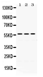
Western blot analysis of ERp57 using anti-ERp57 antibody (PB9772). Electrophoresis was performed on a 5-20% SDS-PAGE gel at 70V (Stacking gel) / 90V (Resolving gel) for 2-3 hours. The sample well of each lane was loaded with 50ug of sample under reducing conditions. Lane 1: Rat Liver Tissue Lysate, Lane 2: Mouse Liver Tissue Lysate, Lane 3: A549 Whole Cell Lysate. After Electrophoresis, proteins were transferred to a Nitrocellulose membrane at 150mA for 50-90 minutes. Blocked the membrane with 5% Non-fat Milk/ TBS for 1.5 hour at RT. The membrane was incubated with rabbit anti-ERp57 antigen affinity purified polyclonal antibody (Catalog # PB9772) at 0.5 μg/mL overnight at 4°C, then washed with TBS-0.1%Tween 3 times with 5 minutes each and probed with a goat anti-rabbit IgG-HRP secondary antibody at a dilution of 1:10000 for 1.5 hour at RT. The signal is developed using an Enhanced Chemiluminescent detection (ECL) kit (Catalog # EK1002) with Tanon 5200 system. A specific band was detected for ERp57 at approximately 57KD. The expected band size for ERp57 is at 57KD.

Click image to see more details
IHC analysis of PB9771 using anti-PB9771 antibody (PB9772). PB9771 was detected in paraffin-embedded section of Human Lung Cancer Tissue. Heat mediated antigen retrieval was performed in citrate buffer (pH6, epitope retrieval solution) for 20 mins. The tissue section was blocked with 10% goat serum. The tissue section was then incubated with 1μg/ml rabbit anti-PB9771 Antibody (PB9772) overnight at 4°C. Biotinylated goat anti-rabbit IgG was used as secondary antibody and incubated for 30 minutes at 37°C. The tissue section was developed using Strepavidin-Biotin-Complex (SABC)(Catalog # SA1022) with DAB as the chromogen.

Click image to see more details
IHC analysis of ERp57 using anti-ERp57 antibody (PB9772).
ERp57 was detected in frozen section of human placenta tissue . The tissue section was blocked with 10% goat serum. The tissue section was then incubated with 1μg/ml rabbit anti-ERp57 Antibody (PB9772) overnight at 4°C. Biotinylated goat anti-rabbit IgG was used as secondary antibody and incubated for 30 minutes at 37°C. The tissue section was developed using Strepavidin-Biotin-Complex (SABC)(Catalog # SA1022) with DAB as the chromogen.

Click image to see more details
IF analysis of ERp57 using anti-ERp57 antibody (PB9772).
ERp57 was detected in immunocytochemical section of U20S cells. Enzyme antigen retrieval was performed using IHC enzyme antigen retrieval reagent (AR0022) for 15 mins. The cells were blocked with 10% goat serum. And then incubated with 2μg/mL rabbit anti-ERp57 Antibody (PB9772) overnight at 4°C. DyLight®488 Conjugated Goat Anti-Rabbit IgG (BA1127) was used as secondary antibody at 1:100 dilution and incubated for 30 minutes at 37°C. The section was counterstained with DAPI. Visualize using a fluorescence microscope and filter sets appropriate for the label used.

Click image to see more details
Flow Cytometry analysis of PC-3 cells using anti-ERp57 antibody (PB9772).
Overlay histogram showing PC-3 cells stained with PB9772 (Blue line). To facilitate intracellular staining, cells were fixed with 4% paraformaldehyde and permeabilized with permeabilization buffer. The cells were blocked with 10% normal goat serum. And then incubated with rabbit anti-ERp57 Antibody (PB9772, 1μg/1x106 cells) for 30 min at 20°C. DyLight®488 conjugated goat anti-rabbit IgG (BA1127, 5-10μg/1x106 cells) was used as secondary antibody for 30 minutes at 20°C. Isotype control antibody (Green line) was rabbit IgG (1μg/1x106) used under the same conditions. Unlabelled sample without incubation with primary antibody and secondary antibody (Red line) was used as a blank control.
Specific Publications For Anti-ERp57/PDIA3 Antibody Picoband® (PB9772)
Loading publications
Recommended Resources
Here are featured tools and databases that you might find useful.
- Boster's Pathways Library
- Protein Databases
- Bioscience Research Protocol Resources
- Data Processing & Analysis Software
- Photo Editing Software
- Scientific Literature Resources
- Research Paper Management Tools
- Molecular Biology Software
- Primer Design Tools
- Bioinformatics Tools
- Phylogenetic Tree Analysis
Customer Reviews
Have you used Anti-ERp57/PDIA3 Antibody Picoband®?
Share your experimental results or join a short interview to earn up to $1,000 in product credits or other rewards.
0 Reviews For Anti-ERp57/PDIA3 Antibody Picoband®
Customer Q&As
Have a question?
Find answers in Q&As, reviews.
Can't find your answer?
Submit your question
7 Customer Q&As for Anti-ERp57/PDIA3 Antibody Picoband®
Question
Thanks for helping with my inquiry over the phone. Here are the WB image, lot number and protocol we used for keratinocyte using anti-ERp57/PDIA3 antibody PB9772. Let me know if you need anything else.
Verified Customer
Verified customer
Asked: 2019-07-29
Answer
Thanks for the data. You have provided everything we needed. Our lab team are working to resolve your inquiry as quickly as possible, and we appreciate your patience and understanding! Please let me know if there is anything you need in the meantime.
Boster Scientific Support
Answered: 2019-07-29
Question
Is there a BSA free version of anti-ERp57/PDIA3 antibody PB9772 available?
Verified Customer
Verified customer
Asked: 2019-07-23
Answer
We appreciate your recent telephone inquiry. I can confirm that some lots of this anti-ERp57/PDIA3 antibody PB9772 are BSA free. For now, these lots are available and we can make a BSA free formula for you free of charge. It will take 3 extra days to prepare. If you require this antibody BSA free again in future, please do not hesitate to contact me and I will be pleased to check which lots we have in stock that are BSA free.
Boster Scientific Support
Answered: 2019-07-23
Question
I was wanting to use your anti-ERp57/PDIA3 antibody for WB for mouse keratinocyte on frozen tissues, but I want to know if it has been validated for this particular application. Has this antibody been validated and is this antibody a good choice for mouse keratinocyte identification?
M. Yang
Verified customer
Asked: 2018-10-08
Answer
It shows on the product datasheet, PB9772 anti-ERp57/PDIA3 antibody has been tested for IHC, WB on human, mouse, rat tissues. We have an innovator award program that if you test this antibody and show it works in mouse keratinocyte in IHC-frozen, you can get your next antibody for free.
Boster Scientific Support
Answered: 2018-10-08
Question
Would anti-ERp57/PDIA3 antibody PB9772 work on zebrafish IHC with corpus epididymis?
K. Miller
Verified customer
Asked: 2018-06-12
Answer
Our lab technicians have not tested anti-ERp57/PDIA3 antibody PB9772 on zebrafish. You can run a BLAST between zebrafish and the immunogen sequence of anti-ERp57/PDIA3 antibody PB9772 to see if they may cross-react. If the sequence homology is close, then you can perform a pilot test. Keep in mind that since we have not validated zebrafish samples, this use of the antibody is not covered by our guarantee. However we have an innovator award program that if you test this antibody and show it works in zebrafish corpus epididymis in IHC, you can get your next antibody for free.
Boster Scientific Support
Answered: 2018-06-12
Question
Is a blocking peptide available for product anti-ERp57/PDIA3 antibody (PB9772)?
S. Walker
Verified customer
Asked: 2018-05-24
Answer
We do provide the blocking peptide for product anti-ERp57/PDIA3 antibody (PB9772). If you would like to place an order for it please contact support@bosterbio.com and make a special request.
Boster Scientific Support
Answered: 2018-05-24
Question
I have a question about product PB9772, anti-ERp57/PDIA3 antibody. I was wondering if it would be possible to conjugate this antibody with biotin. I would need it to be without BSA or sodium azide. I am planning on using a buffer exchange of sodium azide with PBS only. Would there be problems for me to conjugate the antibody and store it in -20 degrees in small aliquots?
Verified Customer
Verified customer
Asked: 2018-03-02
Answer
It is not recommended storing this antibody with PBS buffer only in -20 degrees. If you want to store it in -20 degrees it is best to add some cryoprotectant like glycerol. If you want carrier free PB9772 anti-ERp57/PDIA3 antibody, we can provide it to you in a special formula with trehalose and/or glycerol. These molecules will not interfere with conjugation chemistry and provide a good level of protection for the antibody from degradation. Please be sure to specify this in your purchase order.
Boster Scientific Support
Answered: 2018-03-02
Question
We are currently using anti-ERp57/PDIA3 antibody PB9772 for mouse tissue, and we are happy with the WB results. The species of reactivity given in the datasheet says human, mouse, rat. Is it likely that the antibody can work on zebrafish tissues as well?
N. Johnson
Verified customer
Asked: 2015-08-21
Answer
The anti-ERp57/PDIA3 antibody (PB9772) has not been tested for cross reactivity specifically with zebrafish tissues, though there is a good chance of cross reactivity. We have an innovator award program that if you test this antibody and show it works in zebrafish you can get your next antibody for free. Please contact me if I can help you with anything.
Boster Scientific Support
Answered: 2015-08-21
