Click image to see more details
Product Info Summary
| SKU: | A00786 |
|---|---|
| Size: | 80 µl |
| Reactive Species: | Human |
| Host: | Rabbit |
| Application: | Flow Cytometry, WB |
Customers Who Bought This Also Bought

Product info
Product Name
Anti-ESR2 Antibody (Center)
SKU/Catalog Number
A00786
Size
80 µl
Form
Liquid
Description
Boster Bio Anti-ESR2 Antibody (Center) (Catalog # A00786). Tested in WB, Flow Cytometry application(s). This antibody reacts with Human.
Storage & Handling
Maintain refrigerated at 2-8°C for up to 2 weeks. For long-term storage, store at -20°C in small aliquots to prevent freeze-thaw cycles.
Cite This Product
Anti-ESR2 Antibody (Center) (Boster Biological Technology, Pleasanton CA, USA, Catalog # A00786)
Host
Rabbit
Contents
Purified polyclonal antibody supplied in PBS with 0.09% (W/V) sodium azide.
Clonality
Polyclonal
Isotype
Rabbit IgG
Immunogen
This ESR2 antibody is generated from rabbits immunized with a KLH conjugated synthetic peptide between 369-397 amino acids from the Central region of human ESR2.
Cross-reactivity
No cross reactivity with other proteins.
Reactive Species
A00786 is reactive to ESR2 in Human
Calculated molecular weight
59.2 kDa
Background of ESR2
ESR2 is a nuclear hormone receptor. The protein binds estrogens with an affinity similar to that of ESR1, and activates expression of reporter genes containing estrogen response elements (ERE) in an estrogen-dependent manner.
Antibody Validation
Boster validates all antibodies on WB, IHC, ICC, Immunofluorescence, and ELISA with known positive control and negative samples to ensure specificity and high affinity, including thorough antibody incubations.
Application & Images
Applications
A00786 is guaranteed for Flow Cytometry, WB Boster Guarantee
Assay Dilutions Recommendation
The recommendations below provide a starting point for assay optimization. The actual working concentration varies and should be decided by the user.
WB: 1:1000
FC: 1:10-1:50
Validation Images & Assay Conditions
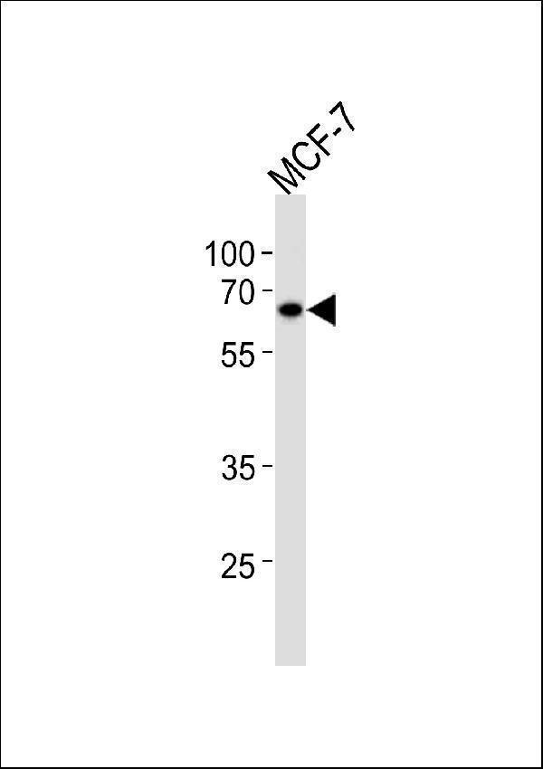
Click image to see more details
ESR2 Antibody (Center) western blot analysis in MCF-7 cell line lysates (35ug/lane). This demonstrates the ESR2 antibody detected the ESR2 protein (arrow).

Click image to see more details
Flow cytometric analysis of HepG2 cells using ESR2 Antibody (Center) (bottom histogram) compared to a negative control cell (top histogram). FITC-conjugated goat-anti-rabbit secondary antibodies were used for the analysis.
Specific Publications For Anti-ESR2 Antibody (Center) (A00786)
Loading publications
Recommended Resources
Here are featured tools and databases that you might find useful.
- Boster's Pathways Library
- Protein Databases
- Bioscience Research Protocol Resources
- Data Processing & Analysis Software
- Photo Editing Software
- Scientific Literature Resources
- Research Paper Management Tools
- Molecular Biology Software
- Primer Design Tools
- Bioinformatics Tools
- Phylogenetic Tree Analysis
Customer Reviews
Have you used Anti-ESR2 Antibody (Center)?
Share your experimental results or join a short interview to earn up to $1,000 in product credits or other rewards.
0 Reviews For Anti-ESR2 Antibody (Center)
Customer Q&As
Have a question?
Find answers in Q&As, reviews.
Can't find your answer?
Submit your question
