This website uses cookies to ensure you get the best experience on our website.
- Table of Contents
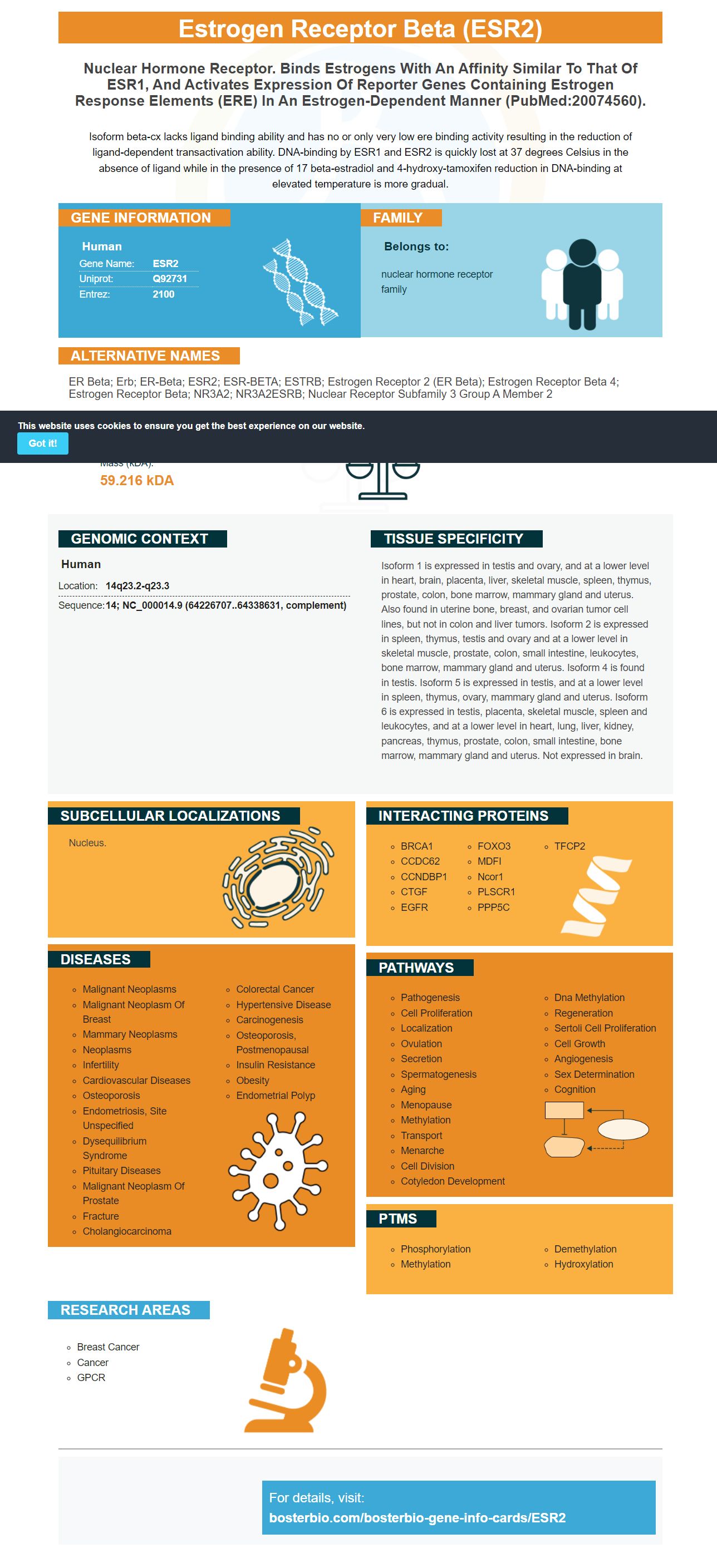
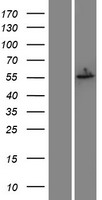
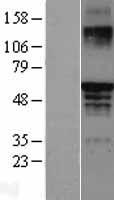
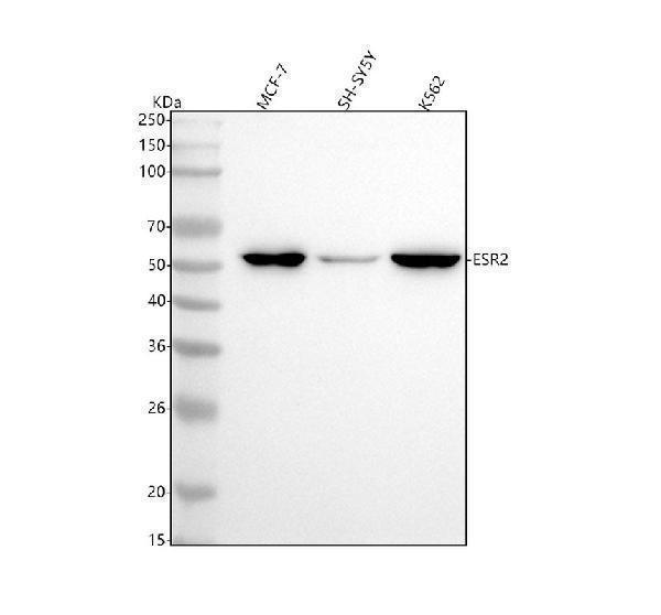
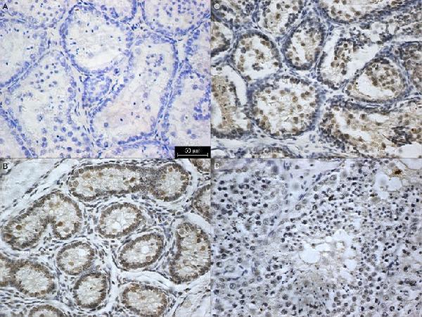
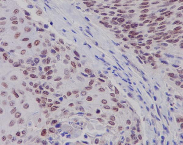
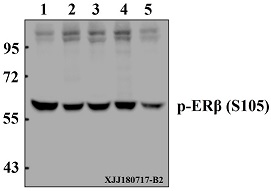
Facts about Estrogen receptor beta.
Isoform beta-cx lacks ligand binding ability and has no or only very low ere binding activity resulting in the reduction of ligand-dependent transactivation ability. DNA-binding by ESR1 and ESR2 is quickly lost at 37 degrees Celsius in the absence of ligand while in the presence of 17 beta-estradiol and 4-hydroxy-tamoxifen reduction in DNA-binding at elevated temperature is more gradual.
Human | |
---|---|
Gene Name: | ESR2 |
Uniprot: | Q92731 |
Entrez: | 2100 |
Belongs to: |
---|
nuclear hormone receptor family |
ER beta; Erb; ER-beta; ESR2; ESR-BETA; ESTRB; estrogen receptor 2 (ER beta); estrogen receptor beta 4; estrogen receptor beta; NR3A2; NR3A2ESRB; Nuclear receptor subfamily 3 group A member 2
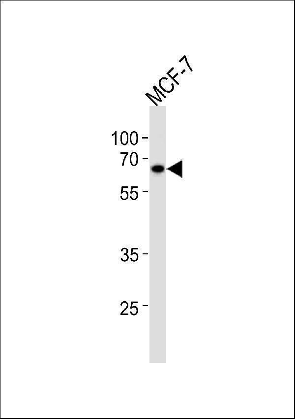
Mass (kDA):
59.216 kDA
Human | |
---|---|
Location: | 14q23.2-q23.3 |
Sequence: | 14; NC_000014.9 (64226707..64338631, complement) |
Isoform 1 is expressed in testis and ovary, and at a lower level in heart, brain, placenta, liver, skeletal muscle, spleen, thymus, prostate, colon, bone marrow, mammary gland and uterus. Also found in uterine bone, breast, and ovarian tumor cell lines, but not in colon and liver tumors. Isoform 2 is expressed in spleen, thymus, testis and ovary and at a lower level in skeletal muscle, prostate, colon, small intestine, leukocytes, bone marrow, mammary gland and uterus. Isoform 4 is found in testis. Isoform 5 is expressed in testis, and at a lower level in spleen, thymus, ovary, mammary gland and uterus. Isoform 6 is expressed in testis, placenta, skeletal muscle, spleen and leukocytes, and at a lower level in heart, lung, liver, kidney, pancreas, thymus, prostate, colon, small intestine, bone marrow, mammary gland and uterus. Not expressed in brain.
Nucleus.
What are the best applications of the ESR2 marker? This article will discuss the Anti-ESR2 Beta 3 antibody, CYBB activation and Autophagic flux impairment as and the advantages of the ESR2 marker. Here are some methods scientists can start. This guide is for scientists from all over the world.
The Anti-ESR2 beta-3 antibody from Boster Bio is able to recognize Human ESR2 antigen. It has been tested for use in Western Blot experiments. Boster Bio sells anti-ESR2 beta3 antibody as well as ESR2 antibodies. The results can then be compared to other studies. Visit the Boster Bio website for more information. Follow the directions on the label to purchase the antibody.
SuperSignal West Pico Chemiluminescent Substrate was used to produce secondary antibodies. Secondary antibodies were conjugated to HRP. After 1 h of incubation secondary antibodies were detected using an Cy3 conjugated secondary antibody. The immunofluorescent chemistry method was used to observe the signal. The antibodies were found to be extremely sensitive and specific to ESR2.
ESR1 is recognized by rabbit polyclonal antibodies HC-20 as well as mouse monoclonal antibodies MA1-310. Both peptides were generated by a synthetic peptide located within the DNA-binding region of human ESR2. The anti-ESR2 Beta-3 antibody is made from a bioreactor concentrate that contains 0.05% sodium azide. It is then dissolved in a solution that contains 0.09 percent sodium zide. The preparation process includes Saturated Ammonium Sulfate precipitation , as well as dialysis using PBS. Boster Bio's antisero2 beta3 antibodies originate from rabbits who have been immunized by a KLH-conjugated synthetic propeptide that contains amino acid one-153.
The Anti-ESR2 beta-3 antibody used in this study is known as A00786-1. The antibody was dispersed in PBS to an end-to-end concentration of 2 to 5percent. Under reducing conditions the samples were then loaded into sample wells. The samples in the second lane were rats ovary tissue lysate. one lane contained human Hela whole cell and lane three was a human MCF-7 whole Cell.
The cytokine IL-12 is well-known to play important roles in the disease process and activation of CYBB is one of them. Recently, Eissa et al. Eissa et al. recently identified a novel somatic mutation within the CYBB gene, and elucidated its effects on inflammatory conditions. The mutation was found to affect the protein's synthesis and function. The research has significant implications for the treatment of this disease.
The beta chain of cytochrome b-245, also known as the cytochrome is a vital component of the NADPH oxidase enzyme complex. It plays a crucial role in the immune system. It is beta chain partner. Both chains are required for NADPH oxidation. It is present in immune system cells, also known as phagocytes. When activated, it breaks down poisons and allows the cells to fight foreign invaders.
Activation by ESR2 of CYBB suppresses tumor cell viability. It also has the ability to affect cancer cells in vivo through non-autonomous mechanisms. Zhao et al. examined the effects of LY500307 on lung metastasis among mice that have a TNBC xenograft. Their research revealed that the compound inhibited lung metastasis and increased neutrophil recruitment to the tumor. This suggests that ESR2 activation could attract immune cells to the tumor microenvironment.
The activation of CYBB by ECR2 marker is a result of phosphorylation of the ERb gene. This allows the E3 ubiquitin-ligase E6AP (unliganded receptor) to be recruited. Additionally, the phosphorylation of ERb by ESR2 enhances its ubiquitination as well as degradation by the 26S proteasome.
ESR2 is a marker of autophagy and can be used to determine the level of function of cells. This marker measures the flux through the entire autophagy pathway starting from induction until the breakdown of the autophagic cargo. The probe showed high sensitivity when EBSS was treated in cell lines and increased puncta when CQ was treated cells. Yellow puncta could still be observed even without Ro however, puncta with red were not. The probe is also able to measure the size of autophagosomes and smaller ones are detected within the group that is starved.
The ESR2 marker can be utilized in conjunction with a multiplex immunophenotyping panel to evaluate the amount of autophagy under a variety of situations. Its use in autophagy studies is extremely flexible, and its application in cancer research could be a beneficial supplement to clinical research. The ESR2 marker's sensitivity ESR2 marker has been reported to be high in cancerous cells, specifically those with inflammation bowel disease.
This marker can also be used to track adiponectin (APC) Adiponectin (APC) is the precursor to apoptosis. It hinders autophagic flux by combining with apocynin. It also inhibits NCF1ROS, an important upstream marker of autophagy. This protein is downstream from autophagy, and it inhibits it, whereas 5-Fu encourages prosurvival.
The ESR2 marker for inhibition of autophagic flux has been used to distinguish between autophagosomes that are generated and blocked. The assay can determine the level of autophagosomes by analyzing the amount of SQSTM1/p62 that is found in cells. The assay was verified using an assay that uses real-time PCR.
In an earlier study, a simple gating strategy excluded dead cells as well as cytotoxic T cells. It prevented the decrease in levels of pCHEK1 by decreasing autophagy in B cells. It also slowed cell cycle progress and increased DNA damage. This approach has the potential to determine the mechanisms responsible for this effect and to optimize treatment options.
The ESR2 gene is expressed in the soleus muscles and myotubes made from C2C12 cell. A probe for hydrolysis, ESR2 PrimePCR, was used to determine the mRNA levels of the gene. The reference gene used in the experiments was hypoxanthine guanine phosphoribosyltransferase (HPRT). The reaction mix contained 5 ul cDNA and 1 LightCycler FastStart TaqMan probe master ESR2 PrimePCR and nuclease free water. LightCycler 2.0 carousel glass capillary-based system were used for the QPCR reactions.
PMID: 9473491 by Ogawa S., et al. The complete primary structure of human estrogen receptor beta (hERbeta) and its heterodimerization with ER alpha in vivo and in vitro.
PMID: 9636657 by Moore J.T., et al. Cloning and characterization of human estrogen receptor beta isoforms.
*More publications can be found for each product on its corresponding product page