Click image to see more details
-

-
-
-
-
+3
Product Info Summary
| SKU: | M02401 |
|---|---|
| Size: | 100 μl |
| Reactive Species: | Human, Mouse, Rat |
| Host: | Rabbit |
| Application: | IF, ICC, WB |
Customers Who Bought This Also Bought

Product info
Product Name
Anti-Ferritin FTH1 Rabbit Monoclonal Antibody
SKU/Catalog Number
M02401
BM4487 is an alternative SKU for this antibody, used in previous lots.
Size
100 μl
Form
Liquid
Description
Boster Bio Anti-Ferritin FTH1 Rabbit Monoclonal Antibody catalog # M02401. Tested in WB, ICC/IF applications. This antibody reacts with Human, Mouse, Rat.
Storage & Handling
Store at -20°C for one year. For short term storage and frequent use, store at 4°C for up to one month. Avoid repeated freeze-thaw cycles.
Cite This Product
Anti-Ferritin FTH1 Rabbit Monoclonal Antibody (Boster Biological Technology, Pleasanton CA, USA, Catalog # M02401)
Host
Rabbit
Contents
Rabbit IgG in stabilizing components, phosphate buffered saline, pH 7.4, 150mM NaCl, 0.02% sodium azide and 50% glycerol.
*This antibody is supplied in a stabilized formulation.
Compatibility with conjugation reactions depends on the chemistry of the conjugation method used.
For conjugation methods that are not compatible with the stabilizing components present in this formulation, a carrier-free antibody format is required.
Clonality
Monoclonal
Clone Number
FBA-6
Isotype
Rabbit IgG
Immunogen
A synthesized peptide derived from human Ferritin
Reactive Species
M02401 is reactive to FTH1 in Human, Mouse, Rat
Observed Molecular Weight
19 kDa
Antibody Validation
Boster validates all antibodies on WB, IHC, ICC, Immunofluorescence, and ELISA with known positive control and negative samples to ensure specificity and high affinity, including thorough antibody incubations.
Application & Images
Applications
M02401 is guaranteed for IF, ICC, WB Boster Guarantee
Recommend Dilution
WB 1:500-2000
ICC/IF 1:50-200
Tested application
Suggested blocking solution with 5% non-fat milk or BSA; (*)Recommended protein loading: 20-40 µg per lane
Validation Images & Assay Conditions
Click image to see more details
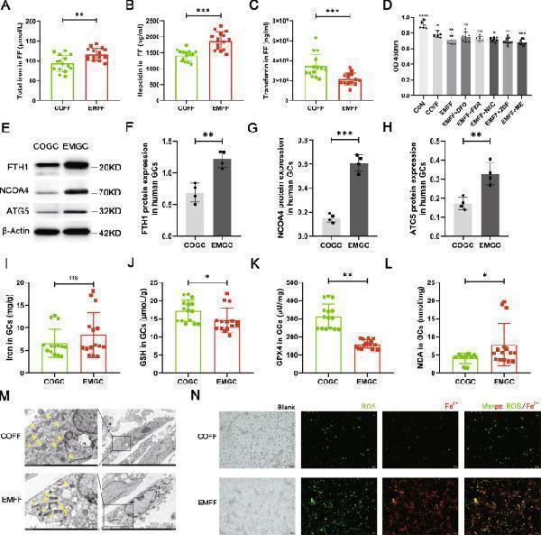
Iron-overloaded EMFF induced ferritinophagy-dependent ferroptosis in granulosa cells. A – C Levels of total iron, hepcidin, and transferrin in EMFF ( n = 15) and COFF ( n = 15). Data are expressed as means ± SD and analyzed by Student’s t test. D Results of mouse granulosa cells proliferation under different intervention conditions (each group in the figure is compared with COFF group). DFO, iron chelators; FER, ferroptosis inhibitor; NEC, necrosis inhibitor; ZDF, apoptosis inhibitor; ME, autophagy inhibitor. Data are expressed as means ± SD and analyzed by one-way ANOVA. E – H Comparison of ferritinophagy-related proteins FTH1, NCOA4, and ATG5 between human granulosa cells of infertile patients with EMs (EMGC) and of control group (COGC). The expression of β-actin was used as an internal control. Data are expressed as means ± SD and analyzed by Student’s t test. I – L Detection of ferroptosis-related indicators iron, GSH, GPX4, and MDA in COGC and EMGC. Data are expressed as means ± SD and analyzed by Student’s t test. M Representative images of the mitochondrial morphology of mouse granulosa cells intervened by COFF and EMFF were observed under TEM. Yellow arrows indicate mitochondrion. Scale bar = 1.0 µm. Scale bar = 5.0 µm. N Representative images of ROS and ferrous ion fluorescence staining after COFF and EMFF intervention in mouse granulosa cells. Scale bar = 100 µm. * P < 0.05, ** P < 0.01, *** P < 0.001, **** P < 0.0001, and ns, no significance.
Index in PubMed under a CC BY license. PMID: 35787614

Click image to see more details
Construction of an iron overload mouse model. A – C Serum levels of E 2 , FSH, and LH in standard iron (STD), low iron (LID), and high iron (HID) diet feeding groups ( n = 8). D – F Total iron, GSH, and MDA levels in the ovary tissues of mice in each group ( n = 8). G Representative images of ROS fluorescence staining of ovarian mouse granulosa cells in three groups of mice. Scale bar = 20 µm. H – K Western blot analysis of ferritinophagy-related proteins, FTH1, NCOA4, and ATG5 in mouse ovary tissues in the STD, LID, and HID group. The expression of β-actin was used as an internal control. All data are expressed as means ± SD and analyzed by one-way ANOVA. * P < 0.05, ** P < 0.01, *** P < 0.001, **** P < 0.0001, and ns, no significance.
Index in PubMed under a CC BY license. PMID: 35787614

Click image to see more details
Kidney tea attenuated ferroptosis in the kidney. (A) Representative images of transmission electron microscopy of kidney samples (scale bar: 2 μm/500 nm). (B) SOD activity in kidney samples. (C, D) MDA and 4-HNE levels in kidney samples. (E–H) The mRNA expression of NCOA4, FTH1, ACSL4 and GPX4 in kidney samples. (I–M) The protein levels of NCOA4, FTH1, ACSL4 and GPX4 in kidney samples. * p < 0.05, ** p < 0.01, *** p < 0.001 compared with the Mod group.
Index in PubMed under a CC BY license. PMID: 38962302

Click image to see more details
Western blot analysis of Ferritin using anti-Ferritin antibody (M02401).
Electrophoresis was performed on a 5-20% SDS-PAGE gel at 70V (Stacking gel) / 90V (Resolving gel) for 2-3 hours. The sample well of each lane was loaded with 30 ug of sample under reducing conditions.
Lane 1: human Hela whole cell lysates,
Lane 2: human 293T whole cell lysates,
Lane 3: human MCF-7 whole cell lysates,
Lane 4: human K562 whole cell lysates,
Lane 5: rat liver tissue lysates,
Lane 6: rat RH35 whole cell lysates,
Lane 7: mouse liver tissue lysates,
Lane 8: mouse HEPA1-6 whole cell lysates.
After electrophoresis, proteins were transferred to a nitrocellulose membrane at 150 mA for 50-90 minutes. Blocked the membrane with 5% non-fat milk/TBS for 1.5 hour at RT. The membrane was incubated with rabbit anti-Ferritin antigen affinity purified monoclonal antibody (M02401) at 1:500 overnight at 4°C, then washed with TBS-0.1%Tween 3 times with 5 minutes each and probed with a goat anti-rabbit IgG-HRP secondary antibody at a dilution of 1:500 for 1.5 hour at RT. The signal is developed using an Enhanced Chemiluminescent detection (ECL) kit (Catalog # EK1002) with Tanon 5200 system. A specific band was detected for Ferritin at approximately 19 kDa. The expected band size for Ferritin is at 21 kDa.

Click image to see more details
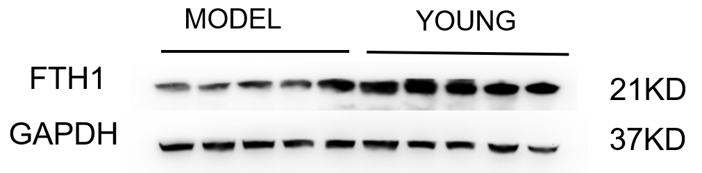
Western blot analysis of FTH1 using anti-FTH1 antibody (M02401).
Electrophoresis was performed on a 5-20% SDS-PAGE gel at 80V (Stacking gel) / 120V (Resolving gel) for 2 hours. The sample well of each lane was loaded with 30 ug of sample under reducing conditions.
Lane 1-5: model group-Human uterine tissue lysates,
Lane 2: young group-Human uterine tissue lysates.
After electrophoresis, proteins were transferred to a nitrocellulose membrane at 150 mA for 50-90 minutes. Blocked the membrane with 5% non-fat milk/TBS for 1.5 hour at RT. The membrane was incubated with rabbit anti-FTH1 antigen affinity purified monoclonal antibody (M02401) at 1:1000 overnight at 4°C, then washed with TBS-0.1%Tween 3 times with 5 minutes each and probed with a goat anti-rabbit IgG-HRP secondary antibody at a dilution of 1:5000 for 1 hour at RT. The signal is developed using an ECL Plus Western Blotting Substrate (Catalog # AR1196-200) with Tanon 5200 system. A specific band was detected for FTH1 at approximately 21 kDa. The expected band size for FTH1 is at 21 kDa.

Click image to see more details
Immunofluorescent analysis of Jurkat cells, using Ferritin Antibody.

Click image to see more details
Immunofluorescent analysis using the Antibody at 1:50 dilution.
Specific Publications For Anti-Ferritin FTH1 Rabbit Monoclonal Antibody (M02401)
Loading publications
Recommended Resources
Here are featured tools and databases that you might find useful.
- Boster's Pathways Library
- Protein Databases
- Bioscience Research Protocol Resources
- Data Processing & Analysis Software
- Photo Editing Software
- Scientific Literature Resources
- Research Paper Management Tools
- Molecular Biology Software
- Primer Design Tools
- Bioinformatics Tools
- Phylogenetic Tree Analysis
Customer Reviews
Have you used Anti-Ferritin FTH1 Rabbit Monoclonal Antibody?
Share your experimental results or join a short interview to earn up to $1,000 in product credits or other rewards.
1 Reviews For Anti-Ferritin FTH1 Rabbit Monoclonal Antibody
The FTH1 antibody was used to detect the expression of the target protein in human uterine tissue. The WB bands were single and clear, and compared with other domestic and international brands, this antibody offers excellent cost-performance.
Excellent
| SKU | M02401 |
|---|---|
| Application | Western Blot |
| Sample | Mouse hippocampus tissue |
| Sample Processing Description | The tissue was minced and sonicated, then lysed on ice for 1 hour using RIPA buffer. After centrifugation to collect the supernatant and protein quantification by BCA, samples were mixed with loading buffer at the appropriate ratio and denatured by boiling in a water bath. Fifteen microliters of each protein sample were loaded per lane onto SDS-PAGE gel. |
| Primary Antibody | Anti-Ferritin FTH1 Rabbit Monoclonal Antibody |
| Primary Incubation | overnight at 4 ℃ |
| Secondary Antibody | HRP-conjugated Anti-Rabbit IgG Secondary Antibody |
| Secondary Incubation | 1 hour in room temperature |
| Detection | Substrate: Ultra-sensitive ECL luminescent reagent (Cat# AR1191), Imaging system:Tanon |
| Results Summary | The FTH1 antibody was used to detect the expression of the target protein in human uterine tissue. The WB bands were single and clear, and compared with other domestic and international brands, this antibody offers excellent cost-performance. |
Anfeng Ning, Peking University Third Hospital
Verified customer
Submitted 2025-11-06
Customer Q&As
Have a question?
Find answers in Q&As, reviews.
Can't find your answer?
Submit your question
7 Customer Q&As for Anti-Ferritin FTH1 Rabbit Monoclonal Antibody
Question
I have a question about product M02401, anti-Ferritin Rabbit Monoclonal antibody. I was wondering if it would be possible to conjugate this antibody with biotin. I would need it to be without BSA or sodium azide. I am planning on using a buffer exchange of sodium azide with PBS only. Would there be problems for me to conjugate the antibody and store it in -20 degrees in small aliquots?
Verified Customer
Verified customer
Asked: 2019-08-05
Answer
It is not recommended storing this antibody with PBS buffer only in -20 degrees. If you want to store it in -20 degrees it is best to add some cryoprotectant like glycerol. If you want carrier free M02401 anti-Ferritin Rabbit Monoclonal antibody, we can provide it to you in a special formula with trehalose and/or glycerol. These molecules will not interfere with conjugation chemistry and provide a good level of protection for the antibody from degradation. Please be sure to specify this in your purchase order.
Boster Scientific Support
Answered: 2019-08-05
Question
I was wanting to use your anti-Ferritin Rabbit Monoclonal antibody for ICC for mouse tibial nerve on frozen tissues, but I want to know if it has been tested for this particular application. Has this antibody been tested and is this antibody a good choice for mouse tibial nerve identification?
Verified Customer
Verified customer
Asked: 2019-05-21
Answer
You can see on the product datasheet, M02401 anti-Ferritin Rabbit Monoclonal antibody has been validated for IF, ICC, WB on human, mouse, rat tissues. We have an innovator award program that if you test this antibody and show it works in mouse tibial nerve in IHC-frozen, you can get your next antibody for free.
Boster Scientific Support
Answered: 2019-05-21
Question
Does M02401 anti-Ferritin Rabbit Monoclonal antibody work on parafin embedded sections? If so, which fixation method do you recommend we use (PFA, paraformaldehyde, other)?
Verified Customer
Verified customer
Asked: 2019-05-07
Answer
As indicated on the product datasheet, M02401 anti-Ferritin Rabbit Monoclonal antibody as been validated on ICC. It is best to use PFA for fixation because it has better tissue penetration ability. PFA needs to be prepared fresh before use. Long term stored PFA turns into formalin, as the PFA molecules congregate and become formalin.
Boster Scientific Support
Answered: 2019-05-07
Question
I see that the anti-Ferritin Rabbit Monoclonal antibody M02401 works with ICC, what is the protocol used to produce the result images on the product page?
Verified Customer
Verified customer
Asked: 2018-12-12
Answer
You can find protocols for ICC on the "support/technical resources" section of our navigation menu. If you have any further questions, please send an email to support@bosterbio.com
Boster Scientific Support
Answered: 2018-12-12
Question
Will anti-Ferritin Rabbit Monoclonal antibody M02401 work for ICC with tibial nerve?
P. Krishna
Verified customer
Asked: 2018-01-16
Answer
According to the expression profile of tibial nerve, FTH1 is highly expressed in tibial nerve. So, it is likely that anti-Ferritin Rabbit Monoclonal antibody M02401 will work for ICC with tibial nerve.
Boster Scientific Support
Answered: 2018-01-16
Question
Is there a BSA free version of anti-Ferritin Rabbit Monoclonal antibody M02401 available?
L. Dhar
Verified customer
Asked: 2017-09-25
Answer
Thank you for your recent telephone inquiry. I can confirm that some lots of this anti-Ferritin Rabbit Monoclonal antibody M02401 are BSA free. For now, these lots are available and we can make a BSA free formula for you free of charge. It will take 3 extra days to prepare. If you require this antibody BSA free again in future, please do not hesitate to contact me and I will be pleased to check which lots we have in stock that are BSA free.
Boster Scientific Support
Answered: 2017-09-25
Question
We are currently using anti-Ferritin Rabbit Monoclonal antibody M02401 for rat tissue, and we are well pleased with the ICC results. The species of reactivity given in the datasheet says human, mouse, rat. Is it possible that the antibody can work on goat tissues as well?
Verified Customer
Verified customer
Asked: 2017-08-24
Answer
The anti-Ferritin Rabbit Monoclonal antibody (M02401) has not been validated for cross reactivity specifically with goat tissues, though there is a good chance of cross reactivity. We have an innovator award program that if you test this antibody and show it works in goat you can get your next antibody for free. Please contact me if I can help you with anything.
Boster Scientific Support
Answered: 2017-08-24
