Click image to see more details
-

-
-
-
-
+2
Product Info Summary
| SKU: | M00564-3 |
|---|---|
| Size: | 100 μl/vial |
| Reactive Species: | Human, Mouse, Rat |
| Host: | Rabbit |
| Application: | Flow Cytometry, IF, IHC, ICC, WB |
Customers Who Bought This Also Bought

Product info
Product Name
Anti-Fibronectin Rabbit Monoclonal Antibody
SKU/Catalog Number
M00564-3
Size
100 μl/vial
Form
Liquid
Description
Boster Bio Anti-Fibronectin Rabbit Monoclonal Antibody catalog # M00564-3. Tested in WB, IHC, ICC/IF, Flow Cytometry applications. This antibody reacts with Human, Mouse, Rat.
Storage & Handling
Store at -20°C for one year. For short term storage and frequent use, store at 4°C for up to one month. Avoid repeated freeze-thaw cycles.
Cite This Product
Anti-Fibronectin Rabbit Monoclonal Antibody (Boster Biological Technology, Pleasanton CA, USA, Catalog # M00564-3)
Host
Rabbit
Contents
Rabbit IgG in stabilizing components, phosphate buffered saline, pH 7.4, 150mM NaCl, 0.02% sodium azide and 50% glycerol.
*This antibody is supplied in a stabilized formulation.
Compatibility with conjugation reactions depends on the chemistry of the conjugation method used.
For conjugation methods that are not compatible with the stabilizing components present in this formulation, a carrier-free antibody format is required.
Clonality
Monoclonal
Clone Number
26F78
Isotype
IgG
Immunogen
A synthesized peptide derived from human Fibronectin
Reactive Species
M00564-3 is reactive to FN1 in Human, Mouse, Rat
Observed Molecular Weight
272 kDa
Calculated molecular weight
272.3 kDa
Antibody Validation
Boster validates all antibodies on WB, IHC, ICC, Immunofluorescence, and ELISA with known positive control and negative samples to ensure specificity and high affinity, including thorough antibody incubations.
Application & Images
Applications
M00564-3 is guaranteed for Flow Cytometry, IF, IHC, ICC, WB Boster Guarantee
Recommend Dilution
WB 1:500-2000
IHC 1:50-200
ICC/IF 1:50-200
FC 1:50
Tested application
Suggested blocking solution with 5% non-fat milk or BSA; (*)Recommended protein loading: 20-40 µg per lane
Use TE buffer pH 9.0 for antigen retrieval; (*) citrate buffer pH 6.0 is an alternative.
Validation Images & Assay Conditions
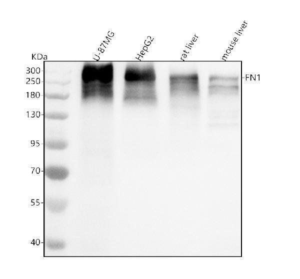
Click image to see more details
Western blot analysis of Fibronectin using anti-Fibronectin antibody (M00564-3).
Electrophoresis was performed on a 5-20% SDS-PAGE gel at 70V (Stacking gel) / 90V (Resolving gel) for 2-3 hours. The sample well of each lane was loaded with 30 ug of sample under reducing conditions.
Lane 1: human U-87MG whole cell lysates,
Lane 2: human HepG2 whole cell lysates,
Lane 3: rat liver tissue lysates,
Lane 4: mouse liver tissue lysates.
After electrophoresis, proteins were transferred to a nitrocellulose membrane at 150 mA for 50-90 minutes. Blocked the membrane with 5% non-fat milk/TBS for 1.5 hour at RT. The membrane was incubated with rabbit anti-Fibronectin antigen affinity purified monoclonal antibody (Catalog # M00564-3) at 1:500 overnight at 4°C, then washed with TBS-0.1%Tween 3 times with 5 minutes each and probed with a goat anti-rabbit IgG-HRP secondary antibody at a dilution of 1:500 for 1.5 hour at RT. The signal is developed using an Enhanced Chemiluminescent detection (ECL) kit (Catalog # EK1002) with Tanon 5200 system. A specific band was detected for Fibronectin at approximately 272 kDa. The expected band size for Fibronectin is at 272 kDa.

Click image to see more details
IHC analysis of Fibronectin using anti-Fibronectin antibody (M00564-3).
Fibronectin was detected in a paraffin-embedded section of human colorectal adenocarcinoma tissue. Heat mediated antigen retrieval was performed in EDTA buffer (pH 8.0, epitope retrieval solution). The tissue section was blocked with 10% goat serum. The tissue section was then incubated with 1:50 rabbit anti-Fibronectin Antibody (M00564-3) overnight at 4°C. Peroxidase Conjugated Goat Anti-rabbit IgG was used as secondary antibody and incubated for 30 minutes at 37°C. The tissue section was developed using HRP Conjugated Rabbit IgG Super Vision Assay Kit (Catalog # SV0002) with DAB as the chromogen.

Click image to see more details
IHC analysis of Fibronectin using anti-Fibronectin antibody (M00564-3).
Fibronectin was detected in a paraffin-embedded section of human liver cancer tissue. Heat mediated antigen retrieval was performed in EDTA buffer (pH 8.0, epitope retrieval solution). The tissue section was blocked with 10% goat serum. The tissue section was then incubated with 1:50 rabbit anti-Fibronectin Antibody (M00564-3) overnight at 4°C. Peroxidase Conjugated Goat Anti-rabbit IgG was used as secondary antibody and incubated for 30 minutes at 37°C. The tissue section was developed using HRP Conjugated Rabbit IgG Super Vision Assay Kit (Catalog # SV0002) with DAB as the chromogen.

Click image to see more details
rFBA and fibronectin interaction analysis by Far-WB and Surface plasmon resonance (SPR) analysis. A Far-WB analysis of rFBA with fibronectin. The first lane: PVDF membrane with transferred rFBA protein incubated with anti- rFBA antibody as a positive control; the second lane: PVDF membrane with transferred rFBA protein incubated with fibronectin and anti-fibronectin antibody; the third lane: PVDF membrane with transferred BSA (negative control) incubated with fibronectin and anti-fibronectin antibody. Protein bands were visualized using ECL substrate. B Sensorgrams depict the binding of immobilised fibronectin to rFBA. Increasing concentrations of rFBA (5, 10, 25, 50 and 100 μg/mL) were injected at a flow rate of 30 μL/min for 180 s over immobilised fibronectin. The arrow indicates the end of the injection period, at which point dissociation of rFBA from fibronectin can be observed. RU resonance units.
Index in PubMed under a CC BY license. PMID: 30454073

Click image to see more details
Representative immunofluorescence images of fibronectin. Fluorescent micrographs of tenocytes after 2 and 5 days of culture on the surface of a culture plate and acellular amniotic membrane. Tenocyte nucleus shape observed under a fluorescence microscope (A,D,G,J) . Fibronectin presented positive after the fluorescent FITC mark was observed under a fluorescence microscope (B,E,H,K) . Tenocyte nucleus and fibronectin merging (C,F,I,L) . The corresponding semi quantitative analysis of fibronectin fluorescence intensity in panels (M,N) (scale bar = 50 um, n = 5, * P < 0.05).
Index in PubMed under a CC BY license. PMID: 32478059

Click image to see more details
Fluorescence images of tenocytes after 5 days of culture on the surface of a culture plate (control group) and acellular amniotic membrane (amnion group). Tenocytes presented a clear cytoskeleton, good biocompatibility with acellular amniotic membrane, and even distribution on the surface of the materials, showing better growth activity than the control group (A,B) . Cell viability was measured by CCK-8, and the proliferation curve of tenocytes in the control and amniotic membrane groups was drawn (C) . Western blot assay for collagen I, fibronectin, TGF-β1, and bFGF expression in the tenocytes of the control and amnion groups for 1 week (D,E) . * p < 0.05; ** p < 0.01; and *** p < 0.001.
Index in PubMed under a CC BY license. PMID: 32478059
Specific Publications For Anti-Fibronectin Rabbit Monoclonal Antibody (M00564-3)
Loading publications
Recommended Resources
Here are featured tools and databases that you might find useful.
- Boster's Pathways Library
- Protein Databases
- Bioscience Research Protocol Resources
- Data Processing & Analysis Software
- Photo Editing Software
- Scientific Literature Resources
- Research Paper Management Tools
- Molecular Biology Software
- Primer Design Tools
- Bioinformatics Tools
- Phylogenetic Tree Analysis
Customer Reviews
Have you used Anti-Fibronectin Rabbit Monoclonal Antibody?
Share your experimental results or join a short interview to earn up to $1,000 in product credits or other rewards.
0 Reviews For Anti-Fibronectin Rabbit Monoclonal Antibody
Customer Q&As
Have a question?
Find answers in Q&As, reviews.
Can't find your answer?
Submit your question
