This website uses cookies to ensure you get the best experience on our website.
- Table of Contents

22 Citations 7 Q&As

10 Citations 5 Q&As
12 Citations 12 Q&As
8 Citations 16 Q&As

6 Citations 16 Q&As


4 Citations 5 Q&As
41 Citations
1 Citations
Facts about Fibronectin.

Involved in osteoblast compaction throughout the fibronectin fibrillogenesis cell-mediated matrix assembly process, essential for osteoblast mineralization. Participates in the regulation of type I collagen deposition by osteoblasts.
| Human | |
|---|---|
| Gene Name: | FN1 |
| Uniprot: | P02751 |
| Entrez: | 2335 |

| Belongs to: |
|---|
| No superfamily |

CIG; ED-B; fibronectin 1; Fibronectin; FINC; FN; FN1; FNZ; GFND; GFND2; LETS; MSF; SMDCF
Mass (kDA):
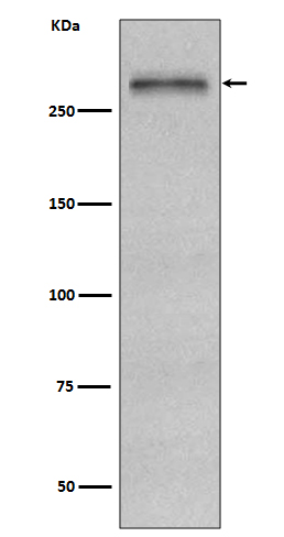
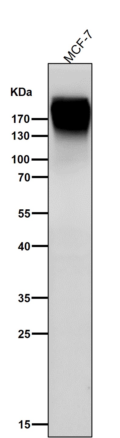
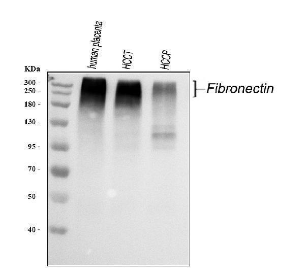
272.32 kDA

| Human | |
|---|---|
| Location: | 2q35 |
| Sequence: | 2; NC_000002.12 (215360440..215436167, complement) |
Plasma FN (soluble dimeric form) is secreted by hepatocytes. Cellular FN (dimeric or cross-linked multimeric forms), made by fibroblasts, epithelial and other cell types, is deposited as fibrils in the extracellular matrix. Ugl-Y1, Ugl-Y2 and Ugl-Y3 are found in urine.
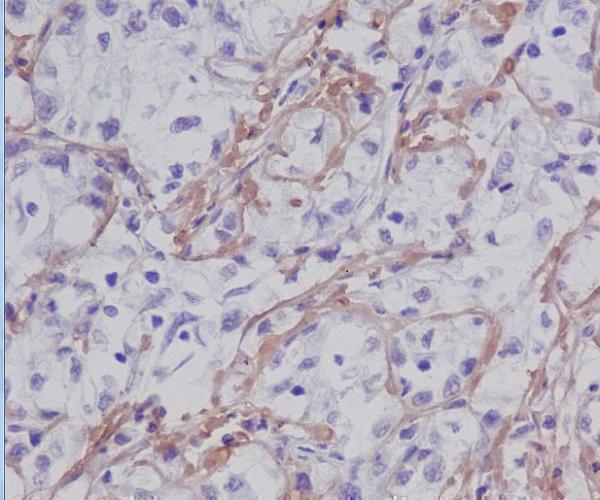
Secreted, extracellular space, extracellular matrix.


PMID: 11737888 by Schor S.L., et al. Phenotypic and genetic alterations in mammary stroma: implications for tumour progression.
PMID: 16322219 by Kay R.A., et al. The expression of migration stimulating factor, a potent oncofetal cytokine, is uniquely controlled by 3'-untranslated region-dependent nuclear sequestration of its precursor messenger RNA.
*Showing only the more recent 20. More publications can be found for each product on its corresponding product page