Click image to see more details
Product Info Summary
| SKU: | A00188-4 |
|---|---|
| Size: | 100 μg/vial |
| Reactive Species: | Mouse, Rat |
| Host: | Rabbit |
| Application: | ELISA, Flow Cytometry, WB |
Customers Who Bought This Also Bought

Product info
Product Name
Anti-Flt3/CD135 Antibody Picoband®
SKU/Catalog Number
A00188-4
Size
100 μg/vial
Form
Lyophilized
Description
Boster Bio Anti-Flt3/CD135 Antibody Picoband® catalog # A00188-4. Tested in ELISA, Flow Cytometry, WB applications. This antibody reacts with Mouse, Rat. The brand Picoband indicates this is a premium antibody that guarantees superior quality, high affinity, and strong signals with minimal background in Western blot applications. Only our best-performing antibodies are designated as Picoband, ensuring unmatched performance.
Storage & Handling
Store at -20˚C for one year from date of receipt. After reconstitution, at 4˚C for one month. It can also be aliquotted and stored frozen at -20˚C for six months. Avoid repeated freeze-thaw cycles.
Cite This Product
Anti-Flt3/CD135 Antibody Picoband® (Boster Biological Technology, Pleasanton CA, USA, Catalog # A00188-4)
Host
Rabbit
Contents
Each vial contains 4 mg Trehalose, 0.9 mg NaCl and 0.2 mg Na2HPO4.
Clonality
Polyclonal
Isotype
Rabbit IgG
Immunogen
E. coli-derived mouse Flt3 / CD135 recombinant protein (Position: E62-E295).
Cross-reactivity
No cross-reactivity with other proteins.
Reactive Species
A00188-4 is reactive to Flt3 in Mouse, Rat
Observed Molecular Weight
150 kDa
Calculated molecular weight
113.5 kDa
Background of Flt3
Cluster of differentiation antigen 135 (CD135) also known as fms like tyrosine kinase 3 (FLT-3), receptor-type tyrosine-protein kinase FLT3, or fetal liver kinase-2 (Flk2) is a protein that in humans is encoded by the FLT3 gene. This gene encodes a class III receptor tyrosine kinase that regulates hematopoiesis. This receptor is activated by binding of the fms-related tyrosine kinase 3 ligand to the extracellular domain, which induces homodimer formation in the plasma membrane leading to autophosphorylation of the receptor. The activated receptor kinase subsequently phosphorylates and activates multiple cytoplasmic effector molecules in pathways involved in apoptosis, proliferation, and differentiation of hematopoietic cells in bone marrow.
Antibody Validation
Boster validates all antibodies on WB, IHC, ICC, Immunofluorescence, and ELISA with known positive control and negative samples to ensure specificity and high affinity, including thorough antibody incubations.
Application & Images
Applications
A00188-4 is guaranteed for ELISA, Flow Cytometry, WB Boster Guarantee
Recommend Dilution
| Application | Dilution | Species |
|---|---|---|
| Western blot | 0.1-0.5μg/ml | Mouse, Rat |
| Flow Cytometry (Fixed) | 1-3μg/1x106 cells | Mouse ELISA, 0.1-0.5μg/ml, - |
Tested application
Suggested blocking solution with 5% non-fat milk or BSA; (*)Recommended protein loading: 20-40 µg per lane
Validation Images & Assay Conditions
Click image to see more details
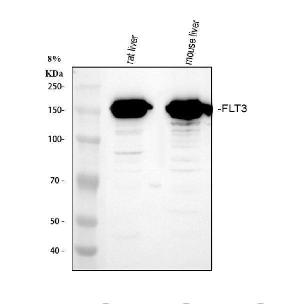
Western blot analysis of Flt3/CD135 using anti-Flt3/CD135 antibody (A00188-4).
Electrophoresis was performed on a 8% SDS-PAGE gel at 80V (Stacking gel) / 120V (Resolving gel) for 2 hours. The sample well of each lane was loaded with 30 ug of sample under reducing conditions.
Lane 1: rat liver tissue lysates,
Lane 2: mouse liver tissue lysates.
After electrophoresis, proteins were transferred to a nitrocellulose membrane at 150 mA for 50-90 minutes. Blocked the membrane with 5% non-fat milk/TBS for 1.5 hour at RT. The membrane was incubated with rabbit anti-Flt3/CD135 antigen affinity purified polyclonal antibody (A00188-4) at 0.5 μg/mL overnight at 4°C, then washed with TBS-0.1%Tween 3 times with 5 minutes each and probed with a goat anti-rabbit IgG-HRP secondary antibody (Catalog # BA1054) at a dilution of 1:5000 for 1.5 hour at RT. The signal is developed using an ECL Plus Western Blotting Substrate (Catalog # AR1196-200) with Tanon 5200 system. A specific band was detected for Flt3/CD135 at approximately 150 kDa. The expected band size for Flt3/CD135 is at 130 kDa.

Click image to see more details
Flow Cytometry analysis of RAW264.7 cells using anti-Flt3/CD135 antibody (A00188-4).
Overlay histogram showing RAW264.7 cells stained with A00188-4 (Blue line). The cells were fixed with 4% paraformaldehyde and blocked with 10% normal goat serum. And then incubated with rabbit anti-Flt3/CD135 Antibody (A00188-4, 1 μg/1x106 cells) for 30 min at 20°C. DyLight®488 conjugated goat anti-rabbit IgG (BA1127, 5-10 μg/1x106 cells) was used as secondary antibody for 30 minutes at 20°C. Isotype control antibody (Green line) was rabbit IgG (1 μg/1x106) used under the same conditions. Unlabelled sample without incubation with primary antibody and secondary antibody (Red line) was used as a blank control.
Specific Publications For Anti-Flt3/CD135 Antibody Picoband® (A00188-4)
Loading publications
Recommended Resources
Here are featured tools and databases that you might find useful.
- Boster's Pathways Library
- Protein Databases
- Bioscience Research Protocol Resources
- Data Processing & Analysis Software
- Photo Editing Software
- Scientific Literature Resources
- Research Paper Management Tools
- Molecular Biology Software
- Primer Design Tools
- Bioinformatics Tools
- Phylogenetic Tree Analysis
Customer Reviews
Have you used Anti-Flt3/CD135 Antibody Picoband®?
Share your experimental results or join a short interview to earn up to $1,000 in product credits or other rewards.
0 Reviews For Anti-Flt3/CD135 Antibody Picoband®
Customer Q&As
Have a question?
Find answers in Q&As, reviews.
Can't find your answer?
Submit your question
4 Customer Q&As for Anti-Flt3/CD135 Antibody Picoband®
Question
Our team were satisfied with the WB result of your anti-Flt3/CD135 antibody. However we have seen positive staining in lymphocyte membrane using this antibody. Is that expected? Could you tell me where is FLT3 supposed to be expressed?
Verified Customer
Verified customer
Asked: 2020-03-20
Answer
According to literature, lymphocyte does express FLT3. Generally FLT3 expresses in membrane. Regarding which tissues have FLT3 expression, here are a few articles citing expression in various tissues:
Bone marrow, Pubmed ID: 7507245
Lymphocyte, Pubmed ID: 8394751, 15057823
Testis, Pubmed ID: 2004790
Boster Scientific Support
Answered: 2020-03-20
Question
We purchased anti-Flt3/CD135 antibody for ELISA on lymphocyte in a previous experiment. I am using mouse, and I plan to use the antibody for WB next. I am interested in examining lymphocyte as well as bone marrow in our next experiment. Could you please give me some suggestion on which antibody would work the best for WB?
Verified Customer
Verified customer
Asked: 2020-01-22
Answer
I took a look at the website and datasheets of our anti-Flt3/CD135 antibody and it appears that A00188-4 has been tested on mouse in both ELISA and WB. Thus A00188-4 should work for your application. Our Boster satisfaction guarantee will cover this product for WB in mouse even if the specific tissue type has not been validated. We do have a comprehensive range of products for WB detection and you can check out our website bosterbio.com to find out more information about them.
Boster Scientific Support
Answered: 2020-01-22
Question
We have observed staining in mouse cerebellar hemisphere. Do you have any suggestions? Is anti-Flt3/CD135 antibody supposed to stain cerebellar hemisphere positively?
K. Williams
Verified customer
Asked: 2017-05-22
Answer
From what I have seen in literature cerebellar hemisphere does express FLT3. From what I have seen in Uniprot.org, FLT3 is expressed in cerebellar hemisphere, bone marrow, lymphocyte, testis, among other tissues. Regarding which tissues have FLT3 expression, here are a few articles citing expression in various tissues:
Bone marrow, Pubmed ID: 7507245
Lymphocyte, Pubmed ID: 8394751, 15057823
Testis, Pubmed ID: 2004790
Boster Scientific Support
Answered: 2017-05-22
Question
We are currently using anti-Flt3/CD135 antibody A00188-4 for rat tissue, and we are content with the ELISA results. The species of reactivity given in the datasheet says mouse, rat. Is it possible that the antibody can work on pig tissues as well?
V. Parker
Verified customer
Asked: 2013-03-18
Answer
The anti-Flt3/CD135 antibody (A00188-4) has not been validated for cross reactivity specifically with pig tissues, though there is a good chance of cross reactivity. We have an innovator award program that if you test this antibody and show it works in pig you can get your next antibody for free. Please contact me if I can help you with anything.
Boster Scientific Support
Answered: 2013-03-18
