Click image to see more details
-

-
-
-
-
+4
Product Info Summary
| SKU: | M01032-2 |
|---|---|
| Size: | 100 µl |
| Reactive Species: | Human, Mouse, Rat |
| Host: | Mouse |
| Application: | Flow Cytometry, IF, IHC, WB |
Customers Who Bought This Also Bought

Product info
Product Name
Anti-FOXA2 Mouse Monoclonal Antibody [Clone ID: OTI3C10]
SKU/Catalog Number
M01032-2
Size
100 µl
Description
Boster Bio Anti-FOXA2 mouse monoclonal antibody, clone OTI3C10 (formerly 3C10). Catalog# M01032-2. Tested in FC, IF, IHC, WB. This antibody reacts with Human, Mouse, Rat.
Storage & Handling
Store at -20°C as received.
Cite This Product
Anti-FOXA2 Mouse Monoclonal Antibody [Clone ID: OTI3C10] (Boster Biological Technology, Pleasanton CA, USA, Catalog # M01032-2)
Host
Mouse
Contents
PBS (pH 7.3) containing 1% stabilizing protein, 50% glycerol and 0.02% sodium azide.
This antibody is supplied in a stabilized formulation.
Compatibility with conjugation reactions depends on the chemistry of the conjugation method used.
For conjugation methods that are not compatible with the stabilizing components present in this formulation, a carrier-free antibody format is required.
Clonality
Monoclonal
Clone Number
OTI3C10
Isotype
IgG1
Immunogen
Recombinant protein expressed in E.coli corresponding to amino acids 300-458 of human FOXA2
Reactive Species
M01032-2 is reactive to FOXA2 in Human, Mouse, Rat
Observed Molecular Weight
48 kDa
Calculated molecular weight
48.3 kDa
Antibody Validation
Boster validates all antibodies on WB, IHC, ICC, Immunofluorescence, and ELISA with known positive control and negative samples to ensure specificity and high affinity, including thorough antibody incubations.
Application & Images
Applications
M01032-2 is guaranteed for Flow Cytometry, IF, IHC, WB Boster Guarantee
Recommend Dilution
| Application | Dilution | Species |
|---|---|---|
| Flow Cytometry: 1:1 | 000 |
Validation Images & Assay Conditions
Click image to see more details
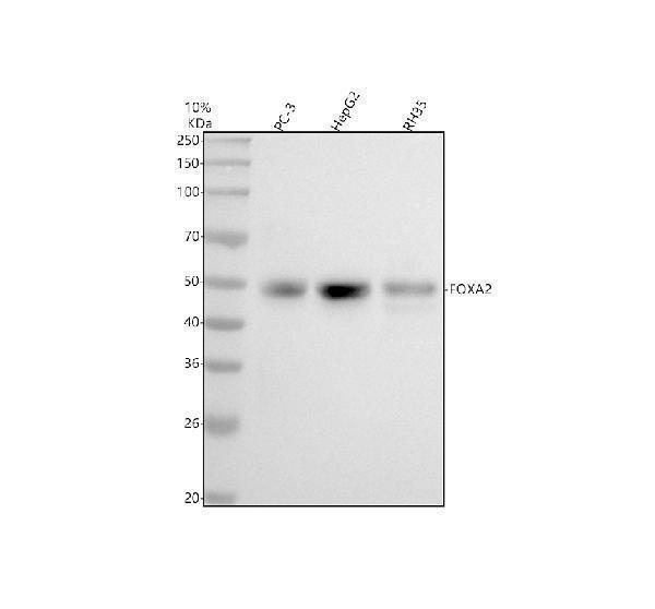
Western blot analysis of FOXA2 using anti-FOXA2 antibody (M01032-2).
Electrophoresis was performed on a 5-20% SDS-PAGE gel at 70V (Stacking gel) / 90V (Resolving gel) for 2-3 hours. The sample well of each lane was loaded with 30 ug of sample under reducing conditions.
Lane 1: human PC-3 whole cell lysates,
Lane 2: human HepG2 whole cell lysates,
Lane 3: rat RH35 whole cell lysates.
After electrophoresis, proteins were transferred to a nitrocellulose membrane at 150 mA for 50-90 minutes. Blocked the membrane with 5% non-fat milk/TBS for 1.5 hour at RT. The membrane was incubated with mouse anti-FOXA2 antigen affinity purified monoclonal antibody (Catalog # M01032-2) at 1:1000 overnight at 4°C, then washed with TBS-0.1%Tween 3 times with 5 minutes each and probed with a goat anti-mouse IgG-HRP secondary antibody at a dilution of 1:5000 for 1.5 hour at RT. The signal is developed using an Enhanced Chemiluminescent detection (ECL) kit (Catalog # EK1001) with Tanon 5200 system. A specific band was detected for FOXA2 at approximately 48 kDa. The expected band size for FOXA2 is at 48 kDa.

Click image to see more details
Immunofluorescent staining of HepG2 cells using anti-FOXA2 mouse monoclonal antibody (M01032-2) at 1:50 dilution.

Click image to see more details
HEK293T cells transfected with either pCMV6-ENTRY FOXA2 (Red) or empty vector control plasmid (Blue) were immunostained with anti-FOXA2 mouse monoclonal (M01032-2

Click image to see more details
Anti-FOXA2 mouse monoclonal antibody (M01032-2) immunofluorescent staining of HeLa cells transiently transfected by pCMV6-ENTRY FOXA2

Click image to see more details
Immunohistochemical staining of paraffin-embedded lung within the normal limits using anti-FOXA2 (M01032-2) mouse monoclonal antibody (Heat-induced epitope retrieval by 10mM citric buffer

Click image to see more details
Immunohistochemical staining of paraffin-embedded Carcinoma of liver using anti-FOXA2 (M01032-2) mouse monoclonal antibody (Heat-induced epitope retrieval by 10mM citric buffer

Click image to see more details
Western blot analysis of extracts (10ug) from a mouse cell line and 3 different mouse tissues by using anti-FOXA2 monoclonal antibody (1:200).

Click image to see more details
HEK293T cells were transfected with the pCMV6-ENTRY control (Left lane) or pCMV6-ENTRY FOXA2 (Right lane) cDNA for 48 hrs and lysed. Equivalent amounts of cell lysates (5 ug per lane) were separated by SDS-PAGE and immunoblotted with anti-FOXA2.
Specific Publications For Anti-FOXA2 Mouse Monoclonal Antibody [Clone ID: OTI3C10] (M01032-2)
Loading publications
Recommended Resources
Here are featured tools and databases that you might find useful.
- Boster's Pathways Library
- Protein Databases
- Bioscience Research Protocol Resources
- Data Processing & Analysis Software
- Photo Editing Software
- Scientific Literature Resources
- Research Paper Management Tools
- Molecular Biology Software
- Primer Design Tools
- Bioinformatics Tools
- Phylogenetic Tree Analysis
Customer Reviews
Have you used Anti-FOXA2 Mouse Monoclonal Antibody [Clone ID: OTI3C10]?
Share your experimental results or join a short interview to earn up to $1,000 in product credits or other rewards.
0 Reviews For Anti-FOXA2 Mouse Monoclonal Antibody [Clone ID: OTI3C10]
Customer Q&As
Have a question?
Find answers in Q&As, reviews.
Can't find your answer?
Submit your question
