Click image to see more details
-

-
-
-
-
+3
Product Info Summary
| SKU: | A00470 |
|---|---|
| Size: | 100 μg/vial |
| Reactive Species: | Human, Mouse, Rat |
| Host: | Rabbit |
| Application: | IF, IHC, ICC, WB, ELISA (Cap) |
Customers Who Bought This Also Bought

Product info
Product Name
Anti-Galectin 1/Lgals1 Antibody Picoband®
SKU/Catalog Number
A00470
Size
100 μg/vial
Form
Lyophilized
Description
Boster Bio Anti-Galectin 1/Lgals1 Antibody Picoband® catalog # A00470. Tested in ELISA, ICC/IF, IHC, WB applications. This antibody reacts with Human, Mouse, Rat. The brand Picoband indicates this is a premium antibody that guarantees superior quality, high affinity, and strong signals with minimal background in Western blot applications. Only our best-performing antibodies are designated as Picoband, ensuring unmatched performance.
Storage & Handling
Store at -20˚C for one year from date of receipt. After reconstitution, at 4˚C for one month. It can also be aliquotted and stored frozen at -20˚C for six months. Avoid repeated freeze-thaw cycles.
Cite This Product
Anti-Galectin 1/Lgals1 Antibody Picoband® (Boster Biological Technology, Pleasanton CA, USA, Catalog # A00470)
Host
Rabbit
Contents
Each vial contains antibody formulated with stabilizing components, 0.9 mg NaCl, 0.2 mg Na2HPO4, and 0.05 mg NaN3.
*This antibody is supplied in a stabilized formulation.
Compatibility with conjugation reactions depends on the chemistry of the conjugation method used.
For conjugation methods that are not compatible with the stabilizing components present in this formulation, a carrier-free antibody format is required.
Clonality
Polyclonal
Isotype
Rabbit IgG
Immunogen
E. coli-derived mouse Galectin 1 recombinant protein (Position: A2-E135).
Cross-reactivity
No cross-reactivity with other proteins.
Reactive Species
A00470 is reactive to Lgals1 in Human, Mouse, Rat
Observed Molecular Weight
15 kDa
Calculated molecular weight
14.9 kDa
Background of Lgals1
Galectin-1 is a protein that in humans is encoded by the LGALS1 gene. The galectins are a family of beta-galactoside-binding proteins implicated in modulating cell-cell and cell-matrix interactions. Galectin-1 may act as an autocrine negative growth factor that regulates cell proliferation. Galectin-1 expression in Hodgkin Lymphoma has also been shown to mediate immunosuppression of CD8+ T-cells. It is thought to play a role in creating immune tolerance in pregnancy. It has been found that Galectin-1-mediated production of IL6 may assist in augmenting the innate immune response against NiV. Galectin-1 may also be a significant factor that augments the efficiency of the HIV-1 infection process.
Antibody Validation
Boster validates all antibodies on WB, IHC, ICC, Immunofluorescence, and ELISA with known positive control and negative samples to ensure specificity and high affinity, including thorough antibody incubations.
Application & Images
Applications
A00470 is guaranteed for IF, IHC, ICC, WB, ELISA (Cap) Boster Guarantee
Recommend Dilution
| Application | Dilution | Species |
|---|---|---|
| Western blot | 0.1-0.5μg/ml | Human, Mouse, Rat |
| Immunohistochemistry (Paraffin-embedded Section) | 2-5μg/ml | Human, Mouse, Rat |
| Immunocytochemistry/Immunofluorescence | 5 μg/ml | Human |
| ELISA (Cap) | 1-5μg/ml | - |
Tested application
Suggested blocking solution with 5% non-fat milk or BSA; (*)Recommended protein loading: 20-40 µg per lane
Use TE buffer pH 9.0 for antigen retrieval; (*) citrate buffer pH 6.0 is an alternative.
Validation Images & Assay Conditions
Click image to see more details
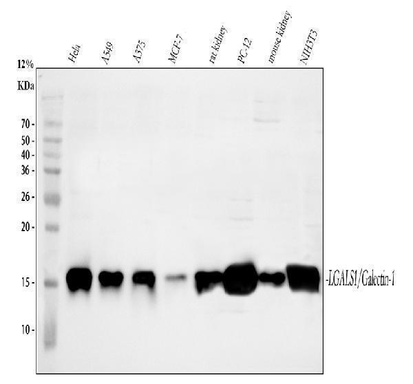
Western blot analysis of Galectin 1 using anti-Galectin 1 antibody (A00470).
Electrophoresis was performed on a 13% SDS-PAGE gel at 80V (Stacking gel) / 120V (Resolving gel) for 2 hours. The sample well of each lane was loaded with 30 ug of sample under reducing conditions.
Lane 1: human Hela whole cell lysates,
Lane 2: human A549 whole cell lysates,
Lane 3: human A375 whole cell lysates,
Lane 4: human MCF-7 whole cell lysates,
Lane 5: rat kidney tissue lysates,
Lane 6: rat PC-12 whole cell lysates,
Lane 7: mouse kidney tissue lysates,
Lane 8: mouse NIH/3T3 whole cell lysates.
After electrophoresis, proteins were transferred to a nitrocellulose membrane at 150 mA for 50-90 minutes. Blocked the membrane with 5% non-fat milk/TBS for 1.5 hour at RT. The membrane was incubated with rabbit anti-Galectin 1 antigen affinity purified polyclonal antibody (A00470) at 0.5 μg/mL overnight at 4°C, then washed with TBS-0.1%Tween 3 times with 5 minutes each and probed with a goat anti-rabbit IgG-HRP secondary antibody at a dilution of 1:5000 for 1.5 hour at RT. The signal is developed using an ECL Plus Western Blotting Substrate (Catalog # AR1196-200) with Tanon 5200 system. A specific band was detected for Galectin 1 at approximately 15 kDa. The expected band size for Galectin 1 is at 15 kDa.

Click image to see more details
IHC analysis of Galectin 1 using anti-Galectin 1 antibody (A00470).
Galectin 1 was detected in a paraffin-embedded section of human colon cancer tissue. Heat mediated antigen retrieval was performed in EDTA buffer (pH 8.0, epitope retrieval solution). The tissue section was blocked with 10% goat serum. The tissue section was then incubated with 2 μg/ml rabbit anti-Galectin 1 Antibody (A00470) overnight at 4°C. Peroxidase Conjugated Goat Anti-rabbit IgG was used as secondary antibody and incubated for 30 minutes at 37°C. The tissue section was developed using HRP Conjugated Rabbit IgG Super Vision Assay Kit (Catalog # SV0002) with DAB as the chromogen.

Click image to see more details
IHC analysis of Galectin 1 using anti-Galectin 1 antibody (A00470).
Galectin 1 was detected in a paraffin-embedded section of mouse intestine tissue. Heat mediated antigen retrieval was performed in EDTA buffer (pH 8.0, epitope retrieval solution). The tissue section was blocked with 10% goat serum. The tissue section was then incubated with 2 μg/ml rabbit anti-Galectin 1 Antibody (A00470) overnight at 4°C. Peroxidase Conjugated Goat Anti-rabbit IgG was used as secondary antibody and incubated for 30 minutes at 37°C. The tissue section was developed using HRP Conjugated Rabbit IgG Super Vision Assay Kit (Catalog # SV0002) with DAB as the chromogen.

Click image to see more details
IHC analysis of Galectin 1 using anti-Galectin 1 antibody (A00470).
Galectin 1 was detected in a paraffin-embedded section of mouse kidney tissue. Heat mediated antigen retrieval was performed in EDTA buffer (pH 8.0, epitope retrieval solution). The tissue section was blocked with 10% goat serum. The tissue section was then incubated with 2 μg/ml rabbit anti-Galectin 1 Antibody (A00470) overnight at 4°C. Peroxidase Conjugated Goat Anti-rabbit IgG was used as secondary antibody and incubated for 30 minutes at 37°C. The tissue section was developed using HRP Conjugated Rabbit IgG Super Vision Assay Kit (Catalog # SV0002) with DAB as the chromogen.

Click image to see more details
IHC analysis of Galectin 1 using anti-Galectin 1 antibody (A00470).
Galectin 1 was detected in a paraffin-embedded section of rat kidney tissue. Heat mediated antigen retrieval was performed in EDTA buffer (pH 8.0, epitope retrieval solution). The tissue section was blocked with 10% goat serum. The tissue section was then incubated with 2 μg/ml rabbit anti-Galectin 1 Antibody (A00470) overnight at 4°C. Peroxidase Conjugated Goat Anti-rabbit IgG was used as secondary antibody and incubated for 30 minutes at 37°C. The tissue section was developed using HRP Conjugated Rabbit IgG Super Vision Assay Kit (Catalog # SV0002) with DAB as the chromogen.

Click image to see more details
IF analysis of Galectin 1 using anti-Galectin 1 antibody (A00470).
Galectin 1 was detected in an immunocytochemical section of A549 cells. Enzyme antigen retrieval was performed using IHC enzyme antigen retrieval reagent (AR0022) for 15 mins. The cells were blocked with 10% goat serum. And then incubated with 5 μg/mL rabbit anti-Galectin 1 Antibody (A00470) overnight at 4°C. Fluoro594 Conjugated Goat Anti-Rabbit IgG (BA1142) was used as secondary antibody at 1:500 dilution and incubated for 30 minutes at 37°C. The section was counterstained with DAPI. Visualize using a fluorescence microscope and filter sets appropriate for the label used.

Click image to see more details
Sandwich ELISA - Recombinant mouse Galectin 1/Lgals1 protein standard curve.
Use in combination with reagents from Mouse Galectin 1/Lgals1 ELISA Kit EZ-Set (DIY Antibody Pairs) (EZ0763).
Specific Publications For Anti-Galectin 1/Lgals1 Antibody Picoband® (A00470)
Loading publications
Recommended Resources
Here are featured tools and databases that you might find useful.
- Boster's Pathways Library
- Protein Databases
- Bioscience Research Protocol Resources
- Data Processing & Analysis Software
- Photo Editing Software
- Scientific Literature Resources
- Research Paper Management Tools
- Molecular Biology Software
- Primer Design Tools
- Bioinformatics Tools
- Phylogenetic Tree Analysis
Customer Reviews
Have you used Anti-Galectin 1/Lgals1 Antibody Picoband®?
Share your experimental results or join a short interview to earn up to $1,000 in product credits or other rewards.
0 Reviews For Anti-Galectin 1/Lgals1 Antibody Picoband®
Customer Q&As
Have a question?
Find answers in Q&As, reviews.
Can't find your answer?
Submit your question
4 Customer Q&As for Anti-Galectin 1/Lgals1 Antibody Picoband®
Question
My colleagues were content with the WB result of your anti-Galectin 1/Lgals1 antibody. However we have been able to see positive staining in placenta skin extracellular space using this antibody. Is that expected? Could you tell me where is LGALS1 supposed to be expressed?
Verified Customer
Verified customer
Asked: 2019-04-12
Answer
From literature, placenta skin does express LGALS1. Generally LGALS1 expresses in secreted, extracellular space, extracellular. Regarding which tissues have LGALS1 expression, here are a few articles citing expression in various tissues:
Hepatoma, Pubmed ID: 2719646
Liver, Pubmed ID: 24275569
Lung, Pubmed ID: 2719964
Lung fibroblast, Pubmed ID: 12761501
Lymphocyte, Pubmed ID: 1988031
Mammary cancer, Pubmed ID: 19497882
Melanoma, Pubmed ID: 1386213
Placenta, Pubmed ID: 3065332, 3611046, 10369126, 18824694
Placenta, and Promyelocytic leukemia, Pubmed ID: 2910856
Placenta, and Skin, Pubmed ID: 15489334
Platelet, Pubmed ID: 12665801
Spleen, Pubmed ID: 2383549
Thalamus, Pubmed ID: 14702039
Boster Scientific Support
Answered: 2019-04-12
Question
We ordered your anti-Galectin 1/Lgals1 antibody for WB on liver in a previous project. I am using mouse, and We are going to use the antibody for IHC next. I am interested in examining liver as well as spleen in our next experiment. Could give a recommendation on which antibody would work the best for IHC?
Verified Customer
Verified customer
Asked: 2017-10-30
Answer
I have checked the website and datasheets of our anti-Galectin 1/Lgals1 antibody and I see that A00470 has been validated on mouse in both WB and IHC. Thus A00470 should work for your application. Our Boster satisfaction guarantee will cover this product for IHC in mouse even if the specific tissue type has not been validated. We do have a comprehensive range of products for IHC detection and you can check out our website bosterbio.com to find out more information about them.
Boster Scientific Support
Answered: 2017-10-30
Question
We have seen staining in rat lung fibroblast. Any tips? Is anti-Galectin 1/Lgals1 antibody supposed to stain lung fibroblast positively?
A. Bhatt
Verified customer
Asked: 2015-12-11
Answer
Based on literature lung fibroblast does express LGALS1. Based on Uniprot.org, LGALS1 is expressed in adipose tissue, lung, placenta promyelocytic leukemia, hepatoma, placenta, lymphocyte, lung fibroblast, thalamus, placenta skin, brain, platelet, brain, cajal-retzius cell fetal brain cortex, melanoma, spleen, mammary cancer, liver, among other tissues. Regarding which tissues have LGALS1 expression, here are a few articles citing expression in various tissues:
Hepatoma, Pubmed ID: 2719646
Liver, Pubmed ID: 24275569
Lung, Pubmed ID: 2719964
Lung fibroblast, Pubmed ID: 12761501
Lymphocyte, Pubmed ID: 1988031
Mammary cancer, Pubmed ID: 19497882
Melanoma, Pubmed ID: 1386213
Placenta, Pubmed ID: 3065332, 3611046, 10369126, 18824694
Placenta, and Promyelocytic leukemia, Pubmed ID: 2910856
Placenta, and Skin, Pubmed ID: 15489334
Platelet, Pubmed ID: 12665801
Spleen, Pubmed ID: 2383549
Thalamus, Pubmed ID: 14702039
Boster Scientific Support
Answered: 2015-12-11
Question
We are currently using anti-Galectin 1/Lgals1 antibody A00470 for rat tissue, and we are happy with the IHC results. The species of reactivity given in the datasheet says mouse, rat. Is it likely that the antibody can work on dog tissues as well?
D. Singh
Verified customer
Asked: 2015-04-01
Answer
The anti-Galectin 1/Lgals1 antibody (A00470) has not been validated for cross reactivity specifically with dog tissues, but there is a good chance of cross reactivity. We have an innovator award program that if you test this antibody and show it works in dog you can get your next antibody for free. Please contact me if I can help you with anything.
Boster Scientific Support
Answered: 2015-04-01
