This website uses cookies to ensure you get the best experience on our website.
- Table of Contents
1 Citations 17 Q&As
17 Citations 9 Q&As
5 Citations 9 Q&As
16 Q&As
2 Citations 4 Q&As
1 Citations 5 Q&As
1 Citations 16 Q&As
1 Citations 8 Q&As
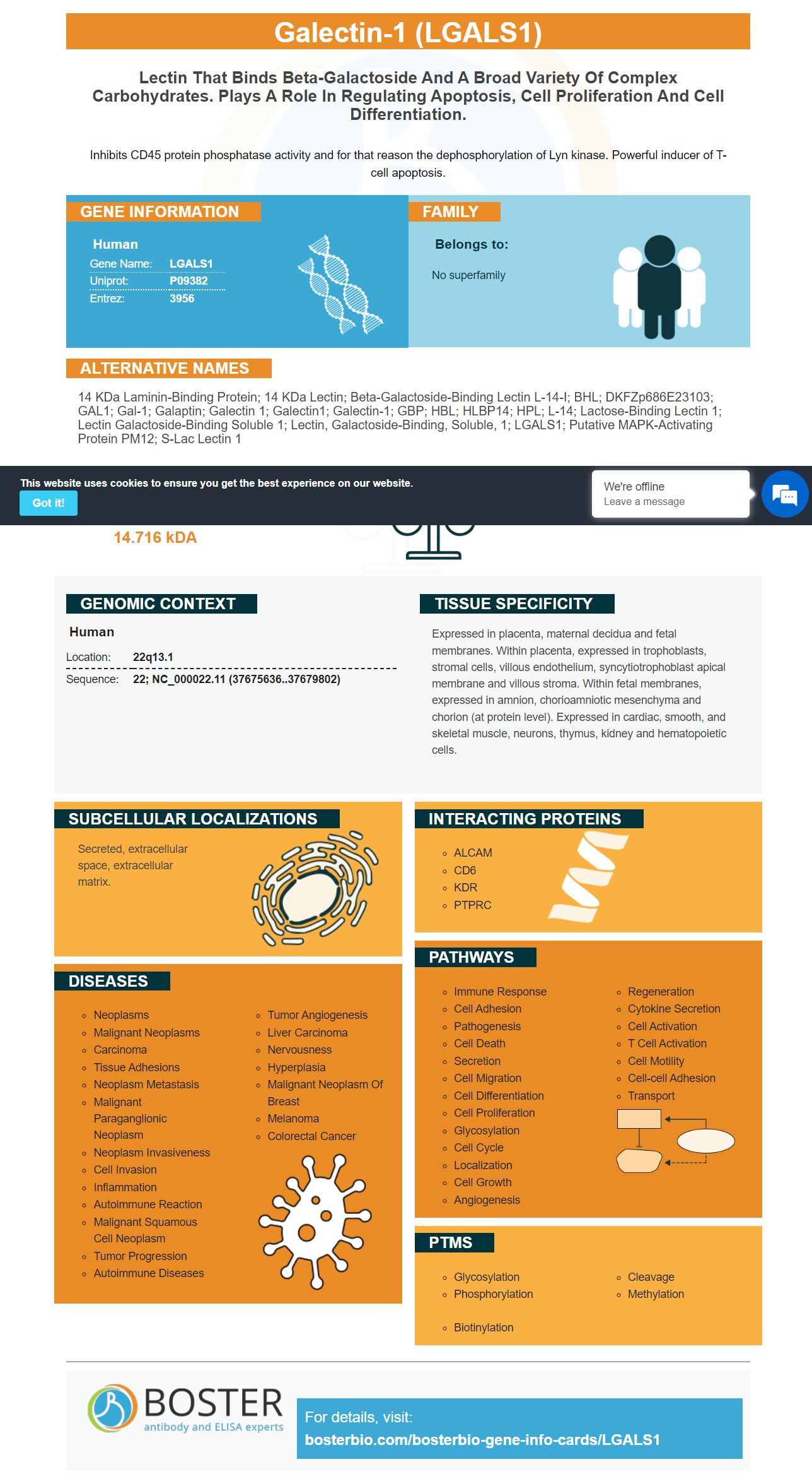
Facts about Galectin-1.
Inhibits CD45 protein phosphatase activity and for that reason the dephosphorylation of Lyn kinase. Powerful inducer of T-cell apoptosis.
Human | |
---|---|
Gene Name: | LGALS1 |
Uniprot: | P09382 |
Entrez: | 3956 |
Belongs to: |
---|
No superfamily |
14 kDa laminin-binding protein; 14 kDa lectin; Beta-galactoside-binding lectin L-14-I; BHL; DKFZp686E23103; GAL1; gal-1; Galaptin; galectin 1; Galectin1; Galectin-1; GBP; HBL; HLBP14; HPL; L-14; Lactose-binding lectin 1; Lectin galactoside-binding soluble 1; lectin, galactoside-binding, soluble, 1; LGALS1; Putative MAPK-activating protein PM12; S-Lac lectin 1
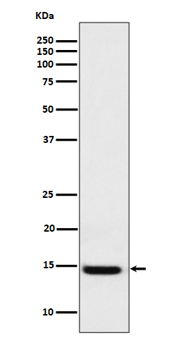
Mass (kDA):
14.716 kDA
Human | |
---|---|
Location: | 22q13.1 |
Sequence: | 22; NC_000022.11 (37675636..37679802) |
Expressed in placenta, maternal decidua and fetal membranes. Within placenta, expressed in trophoblasts, stromal cells, villous endothelium, syncytiotrophoblast apical membrane and villous stroma. Within fetal membranes, expressed in amnion, chorioamniotic mesenchyma and chorion (at protein level). Expressed in cardiac, smooth, and skeletal muscle, neurons, thymus, kidney and hematopoietic cells.
Secreted, extracellular space, extracellular matrix.
There are many advantages when using the boster Bio LGALS1 Marker, no matter if you're a medical researcher or technician. Not only does it measure the presence of LGALS1 protein however, it's also an extremely effective tool for molecular biology research. This article will provide additional details about Boster Bio's biological methods. You'll be taught about picogram sensitivity and guaranteed quality as well as IHC-optimized polyclonal antibodies.
LGALS1 encodes a protein called galectin-1. Galectins belong to the family of beta-galactoside-binding lectins and have various roles in the body. This gene is located on the chromosome at 22q12-q13. It's expressed primarily by subventricular zone astrocytes. It is an autocrine growth factor. Boster Bio's molecular biological methods using the LGALS1 marker and various molecular markers have allowed researchers to utilize these markers in their research and development.
The MBS765403 ELISA Kit analyzes LGALS1 concentrations in biological samples by using the HRP colorimetric detection method. This kit can detect recombinant or native LGALS1 antigens in a variety of sample types including tissues as well as undiluted body fluids. It comes with reproducibility tests. It can measure native and recombinant human Galectin-1 from various samples.
This sandwich ELISA kit analyzes GAL1 in plasma or serum of a human. It can also measure GAL1 from human tissue homogenates. The sandwich method is extremely sensitive and accurate. Its sensitivity is generally determined by the picogram of the target. The sensitivity is a crucial factor in the quality of the results, which is why it is important to choose a suitable instrument for your testing needs.
ELISA kits come with specific instructions for using. Each kit comes with a specific range of sensitivity. The sensitivity of the picograms of the LGALS1 marker ELISA kits picogram is higher than 0.5 mg/mL. Higher results can be obtained if the sample is diluted. It is crucial to follow the instructions in the kit to ensure the results are reliable. It is also possible to refer to the manufacturer's website or a manual for more details.
There are ELISA kits for a variety of analytes which include the LGALS1 gene. The kits include detection antibodies, buffers, diluentsand standards and matched pairs of antibodies. The LGALS1 ELISA kit also includes an elongated strip well that has standard curves that maximize 96-well plate utilization. These kits are extremely sensitive and reliable.
IHC-optimized antibodies that use the LGALS1 marker are extremely specific and sensitive to small changes in the antigen. For optimal staining antibodies should be maintained at specific conditions for a short period of time. Incubation time and temperature should be altered to ensure specific staining, especially with a high-affinity polyclonal antibody. Polyclonal antibodies are made by a DNA sequence. They have higher specificity which means they have less variability from batch to batch.
Monoclonal antibody are less specific than polyclonal and have weaker signaling from the sample. They also are less effective in staining because of the possibility that their epitope structure could change during an experiment. This could result in a dramatic change of staining. A small amount of antigen is in a host animal like donkey, mouse, to create polyclonal antibodies.
The success of an experiment is dependent on the choice of primary antibody. The quality of the primary antibody depends on its specificity to the epitope of interest. To show specific staining and minimize background signal, it is important to optimize each step of the IHC/ICC experiment. Since monoclonal and polyclonal antibodies have different conditions for incubation it is essential to establish them experimentally.
IHC-optimized polyclonals that contain the LGALS1 marker have many advantages. Because of the amount of binding events that surround the target molecule, their affinity is high, however the background is very low. Furthermore, they are potent, and the amount of binding events surrounding the target molecule is greater than background noise. For small antigens, chemistry of the antigen-hapten connection is important.
Galectin-1 is encoded by LGALS1 gene. Galectins belong to the beta-galactoside-binding lectin family. It is expressed in the subventricular zone of astrocytes. It is described as an autocrine negative growth factor. The marker is produced by boster antibodies that have been tested against known positive and negative samples. These antibodies are available for purchase.
PMID: 2719964 by Hirabayashi J., et al. Cloning and nucleotide sequence of a full-length cDNA for human 14 kDa beta-galactoside-binding lectin.
PMID: 2910856 by Couraud P.-O., et al. Molecular cloning, characterization, and expression of a human 14-kDa lectin.
*More publications can be found for each product on its corresponding product page