Click image to see more details
Product Info Summary
| SKU: | A00701 |
|---|---|
| Size: | 100ug |
| Reactive Species: | Mouse |
| Host: | Rabbit |
| Application: | ELISA, IHC, WB |
Customers Who Bought This Also Bought

Product info
Product Name
Anti-Gli-2 Antibody
SKU/Catalog Number
A00701
Size
100ug
Form
Liquid (sterile filtered)
Description
Boster Bio Anti-Gli-2 Antibody (Catalog # A00701). Tested in ELISA, IHC, WB applications. This antibody reacts with Mouse.
Storage & Handling
Store vial at -20°C prior to opening. Aliquot contents and freeze at -20°C or below for extended storage. Avoid cycles of freezing and thawing. Centrifuge product if not completely clear after standing at room temperature. This product is stable for several weeks at 4°C as an undiluted liquid. Dilute only prior to immediate use. Expiration date is one (1) year from date of opening. (Ship on dry ice.)
Cite This Product
Anti-Gli-2 Antibody (Boster Biological Technology, Pleasanton CA, USA, Catalog # A00701)
Host
Rabbit
Contents
0.02 M Potassium Phosphate, 0.15 M Sodium Chloride, pH 7.2, 0.01% (w/v) Sodium Azide
Clonality
Polyclonal
Isotype
IgG
Immunogen
This affinity purified antibody was prepared from whole rabbit serum produced by repeated immunizations with a synthetic peptide corresponding to amino acids from an internal region of Mouse Gli-2.
Reactive Species
A00701 is reactive to GLI2 in Mouse
Observed Molecular Weight
42 kDa
Background of GLI2
Gli-2 (also known as Zinc Finger Protein Gli-2, GLI-Kruppel family member GLI-2 or Tax helper protein) belongs to the C2H2-type zinc finger protein subclass of the Gli family. Members of this subclass are characterized as transcription factors that bind DNA through zinc finger motifs. These motifs contain conserved H-C links. Gli family zinc finger proteins are mediators of Sonic hedgehog (Shh) signaling, and they are implicated as potent oncogenes in the embryonal carcinoma cell. The protein encoded by this gene localizes to the cytoplasm and activates patched Drosophila homolog (PTCH) gene expression. Gli-2 is also thought to play a role during embryogenesis. The encoded protein is associated with several phenotypes: Greig cephalopolysyndactyly syndrome, Pallister-Hall syndrome, pre-axial polydactyly type IV, and postaxial polydactyly types A1 and B. Anti-Gli 2 Antibody is useful for researchers interested in transcription factor activities, DNA binding, and chromatin binding research.
Antibody Validation
Boster validates all antibodies on WB, IHC, ICC, Immunofluorescence, and ELISA with known positive control and negative samples to ensure specificity and high affinity, including thorough antibody incubations.
Application & Images
Applications
A00701 is guaranteed for ELISA, IHC, WB Boster Guarantee
Recommend Dilution
| Application | Dilution | Species |
|---|---|---|
| ELISA: 1:15 | 000 - 1:60 | 000 |
| WB: 1:500 - 1:2 | 000 | |
| This antibody has been tested for use in ELISA | immunohistochemistry and western blot. Specific conditions for reactivity should be optimized by the end user. See figure legend for expectations by WB and IHC. Multiple splice variants have been reported for this protein. |
Validation Images & Assay Conditions
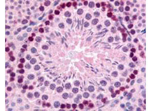
Click image to see more details
Boster's Affinity Purified anti-mouse Gli-2 antibody was used at 10 µg/ml to evaluate staining on several mouse tissues. Moderate to strong staining was seen on many tissues, with low background staining. This image shows Gli-2 staining of mouse testis. Tissue was formalin-fixed and paraffin embedded.

Click image to see more details
Western blot using Boster's Affinity Purified anti-Gli-2 antibody shows detection of a predominant band at ~190 kDa corresponding to Gli-2 (arrowhead) in mouse brain whole cell lysate (lane 1). Pre-incubation of antibody with immunizing peptide completely blocks staining of this band (lane 2). Load 25µg of lysate was resolved on a 4-8% Tris-glycine gel by SDS-PAGE and transferred onto nitrocellulose. After blocking with 5% goat serum and 0.5% BLOTTO in PBS, the membrane was probed with the primary antibody diluted to 1:750. Incubation was at room temperature for 2 h followed by washes and reaction with a 1:10,000 dilution of IRDye® 800 conjugated Gt-a-Rabbit IgG (H&L) MX10 for 45 min at room temperature. Molecular weight markers are shown (M) using the 700 nm channel (red). IRDye® 800 fluorescence image was captured using the Odyssey® Infrared Imaging System developed by LI-COR. IRDye is a trademark of LI-COR, Inc. Other detection systems will yield similar results.

Click image to see more details
Boster's Affinity Purified anti-mouse Gli-2 antibody was used at 10 µg/ml to evaluate staining on several mouse tissues. Moderate to strong staining was seen on many tissues with low background staining. This image shows Gli-2 staining of mouse brain. Tissue was formalin-fixed and paraffin embedded.
Specific Publications For Anti-Gli-2 Antibody (A00701)
Loading publications
Recommended Resources
Here are featured tools and databases that you might find useful.
- Boster's Pathways Library
- Protein Databases
- Bioscience Research Protocol Resources
- Data Processing & Analysis Software
- Photo Editing Software
- Scientific Literature Resources
- Research Paper Management Tools
- Molecular Biology Software
- Primer Design Tools
- Bioinformatics Tools
- Phylogenetic Tree Analysis
Customer Reviews
Have you used Anti-Gli-2 Antibody?
Share your experimental results or join a short interview to earn up to $1,000 in product credits or other rewards.
0 Reviews For Anti-Gli-2 Antibody
Customer Q&As
Have a question?
Find answers in Q&As, reviews.
Can't find your answer?
Submit your question
