Click image to see more details
-

-
-
-
-
+1
Product Info Summary
| SKU: | A01171 |
|---|---|
| Size: | 100ug |
| Reactive Species: | Human |
| Host: | Rabbit |
| Application: | ELISA, IF, IHC, WB |
Customers Who Bought This Also Bought

Product info
Product Name
Anti-Gli-3 Antibody
SKU/Catalog Number
A01171
Size
100ug
Form
Liquid (sterile filtered)
Description
Boster Bio Anti-Gli-3 Antibody (Catalog # A01171). Tested in ELISA, IF, IHC, WB applications. This antibody reacts with Human.
Storage & Handling
Store vial at -20°C prior to opening. Aliquot contents and freeze at -20°C or below for extended storage. Avoid cycles of freezing and thawing. Centrifuge product if not completely clear after standing at room temperature. This product is stable for several weeks at 4°C as an undiluted liquid. Dilute only prior to immediate use. Expiration date is one (1) year from date of opening. (Ship on dry ice.)
Cite This Product
Anti-Gli-3 Antibody (Boster Biological Technology, Pleasanton CA, USA, Catalog # A01171)
Host
Rabbit
Contents
0.02 M Potassium Phosphate, 0.15 M Sodium Chloride, pH 7.2, 0.01% (w/v) Sodium Azide
Clonality
Polyclonal
Isotype
IgG
Immunogen
This affinity purified antibody was produced from monospecific rabbit serum by repeated immunizations with a synthetic peptide corresponding to an internal region near amino acids 30-60 of human Gli-3 protein.
Reactive Species
A01171 is reactive to GLI3 in Human
Observed Molecular Weight
42 kDa
Calculated molecular weight
169.9 kDa
Background of GLI3
Gli-3 (also known as Zinc Finger Protein Gli-3 or GLI-Kruppel family member GLI-3) belongs to the GLI C2H2-type zinc-finger protein family and contains 5 C2H2-type zinc fingers. Gli-3 is very important for normal limb and brain development and is implicated in the transduction of Shh signal. Gli-3 is a nuclear protein expressed in a wide variety of normal adult tissues, including lung, colon, spleen, placenta, testis, and myometrium. Defects in Gli-3 are the cause of Greig cephalo-poly-syndactyly syndrome (GCPS); an autosomal dominant disorder-affecting limb and cranio-facial development. Two isoforms of human Gli-3 have been reported. One is the full-length protein at ~170-190kDa and the other is a truncated isoform at ~80kDa.
Antibody Validation
Boster validates all antibodies on WB, IHC, ICC, Immunofluorescence, and ELISA with known positive control and negative samples to ensure specificity and high affinity, including thorough antibody incubations.
Application & Images
Applications
A01171 is guaranteed for ELISA, IF, IHC, WB Boster Guarantee
Recommend Dilution
| Application | Dilution | Species |
|---|---|---|
| ELISA: 1:6 | 000 - 1:30 | 000 |
| WB: 1:500 - 1:2 | 000 | |
| This antibody has been tested for use in ELISA | immunohistochemistry | immunofluorescence, and western blot. Specific conditions for reactivity should be optimized by the end user. Detection of Gli-3 by western blot may be enhanced if nuclear extracts are used instead of whole cell lysates as the expression/abundance of Gli-3 is likely to be low. Furthermore, Gli3 expression is likely to be developmentally regulated and induced, making it difficult to detect in whole tissue homogenates. |
Validation Images & Assay Conditions
Click image to see more details
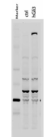
Western Blot of Rabbit anti-Gli-3 antibody. Lane 1: 50 kDa molecular weight marker. Lane 2: 293T cells transfected with CrkL-Flag. Lane 3: 293T cells transfected with human Gli-3. Load: 35 µg per lane. Primary antibody: Gli-3 antibody at 1:400 for overnight at 4°C. Secondary antibody: IRDye800™ rabbit secondary antibody at 1:10,000 for 45 min at RT. Block: 5% BLOTTO overnight at 4°C. Predicted/Observed size: 170-190 kDa for hGli-3. Other band(s): Non specific background ~60kDa.

Click image to see more details
Immunofluorescence Microscopy of Rabbit anti-Gli-3 antibody. Tissue: MCF-7 cell. Antigen retrieval: not required. Primary antibody: Gli-3 antibody and Anti-alpha-Tubulin at 5 µg/mL for 1 h at RT. Secondary antibody: Fluorescein secondary antibody at 1:10,000 for 45 min at RT. Localization: Gli-3 is nuclear. Staining: Gli-3 staining as red fluorescent signal and Anti-alpha-Tubulin staining as green fluorescent signal using STED.

Click image to see more details
Immunofluorescence Microscopy of Rabbit anti-Gli-3 antibody. Tissue: MCF-7 cell. Antigen retrieval: not required. Primary antibody: Gli-3 antibody and Anti-alpha-Tubulin at 5 µg/mL for 1 h at RT. Secondary antibody: Fluorescein secondary antibody at 1:10,000 for 45 min at RT. Localization: Gli-3 is nuclear. Staining: Image (1) shows alpha-Tubulin staining as green fluorescent signal. Image (2) shows Gli-3 staining as red fluorescent signal and Images (3) shows both antibodies fluorescing using STED microscopy.

Click image to see more details
Immunohistochemistry of Rabbit anti-Gli-3 antibody. This image tissue: human glioblastoma. Specific staining was also noted in tissue from adrenal, brain, glioblastoma, colon, heart, kidney, lung, liver, skeletal muscle, ovary, pancreas, placenta, skin, spleen, stomach, testes, thymus, thyroid, tonsil and uterus. Fixation: formalin fixed paraffin embedded. Antigen retrieval: not required. Primary antibody: Gli-3 antibody at 0.625 µg/ml for 1 h at RT. Secondary antibody: Peroxidase rabbit secondary antibody at 1:10,000 for 45 min at RT. Localization: Gli-3 is nuclear and smooth muscle. Staining: Gli-3 as precipitated red signal with hematoxylin purple nuclear counterstain.

Click image to see more details
Western Blot of Rabbit anti-Gli-3 antibody. Lane 1: human brain whole cell lysate. Lane 2: human lung whole cell lysate. Lane 3: human spleen whole cell lysate. Lane 4: mouse brain whole cell lysate . Lane 5: mouse lung whole cell lysate . Load: 20 µg per lane. Primary antibody: Gli-3 antibody at 1:500 for overnight at 4°C. Secondary antibody: IRDye800™ rabbit secondary antibody at 1:10,000 for 45 min at RT. Block: 5% BLOTTO overnight at 4°C. Predicted/Observed size: Isoforms at ~170-190kDa and ~80kDa. Lane 2 shows what may be truncated Gli-3 (~80kDa). Other band(s): The strong band at ~50 kDa is unknown.
Specific Publications For Anti-Gli-3 Antibody (A01171)
Loading publications
Recommended Resources
Here are featured tools and databases that you might find useful.
- Boster's Pathways Library
- Protein Databases
- Bioscience Research Protocol Resources
- Data Processing & Analysis Software
- Photo Editing Software
- Scientific Literature Resources
- Research Paper Management Tools
- Molecular Biology Software
- Primer Design Tools
- Bioinformatics Tools
- Phylogenetic Tree Analysis
Customer Reviews
Have you used Anti-Gli-3 Antibody?
Share your experimental results or join a short interview to earn up to $1,000 in product credits or other rewards.
0 Reviews For Anti-Gli-3 Antibody
Customer Q&As
Have a question?
Find answers in Q&As, reviews.
Can't find your answer?
Submit your question
