Click image to see more details
-

-
-
-
-
+19
Product Info Summary
| SKU: | M02059 |
|---|---|
| Size: | 100 μl |
| Reactive Species: | Human, Mouse, Rat |
| Host: | Rabbit |
| Application: | IF, IHC, ICC, WB |
Customers Who Bought This Also Bought

Product info
Product Name
Anti-GPX4 Rabbit Monoclonal Antibody
SKU/Catalog Number
M02059
BM5231 is an alternative SKU for this antibody, used in previous lots.
Size
100 μl
Form
Liquid
Description
Boster Bio Anti-GPX4 Rabbit Monoclonal Antibody catalog # M02059. Tested in WB, IHC, ICC/IF applications. This antibody reacts with Human, Mouse, Rat.
Storage & Handling
Store at -20°C for one year. For short term storage and frequent use, store at 4°C for up to one month. Avoid repeated freeze-thaw cycles.
Cite This Product
Anti-GPX4 Rabbit Monoclonal Antibody (Boster Biological Technology, Pleasanton CA, USA, Catalog # M02059)
Host
Rabbit
Contents
Rabbit IgG in stabilizing components, phosphate buffered saline, pH 7.4, 150mM NaCl, 0.02% sodium azide and 50% glycerol.
*This antibody is supplied in a stabilized formulation.
Compatibility with conjugation reactions depends on the chemistry of the conjugation method used.
For conjugation methods that are not compatible with the stabilizing components present in this formulation, a carrier-free antibody format is required.
Clonality
Monoclonal
Clone Number
ACCO-7
Isotype
Rabbit IgG
Immunogen
A synthesized peptide derived from human GPX4
Reactive Species
M02059 is reactive to GPX4 in Human, Mouse, Rat
Observed Molecular Weight
19 kDa
Calculated molecular weight
22.2 kDa
Antibody Validation
Boster validates all antibodies on WB, IHC, ICC, Immunofluorescence, and ELISA with known positive control and negative samples to ensure specificity and high affinity, including thorough antibody incubations.
Application & Images
Applications
M02059 is guaranteed for IF, IHC, ICC, WB Boster Guarantee
Assay Dilutions Recommendation
The recommendations below provide a starting point for assay optimization. The actual working concentration varies and should be decided by the user.
WB 1:500-2000
IHC 1:50-200
ICC/IF 1:50-200
Positive Control
WB: mouse RAW264.7(WT) whole cell, mouse RAW264.7(KO) whole cell, human MCF-7 whole cell, human HepG2 whole cell, human CACO-2 whole cell, human 293T whole cell, human K562 whole cell, rat PC-12 whole cell, rat NRK whole cell, mouse RAW264.7 whole cell, mouse NIH/3T3 whole cell, rat kidney tissue, rat testis tissue, mouse kidney tissue, mouse testis tissue, human HCT116- WT whole cell, human HCT116-GPX4 KO whole cell
Validation Images & Assay Conditions
Click image to see more details
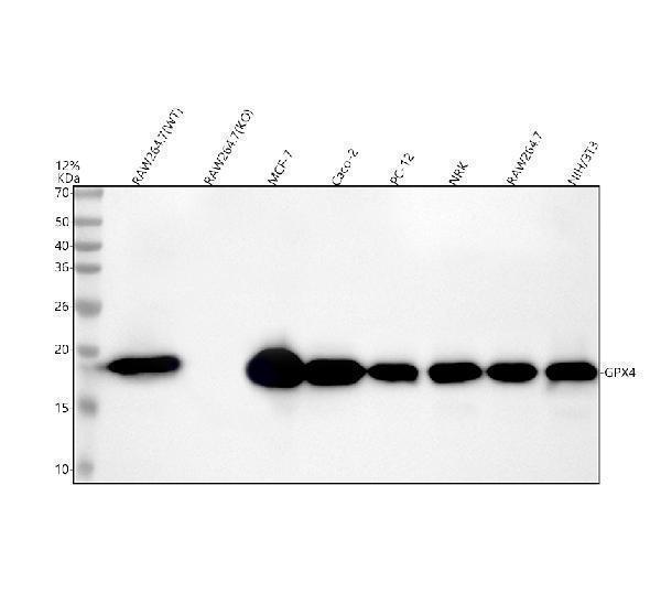
Western blot analysis of GPX4 using anti-GPX4 antibody (M02059).
Electrophoresis was performed on a 5-20% SDS-PAGE gel at 70V (Stacking gel) / 90V (Resolving gel) for 2-3 hours. The sample well of each lane was loaded with 30 ug of sample under reducing conditions.
Lane 1: mouse RAW264.7(WT) whole cell lysates,
Lane 2: mouse RAW264.7(KO) whole cell lysates,
Lane 3: human MCF-7 whole cell lysates,
Lane 4: human Caco-2 whole cell lysates,
Lane 5: rat PC-12 whole cell lysates,
Lane 6: rat NRK whole cell lysates,
Lane 7: mouse RAW264.7 whole cell lysates,
Lane 8: mouse NIH/3T3 whole cell lysates.
After electrophoresis, proteins were transferred to a nitrocellulose membrane at 150 mA for 50-90 minutes. Blocked the membrane with 5% non-fat milk/TBS for 1.5 hour at RT. The membrane was incubated with rabbit anti-GPX4 antigen affinity purified monoclonal antibody (M02059) at 1:500 overnight at 4°C, then washed with TBS-0.1%Tween 3 times with 5 minutes each and probed with a goat anti-rabbit IgG-HRP secondary antibody at a dilution of 1:500 for 1.5 hour at RT. The signal is developed using an Enhanced Chemiluminescent detection (ECL) kit (Catalog # EK1002) with Tanon 5200 system. A specific band was detected for GPX4 at approximately 19 kDa. The expected band size for GPX4 is at 22 kDa.

Click image to see more details
Western blot analysis of GPX4 using anti-GPX4 antibody (M02059).
Electrophoresis was performed on a 5-20% SDS-PAGE gel at 70V (Stacking gel) / 90V (Resolving gel) for 2-3 hours. The sample well of each lane was loaded with 30 ug of sample under reducing conditions.
Lane 1: human HepG2 whole cell lysates,
Lane 2: human CACO-2 whole cell lysates,
Lane 3: human 293T whole cell lysates,
Lane 4: human K562 whole cell lysates,
Lane 5: rat kidney tissue lysates,
Lane 6: rat testis tissue lysates,
Lane 7: mouse kidney tissue lysates,
Lane 8: mouse testis tissue lysates.
After electrophoresis, proteins were transferred to a nitrocellulose membrane at 150 mA for 50-90 minutes. Blocked the membrane with 5% non-fat milk/TBS for 1.5 hour at RT. The membrane was incubated with rabbit anti-GPX4 antigen affinity purified monoclonal antibody (Catalog # M02059) at 1:500 overnight at 4°C, then washed with TBS-0.1%Tween 3 times with 5 minutes each and probed with a goat anti-rabbit IgG-HRP secondary antibody at a dilution of 1:1000 for 1.5 hour at RT. The signal is developed using an Enhanced Chemiluminescent detection (ECL) kit (Catalog # EK1002) with Tanon 5200 system. A specific band was detected for GPX4 at approximately 19 kDa. The expected band size for GPX4 is at 22 kDa.

Click image to see more details
Immunofluorescent analysis using the Antibody at 1:500 dilution.

Click image to see more details
Western blot analysis of GPX4 using anti-GPX4 antibody (M02059).
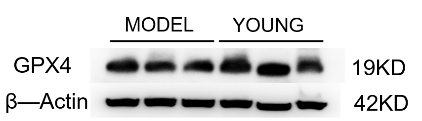
Electrophoresis was performed on a 5-20% SDS-PAGE gel at 80V (Stacking gel) / 120V (Resolving gel) for 2 hours. The sample well of each lane was loaded with 30 ug of sample under reducing conditions.
Lane 1-3: model group-mouse uterine tissue lysates,
Lane 4-6: young group-mouse uterine tissue lysates.
After electrophoresis, proteins were transferred to a nitrocellulose membrane at 150 mA for 50-90 minutes. Blocked the membrane with 5% non-fat milk/TBS for 1.5 hour at RT. The membrane was incubated with rabbit anti-GPX4 antigen affinity purified monoclonal antibody (M02059) at 1:1000 overnight at 4°C, then washed with TBS-0.1%Tween 3 times with 5 minutes each and probed with a goat anti-rabbit IgG-HRP secondary antibody (Catalog # BA1054) at a dilution of 1:5000 for 1 hour at RT. The signal is developed using an ECL Plus Western Blotting Substrate (Catalog # AR1196-200) with Tanon 5200 system. A specific band was detected for GPX4 at approximately 19 kDa. The expected band size for GPX4 is at 19 kDa.

Click image to see more details
Western blot analysis of GPX4 using anti-GPX4 antibody (M02059).
Electrophoresis was performed on a 12% SDS-PAGE gel at 80V (Stacking gel) / 120V (Resolving gel) for 2 hours. The sample well of each lane was loaded with 30 ug of sample under reducing conditions.
Lane 1: human HCT116- WT whole cell lysates,
Lane 2: human HCT116-GPX4 KO whole cell lysates.
After electrophoresis, proteins were transferred to a nitrocellulose membrane at 150 mA for 50-90 minutes. Blocked the membrane with 5% non-fat milk/TBS for 1.5 hour at RT. Then the membrane was incubated with rabbit anti-GPX4 antigen affinity purified monoclonal antibody (M02059) at 0.5 μg/mL overnight at 4°C, then washed with TBS-0.1%Tween 3 times with 5 minutes each and probed with a goat anti-rabbit IgG-HRP secondary antibody (Catalog # BA1054) at a dilution of 1:5000 for 1.5 hour at RT. The signal is developed using an ECL Plus Western Blotting Substrate (Catalog # AR1196-200) with Tanon 5200 system. A specific band was detected for GPX4 at approximately 19 kDa. The expected band size for GPX4 is at 22 kDa.

Click image to see more details
Effects of Vitamin C intervention on the IL-17A/IL-17RA/ACT1 signaling pathway and ferroptosis in FRTL5 cells stimulated by PM2.5. (A-D) qPCR was used to detect the transcription levels of IL-17A/IL-17RA/ACT1 signaling pathway-related factors. (E-I) The protein expression levels of the IL-17A/IL-17RA/ACT1 signaling pathway were detected by western blot. The data are presented as mean ± SD; n = 3/group. Compared with the Con group, * P < 0.05 or ** P < 0.01; Compared with the Mod group, # P < 0.05 or ## P < 0.01. Con: Control group, normal medium for 24 h; Vc: medium containing 50 μmol/L Vc for 24 h; Mod: medium containing 400 μg/mL PM2.5 for 24 h; Mod + Vc: medium containing 400 μg/mL PM2.5 and 50 μmol/L Vc for 24 h; Mod + Y320: medium containing 400 μg/mL PM2.5 and 0.08 μmol/L Y320 for 24 h.
Index in PubMed under a CC BY license. PMID: 40753780

Click image to see more details
Effect of Vitamin C on FRTL5 cell function and IL-17A signaling pathway and ferroptosis-related factors stimulated by PM2.5. (A-C) The levels of (A, TSH; B, FT4; and C, FT3) in the supernatant of FRTL5 cells were detected by ELISA. (D-G) ELISA was used to measure the levels of IL-17A (D), IL-17RA (E), ACT1 (F), and GPX4 (G) in the supernatant of FRTL5 cells. The data are presented as mean ± SD; n = 5/group). Compared with the Con group, ** P < 0.01; Compared with the Mod group, # P < 0.05 or ## P < 0.01. Con: Control group, normal medium for 24 h; Vc: medium containing 50 μmol/L Vc for 24 h; Mod: medium containing 400 μg/mL PM2.5 for 24 h; Mod + Vc: medium containing 400 μg/mL PM2.5 and 50 μmol/L Vc for 24 h; Mod + Y320: medium containing 400 μg/mL PM2.5 and 0.08 μmol/L Y320 for 24 h.
Index in PubMed under a CC BY license. PMID: 40753780

Click image to see more details
Vitamin C regulated the protein expression levels of IL-17A signaling pathway and ferroptosis-related factors. (A) The expression levels of the IL-17A pathway (B, IL-17A; C, IL-17RA; D, ACT1) and ferroptosis (E, GPX4)-related genes in the thyroid tissues were determined by western blot (n = 3/group).The data are presented as mean ± SD. Compared with the Con group, *P < 0.05, **P < 0.01, or ***P < 0.001; Compared with the Mod group, # P < 0.05 or ## P < 0.01. Con: Control group, normal environment for eight weeks; Vc: vitamin C was administered by gavage at 120 mg/kg for eight weeks; Mod: PM2.5 exposure for eight weeks; Mod + Vc: after PM2.5 exposure, vitamin C was administered by gavage at 120 mg/kg for eight weeks.
Index in PubMed under a CC BY license. PMID: 40753780

Click image to see more details
Vitamin C improved PM2.5-induced female rats’ thyroid ferroptosis. (A) Immunofluorescence assay was used to detect the expression of GPX4 protein in thyroid tissues. (B) Quantitative immunofluorescence analysis (n = 3/group). (C) Serum malondialdehyde (MDA) content. (D) Serum ferrous ion (Fe2+) content (n = 10/group). The data are presented as mean ± SD. Compared with the Con group, *P < 0.05 or ** P < 0.01; Compared with the Mod group, # P < 0.05, ## P < 0.01, or ### P < 0.001. Con: Control group, normal environment for eight weeks; Vc: vitamin C was administered by gavage at 120 mg/kg for eight weeks; Mod: PM2.5 exposure for eight weeks; Mod + Vc: after PM2.5 exposure, vitamin C was administered by gavage at 120 mg/kg for eight weeks.
Index in PubMed under a CC BY license. PMID: 40753780

Click image to see more details
Immunohistochemically stained pancreas tissue ( n = 6). Protein levels of ( A ) GPX4, ( B ) ACSL4, and ( C ) LPCAT3 in rats (×400). GPX4, glutathione peroxidase 4; xCT, cysteine/glutamate transporter; ACSL4, acyl-CoA synthetase long-chain family member 4.
Index in PubMed under a CC BY license. PMID: 38664508

Click image to see more details
Ferroptosis was observed and alleviated in the pancreas of rats ( n = 6). ( A ) Ultrastructure of the mitochondria in the pancreas obtained by TEM (×20,000); ( B – F ) Western blot analysis of ferroptosis-related proteins, GPX4, xCT, ACSL4, and LPCAT3. GPX4, glutathione peroxidase 4; xCT, cysteine/glutamate transporter; ACSL4, acyl-CoA synthetase long-chain family member 4; LPCAT3, lysophosphatidylcholine acyltransferase 3; C, control; AP, acute pancreatitis; HTG, hypertriglyceridemic; HTGP, HTG pancreatitis; *b vs. the C group, *c vs. the AP group, *B vs. the HTG group, *C vs. the HTGP group, * P < 0.05, # P < 0.01, • P < 0.001, ns: no significance.
Index in PubMed under a CC BY license. PMID: 38664508

Click image to see more details
SS attenuated the oxidation resistance of LUAD cells. (A, B) GSH or Cys was detected in Calu-1 or A549 cells, which were respectively treated with 10, 15, 20 μM or 20, 25, 30 μM SS for 6 h, and pretreated with or without Fer-1 (1 μM), the data statistic was shown in a histogram (*P < 0.05, **P < 0.01, ***P < 0.001). (C) Western blot analysis was used to detect the expressions of SLC7A11 and GPX4 in Calu-1 cells, which were treated with 10, 15, 20 μM SS for 6 h. (D) Quantitative analysis of gray value of the SLC7A11 and GPX4 blots. (E) Western blotting analysis was used to detect the expressions of SLC7A11 and GPX4 in Calu-1 cells, which were treated with 20 μM SS or 4 μM erastin, with or without Fer-1 (1 μM) for 6 h. (F) Quantitative analysis of gray value of the SLC7A11 and GPX4 blots.
Index in PubMed under a CC BY license. PMID: 35664792

Click image to see more details
PCTR1 activates CREB by the ALX/PKA pathway to increase GPX4 expression in vitro. BOC-2 (ALX receptor inhibitor, 10 μM), H89 (PKA inhibitor, 10 μM), 666-15 (CREB inhibitor, 1 μM) or an equivalent volume of DMSO was administered to H1299 cells for 30 min in advance, and then LPS and PCTR1 were co-administered for 48 h. A , B The protein level of P- PKA was measured by western blot. C , D The protein level of P-CREB was measured by western blot. E , F The protein level of GPX4 was measured by western blot. Data are presented as the mean ± SD, n = 5–6. * p < 0.05, ** p < 0.01, *** p < 0.001, **** p < 0.0001 and ns: p > 0.05
Index in PubMed under a CC BY license. PMID: 37121999

Click image to see more details
PCTR1 activates CREB via the ALX/PKA pathway to increase GPX4 expression in vivo. PCTR1 at a dose of 200 ng was injected into each mouse via the caudal vein 6 h after LPS (15 mg/kg, ip) administration. BOC‐2 (ALX receptor inhibitor, 600 ng/kg), H89 (PKA inhibitor, 10 mg/kg), 666-15 (CREB inhibitor, 10 mg/kg) or an equivalent volume of DMSO was injected into the caudal vein 1 h before PCTR1 treatment. The mice were sacrificed 24 h after LPS stimulation. A , B The protein expression level of P-PKA was determined by western blotting. C , D The protein expression level of P-CREB was determined by western blotting. E , F The protein expression level of GPX4 was determined by western blotting. Data are presented as the mean ± SD, n = 4–6. * p < 0.05, ** p < 0.01, *** p < 0.001, **** p < 0.0001 and ns: p > 0.05. ip: intraperitoneal
Index in PubMed under a CC BY license. PMID: 37121999

Click image to see more details
M2c macrophages increase ferroptosis resistance in gastric cancer cells. a The CCK-8 method was used to detect the survival of gastric cancer cells (Hgc27 and MKN45) intervened with RSL3 for 24 h. b The CCK-8 method was used to detect the survival of gastric cancer cells (Hgc27 and MKN45) intervened with Fer-1 for 24 h. c The expression of SOD in different intervention groups. d The expression of MDA in different intervention groups. e The expression of GSH in different intervention groups. f The expression of TGFβ1 protein WB in different cell lines. g The expression results of TGFβ1 protein. h The expression of key ferroptosis proteins WB in different cell lines. i The expression results of FSP1 protein. j Expression results of DHODH protein. k Expression results of GPX4 protein. l SLC7A11 protein expression results. m The intervention of RSL3 on the expression of key ferroptosis protein WB in different co culture groups. n The expression results of GPX4 protein. o SLC7A11 protein expression results. p The WB expression of key proteins involved in ferroptosis in different intervention groups. q The expression results of GPX4 protein. r The expression results of SLC7A11 protein. s Fluorescence results of mitochondrial membrane potential in different intervention groups. Scale bar=50 μm. *p<0.05, **p<0.01, ***p<0.001.
Index in PubMed under a CC BY license. PMID: 39991579

Click image to see more details
Liver-specific GPX4 knockdown inhibits TA-regulated ER stress in APAP-induced hepatotoxicity. (A) Western blotting assessed hepatic GRP78 expression, with β-actin as a loading control. Band intensities were quantified using ImageJ. (B) q-PCR was conducted to evaluate the transcript levels of the Grp78 gene. (C) Western blotting assessed hepatic phosphorylated-PERK, phosphorylated-eIF2α, PERK, and eIF2α protein expressions. Band intensities were quantified using ImageJ. (D) Western blotting assessed hepatic ATF4 expression, with β-actin as a loading control. Band intensities were quantified using ImageJ. (E) q-PCR was conducted to evaluate the transcript levels of the Atf4 gene. All data were presented as mean ± SD, n = 3 for Western blotting, n = 5 for others. * P < 0.05 vs. corresponding control.
Index in PubMed under a CC BY license. PMID: 40051561

Click image to see more details
Liver-specific GPX4 knockdown abrogates the protective effect of TA against APAP-induced hepatocyte apoptosis. (A) Western blotting assessed hepatic BCL2 and BAX expression, with β-actin as a loading control. Band intensities were quantified using ImageJ. (B) q-PCR was conducted to evaluate the transcript levels of BCL2 and BAX-related genes. (C) Hepatic caspase 3 activity was quantified using a commercially available kit (fold of control). (D) Western blotting assessed hepatic CHOP expression, with β-actin as a loading control. Band intensities were quantified using ImageJ. (E) q-PCR was conducted to evaluate the transcript levels of the Chop gene. All data were presented as mean ± SD, n = 3 for Western blotting, n = 5 for others. * P < 0.05 vs. corresponding control.
Index in PubMed under a CC BY license. PMID: 40051561

Click image to see more details
Liver-specific GPX4 knockdown abrogates the hepatoprotective effects of TA against APAP-induced hepatotoxicity. (A) Western blotting assessed the detected hepatic GPX4 knockdown efficiency in protein levels. Hepatocyte-specific GPX4 knockdown mice were created by AAV8-mediated delivery of a TBG promoter-driven shRNA targeting GPX4. Null-vector-injected mice served as control. (B) Plasma levels of ALT and AST. (C) Liver tissues were subjected to H&E staining for histological examination. (D) Concentrations of hepatic IL-1β, IL-6, TNF-α, and MCP-1 were quantified using commercially available ELISA kits. All data were presented as mean ± SD, n = 3 for Western blotting, n = 5 for others. * P < 0.05 vs. corresponding control.
Index in PubMed under a CC BY license. PMID: 40051561

Click image to see more details
TA mediates the enhancement of hepatic antioxidant function by GPX4. (A) Hepatic concentrations of MDA, alongside the enzymatic activities of SOD and CAT. (B) Hepatic concentrations of GSH-Px, GSH, and the GSH redox ratio (GSH/GSSG) were determined. (C) Western blotting assessed hepatic GPX4 expression, with β-actin as a loading control. Band intensities were quantified using ImageJ. (D) q-PCR was conducted to evaluate the transcript levels of the Gpx4 gene. All data were presented as mean ± SD, n = 3 for Western blotting, n = 6–8 for others. * P < 0.05 vs. corresponding control.
Index in PubMed under a CC BY license. PMID: 40051561

Click image to see more details
Ferroptosis is induced in LPS-induced ALI and associated with lung damage. LPS (15 mg/kg) in saline or an equivalent volume of saline was intraperitoneally injected into mice, and lung tissues were collected at 0, 12, 24, and 48 h, respectively. A–C Representative western blotting and quantification analysis of GPX4 and PTGS2. D–F Relative values of Fe 2+ , GSH and MDA concentrations. Mice were pretreated with ferrostatin-1 (10 mg/kg, ip) 1h1 h before being injected with LPS (15 mg/kg, ip). Lung samples were collected 24 h after LPS injection. G Representative H&E staining of lung tissues (original magnification, ×200; inset, ×400). H Acute lung injury score of each group. I–K The relative mRNA expression levels of the inflammatory cytokines: IL-6, TNF-α and IL-1β. The acute lung injury score data are presented as the median and range (25th–75th percentile), and other data are presented as the mean ± SD. n = 4–6. * p < 0.05, ** p < 0.01, *** p < 0.001, **** p < 0.0001 and ns: p > 0.05. ip: intraperitoneal
Index in PubMed under a CC BY license. PMID: 37121999

Click image to see more details
Effects of PCTR1 on ferroptosis in LPS-induced ALI. PCTR1 (100 or 200 ng) was injected into the caudal vein of mice 6 h after LPS (15 mg/kg, ip) treatment. All lung specimens were harvested at 24 h after LPS stimulation. A–C Representative western blotting and quantification analysis of GPX4 and PTGS2. D–F Relative values of Fe 2+ , GSH and MDA concentrations. G Representative TEM images of each group. The black arrow indicates ferroptotic mitochondria. Magnification ×30,000. H Representative H&E staining of lung tissues (original magnification, ×200; inset, ×400). I Acute lung injury score. J–L The relative mRNA expression levels of the inflammatory cytokines: IL-6, TNF-α and IL-1β. The acute lung injury score data are presented as the median and range (25th–75th percentile), and other data are presented as the mean ± SD. n = 4–6. * p < 0.05, ** p < 0.01, *** p < 0.001, **** p < 0.0001 and ns: p > 0.05. ip: intraperitoneal
Index in PubMed under a CC BY license. PMID: 37121999

Click image to see more details
Effects of PCTR1 on LPS-induced ferroptosis in vitro. A H1299 cells were treated with LPS (10 μg/mL) and different concentrations of PCTR1 for 48 h. Fold change in cell viability. B H1299 cells were stimulated with RSL3 (10 nM) and different concentrations of PCTR1 for 48 h. Fold change in cell viability. C–E Representative western blotting and quantification analysis of GPX4 and PTGS2. F–H Relative levels of Fe 2+ , GSH and MDA. I The level of lipid peroxidation was determined with the C11-BODIPY 581/591 fluorescent probe (original magnification ×400). J Oxidized C11-BODIPY 581/591 probe was quantified by flow cytometry. Data are presented as the mean ± SD, n = 4–6. * p < 0.05, ** p < 0.01, *** p < 0.001, **** p < 0.0001 and ns: p > 0.05
Index in PubMed under a CC BY license. PMID: 37121999

Click image to see more details
CREB mediates the effect of PCTR1 on eliminating lipid peroxides in vitro. 666-15 (CREB inhibitor, 1 μM) or an equivalent volume of DMSO was administered to H1299 cells for 30 min in advance, and then LPS and PCTR1 were co-administered for 48 h. A Immunofluorescence staining images of GPX4 (original magnification ×400). B , C Relative expression levels of GSH, MDA and 4-HNE. D The level of lipid peroxidation was determined with the C11-BODIPY 581/591 fluorescent probe (original magnification ×400). E Oxidized C11-BODIPY 581/591 probe was quantified by flow cytometry. Data are presented as the mean ± SD, n = 5–6. * p < 0.05, ** p < 0.01, *** p < 0.001, **** p < 0.0001 and ns: p > 0.05
Index in PubMed under a CC BY license. PMID: 37121999
Specific Publications For Anti-GPX4 Rabbit Monoclonal Antibody (M02059)
Loading publications
Recommended Resources
Here are featured tools and databases that you might find useful.
- Boster's Pathways Library
- Protein Databases
- Bioscience Research Protocol Resources
- Data Processing & Analysis Software
- Photo Editing Software
- Scientific Literature Resources
- Research Paper Management Tools
- Molecular Biology Software
- Primer Design Tools
- Bioinformatics Tools
- Phylogenetic Tree Analysis
Customer Reviews
Have you used Anti-GPX4 Rabbit Monoclonal Antibody?
Share your experimental results or join a short interview to earn up to $1,000 in product credits or other rewards.
1 Reviews For Anti-GPX4 Rabbit Monoclonal Antibody
The GPX4 antibody was used to detect the expression of the protein in mouse uterine tissue. The WB results showed clear bands, and the antibody could be reused after recovery with good performance, offering excellent cost-effectiveness.
Excellent
| SKU | M02059 |
|---|---|
| Application | Western Blot |
| Sample | Mouse Uterus tissue |
| Sample Processing Description | The GPX4 antibody was used to detect the expression of the protein in mouse uterine tissue. The Western blot results showed clear bands, and the antibody retained good performance after reuse, offering excellent cost-effectiveness. |
| Primary Antibody | Anti-GPX4 Rabbit Monoclonal Antibody |
| Primary Incubation | overnight at 4 ℃ |
| Secondary Antibody | HRP-conjugated Anti-Rabbit IgG Secondary Antibody |
| Secondary Incubation | 1 hour in room temperature |
| Detection | Substrate: Ultra-sensitive ECL luminescent reagent (Cat# AR1191), Imaging system:Tanon |
| The GPX4 antibody was used to detect the protein expression in mouse uterine tissue. The Western blot results showed clear bands, and the antibody maintained good performance after reuse, providing excellent cost-effectiveness. |
Anfeng Ning, Peking University Third Hospital
Verified customer
Submitted 2025-11-06
Customer Q&As
Have a question?
Find answers in Q&As, reviews.
Can't find your answer?
Submit your question
16 Customer Q&As for Anti-GPX4 Rabbit Monoclonal Antibody
Question
Our lab were satisfied with the WB result of your anti-GPX4 Rabbit Monoclonal antibody. However we have seen positive staining in right testis isoform cytoplasmic: cytoplasm using this antibody. Is that expected? Could you tell me where is GPX4 supposed to be expressed?
Verified Customer
Verified customer
Asked: 2020-02-28
Answer
From literature, right testis does express GPX4. Generally GPX4 expresses in isoform mitochondrial: mitochondrion, isoform cytoplasmic: cytoplasm. Regarding which tissues have GPX4 expression, here are a few articles citing expression in various tissues:
Brain, Eye, Lung, Pancreas, and Testis, Pubmed ID: 15489334
Liver, Pubmed ID: 24275569
Testis, Pubmed ID: 8039723
Boster Scientific Support
Answered: 2020-02-28
Question
We are currently using anti-GPX4 Rabbit Monoclonal antibody M02059 for mouse tissue, and we are well pleased with the IHC results. The species of reactivity given in the datasheet says human, mouse, rat. Is it likely that the antibody can work on dog tissues as well?
Verified Customer
Verified customer
Asked: 2020-01-24
Answer
The anti-GPX4 Rabbit Monoclonal antibody (M02059) has not been validated for cross reactivity specifically with dog tissues, though there is a good chance of cross reactivity. We have an innovator award program that if you test this antibody and show it works in dog you can get your next antibody for free. Please contact me if I can help you with anything.
Boster Scientific Support
Answered: 2020-01-24
Question
We ordered your anti-GPX4 Rabbit Monoclonal antibody for WB on right testis a few years ago. I am using mouse, and We intend to use the antibody for IF next. I am interested in examining right testis as well as pancreas testis in our next experiment. Do you have any suggestion on which antibody would work the best for IF?
Verified Customer
Verified customer
Asked: 2019-12-27
Answer
I have checked the website and datasheets of our anti-GPX4 Rabbit Monoclonal antibody and I see that M02059 has been validated on mouse in both WB and IF. Thus M02059 should work for your application. Our Boster satisfaction guarantee will cover this product for IF in mouse even if the specific tissue type has not been validated. We do have a comprehensive range of products for IF detection and you can check out our website bosterbio.com to find out more information about them.
Boster Scientific Support
Answered: 2019-12-27
Question
Will anti-GPX4 Rabbit Monoclonal antibody M02059 work on pig WB with liver?
Z. Collins
Verified customer
Asked: 2019-09-02
Answer
Our lab technicians have not validated anti-GPX4 Rabbit Monoclonal antibody M02059 on pig. You can run a BLAST between pig and the immunogen sequence of anti-GPX4 Rabbit Monoclonal antibody M02059 to see if they may cross-react. If the sequence homology is close, then you can perform a pilot test. Keep in mind that since we have not validated pig samples, this use of the antibody is not covered by our guarantee. However we have an innovator award program that if you test this antibody and show it works in pig liver in WB, you can get your next antibody for free.
Boster Scientific Support
Answered: 2019-09-02
Question
See below the WB image, lot number and protocol we used for eye using anti-GPX4 Rabbit Monoclonal antibody M02059. Please let me know if you require anything else.
Verified Customer
Verified customer
Asked: 2019-07-12
Answer
Thank you very much for the data. Our lab team are working to resolve this as quickly as possible, and we appreciate your patience and understanding! You have provided everything we needed. Please let me know if there is anything you need in the meantime.
Boster Scientific Support
Answered: 2019-07-12
Question
Thank you for helping with my inquiry over the phone. Here are the WB image, lot number and protocol we used for eye using anti-GPX4 Rabbit Monoclonal antibody M02059. Let me know if you need anything else.
Verified Customer
Verified customer
Asked: 2019-06-12
Answer
We appreciate the data. You have provided everything we needed. Our lab team are working to resolve your inquiry as quickly as possible, and we appreciate your patience and understanding! Please let me know if there is anything you need in the meantime.
Boster Scientific Support
Answered: 2019-06-12
Question
I was wanting to use your anti-GPX4 Rabbit Monoclonal antibody for IHC for mouse eye on frozen tissues, but I want to know if it has been tested for this particular application. Has this antibody been tested and is this antibody a good choice for mouse eye identification?
Verified Customer
Verified customer
Asked: 2019-06-06
Answer
As indicated on the product datasheet, M02059 anti-GPX4 Rabbit Monoclonal antibody has been validated for IF, IHC, ICC, WB on human, mouse, rat tissues. We have an innovator award program that if you test this antibody and show it works in mouse eye in IHC-frozen, you can get your next antibody for free.
Boster Scientific Support
Answered: 2019-06-06
Question
I am interested in to test anti-GPX4 Rabbit Monoclonal antibody M02059 on mouse eye for research purposes, then I may be interested in using anti-GPX4 Rabbit Monoclonal antibody M02059 for diagnostic purposes as well. Is the antibody suitable for diagnostic purposes?
Verified Customer
Verified customer
Asked: 2019-02-04
Answer
The products we sell, including anti-GPX4 Rabbit Monoclonal antibody M02059, are only intended for research use. They would not be suitable for use in diagnostic work. If you have the means to develop a product into diagnostic use, and are interested in collaborating with us and develop our product into an IVD product, please contact us for more discussions.
Boster Scientific Support
Answered: 2019-02-04
Question
We have observed staining in human lung. Are there any suggestions? Is anti-GPX4 Rabbit Monoclonal antibody supposed to stain lung positively?
Verified Customer
Verified customer
Asked: 2018-11-28
Answer
From what I have seen in literature lung does express GPX4. From what I have seen in Uniprot.org, GPX4 is expressed in right testis, testis, brain, eye, lung, pancreas testis, liver, among other tissues. Regarding which tissues have GPX4 expression, here are a few articles citing expression in various tissues:
Brain, Eye, Lung, Pancreas, and Testis, Pubmed ID: 15489334
Liver, Pubmed ID: 24275569
Testis, Pubmed ID: 8039723
Boster Scientific Support
Answered: 2018-11-28
Question
I have a question about product M02059, anti-GPX4 Rabbit Monoclonal antibody. I was wondering if it would be possible to conjugate this antibody with biotin. I would need it to be without BSA or sodium azide. I am planning on using a buffer exchange of sodium azide with PBS only. Would there be problems for me to conjugate the antibody and store it in -20 degrees in small aliquots?
E. Edwards
Verified customer
Asked: 2018-11-28
Answer
We do not advise storing this antibody with PBS buffer only in -20 degrees. If you want to store it in -20 degrees it is best to add some cryoprotectant like glycerol. If you want carrier free M02059 anti-GPX4 Rabbit Monoclonal antibody, we can provide it to you in a special formula with trehalose and/or glycerol. These molecules will not interfere with conjugation chemistry and provide a good level of protection for the antibody from degradation. Please be sure to specify this in your purchase order.
Boster Scientific Support
Answered: 2018-11-28
Question
I see that the anti-GPX4 Rabbit Monoclonal antibody M02059 works with IHC, what is the protocol used to produce the result images on the product page?
Verified Customer
Verified customer
Asked: 2018-07-20
Answer
You can find protocols for IHC on the "support/technical resources" section of our navigation menu. If you have any further questions, please send an email to support@bosterbio.com
Boster Scientific Support
Answered: 2018-07-20
Question
Do you have a BSA free version of anti-GPX4 Rabbit Monoclonal antibody M02059 available?
Verified Customer
Verified customer
Asked: 2018-04-20
Answer
We appreciate your recent telephone inquiry. I can confirm that some lots of this anti-GPX4 Rabbit Monoclonal antibody M02059 are BSA free. For now, these lots are available and we can make a BSA free formula for you free of charge. It will take 3 extra days to prepare. If you require this antibody BSA free again in future, please do not hesitate to contact me and I will be pleased to check which lots we have in stock that are BSA free.
Boster Scientific Support
Answered: 2018-04-20
Question
Would M02059 anti-GPX4 Rabbit Monoclonal antibody work on parafin embedded sections? If so, which fixation method do you recommend we use (PFA, paraformaldehyde, other)?
K. Brown
Verified customer
Asked: 2017-08-02
Answer
You can see on the product datasheet, M02059 anti-GPX4 Rabbit Monoclonal antibody as been tested on IHC. It is best to use PFA for fixation because it has better tissue penetration ability. PFA needs to be prepared fresh before use. Long term stored PFA turns into formalin, as the PFA molecules congregate and become formalin.
Boster Scientific Support
Answered: 2017-08-02
Question
Is a blocking peptide available for product anti-GPX4 Rabbit Monoclonal antibody (M02059)?
B. Bhatt
Verified customer
Asked: 2016-09-20
Answer
We do provide the blocking peptide for product anti-GPX4 Rabbit Monoclonal antibody (M02059). If you would like to place an order for it please contact support@bosterbio.com and make a special request.
Boster Scientific Support
Answered: 2016-09-20
Question
Is this M02059 anti-GPX4 Rabbit Monoclonal antibody reactive to the isotypes of GPX4?
B. Roberts
Verified customer
Asked: 2014-07-18
Answer
The immunogen of M02059 anti-GPX4 Rabbit Monoclonal antibody is A synthesized peptide derived from human GPX4. Could you tell me which isotype you are interested in so I can help see if the immunogen is part of this isotype?
Boster Scientific Support
Answered: 2014-07-18
Question
Would anti-GPX4 Rabbit Monoclonal antibody M02059 work for IHC with eye?
J. Singh
Verified customer
Asked: 2013-09-27
Answer
According to the expression profile of eye, GPX4 is highly expressed in eye. So, it is likely that anti-GPX4 Rabbit Monoclonal antibody M02059 will work for IHC with eye.
Boster Scientific Support
Answered: 2013-09-27
