Click image to see more details
-

-
-
-
-
+1
Product Info Summary
| SKU: | PA1789 |
|---|---|
| Size: | 100 μg/vial |
| Reactive Species: | Human, Mouse, Rat |
| Host: | Rabbit |
| Application: | IHC, IHC-F, ICC, WB |
Customers Who Bought This Also Bought

Product info
Product Name
Anti-Grp75/HSPA9 Antibody Picoband®
SKU/Catalog Number
PA1789
BA3760 is an alternative SKU for this antibody, used in previous lots.
Size
100 μg/vial
Form
Lyophilized
Description
Boster Bio Anti-Grp75/HSPA9 Antibody catalog # PA1789. Tested in IHC, IHC-F, ICC, WB applications. This antibody reacts with Human, Mouse, Rat. The brand Picoband indicates this is a premium antibody that guarantees superior quality, high affinity, and strong signals with minimal background in Western blot applications. Only our best-performing antibodies are designated as Picoband, ensuring unmatched performance.
Storage & Handling
Store at -20˚C for one year from date of receipt. After reconstitution, at 4˚C for one month. It can also be aliquotted and stored frozen at -20˚C for six months. Avoid repeated freeze-thaw cycles.
Cite This Product
Anti-Grp75/HSPA9 Antibody Picoband® (Boster Biological Technology, Pleasanton CA, USA, Catalog # PA1789)
Host
Rabbit
Contents
Each vial contains antibody formulated with stabilizing components, 0.9mg NaCl, 0.2mg Na2HPO4, 0.05mg Thimerosal, 0.05mg NaN3.
*This antibody is supplied in a stabilized formulation.
Compatibility with conjugation reactions depends on the chemistry of the conjugation method used.
For conjugation methods that are not compatible with the stabilizing components present in this formulation, a carrier-free antibody format is required.
Clonality
Polyclonal
Isotype
Rabbit IgG
Immunogen
A synthetic peptide corresponding to a sequence at the C-terminus of human Grp75, identical to the related rat and mouse sequences.
Cross-reactivity
No cross-reactivity with other proteins
Reactive Species
PA1789 is reactive to HSPA9 in Human, Mouse, Rat
Observed Molecular Weight
75 kDa
Calculated molecular weight
73.7 kDa
Background of HSPA9
HSPA9 (heat shock 70kDa protein 9 (mortalin)),also known as GRP75, mot-2, mthsp75, PBP74, HSPA9B, MORTALIN or MORTALIN, PERINUCLEAR, is a highly conserved member of the HSP70 family of proteins. It functions as a chaperone in the mitochondria, cytoplasm, and centrosome. The HSPA9 gene is mapped to chromosome 5q31.2 based on an alignment of the HSPA9 sequence with the genomic sequence. Knockdown of HSPA9 in erythroid cultures was associated with an increased number of cells in the G0/G1 phase of the cell cycle and accelerated apoptosis. Knockdown of Hspa9 in mouse bone marrow cells, followed by transplantation into wildtype recipients, also resulted in loss of erythroid cell number. Haploinsufficiency for HSPA9 may contribute to abnormal hematopoiesis in myelodysplastic syndromes. This protein plays a role in the control of cell proliferation.
Antibody Validation
Boster validates all antibodies on WB, IHC, ICC, Immunofluorescence, and ELISA with known positive control and negative samples to ensure specificity and high affinity, including thorough antibody incubations.
Application & Images
Applications
PA1789 is guaranteed for IHC, IHC-F, ICC, WB Boster Guarantee
Recommend Dilution
| Application | Dilution | Species |
|---|---|---|
| Western blot | 0.1-0.5μg/ml | Human, Mouse, Rat |
| Immunohistochemistry (Paraffin-embedded Section) | 0.5-1μg/ml | Human, Mouse, Rat |
| Immunohistochemistry (Frozen Section) | 0.5-1μg/ml | Rat |
| Immunocytochemistry | 0.5-1μg/ml | Human |
Tested application
Suggested blocking solution with 5% non-fat milk or BSA; (*)Recommended protein loading: 20-40 µg per lane
Use TE buffer pH 9.0 for antigen retrieval; (*) citrate buffer pH 6.0 is an alternative.
Validation Images & Assay Conditions
Click image to see more details
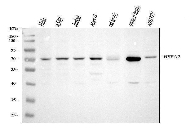
Western blot analysis of Grp75 using anti-Grp75 antibody (PA1789).
Electrophoresis was performed on a 5-20% SDS-PAGE gel at 70V (Stacking gel) / 90V (Resolving gel) for 2-3 hours. The sample well of each lane was loaded with 30 ug of sample under reducing conditions.
Lane 1: human Hela whole cell lysates,
Lane 2: human A549 whole cell lysates,
Lane 3: human Jurkat whole cell lysates,
Lane 4: human HepG2 whole cell lysates,
Lane 5: rat testis tissue lysates,
Lane 6: mouse testis tissue lysates,
Lane 7: mouse NIH/3T3 whole cell lysates.
After electrophoresis, proteins were transferred to a nitrocellulose membrane at 150 mA for 50-90 minutes. Blocked the membrane with 5% non-fat milk/TBS for 1.5 hour at RT. The membrane was incubated with rabbit anti-Grp75 antigen affinity purified polyclonal antibody (Catalog # PA1789) at 0.5 μg/mL overnight at 4°C, then washed with TBS-0.1%Tween 3 times with 5 minutes each and probed with a goat anti-rabbit IgG-HRP secondary antibody at a dilution of 1:5000 for 1.5 hour at RT. The signal is developed using an Enhanced Chemiluminescent detection (ECL) kit (Catalog # EK1002) with Tanon 5200 system. A specific band was detected for Grp75 at approximately 75 kDa. The expected band size for Grp75 is at 73 kDa.

Click image to see more details
IHC analysis of Grp75 using anti-Grp75 antibody (PA1789).
Grp75 was detected in a paraffin-embedded section of human lung cancer tissue. Heat mediated antigen retrieval was performed in EDTA buffer (pH 8.0, epitope retrieval solution). The tissue section was blocked with 10% goat serum. The tissue section was then incubated with 1 μg/ml rabbit anti-Grp75 Antibody (PA1789) overnight at 4°C. Peroxidase Conjugated Goat Anti-rabbit IgG was used as secondary antibody and incubated for 30 minutes at 37°C. The tissue section was developed using HRP Conjugated Rabbit IgG Super Vision Assay Kit (Catalog # SV0002) with DAB as the chromogen.

Click image to see more details
IHC analysis of Grp75 using anti-Grp75 antibody (PA1789).
Grp75 was detected in a paraffin-embedded section of rat liver tissue. Heat mediated antigen retrieval was performed in EDTA buffer (pH 8.0, epitope retrieval solution). The tissue section was blocked with 10% goat serum. The tissue section was then incubated with 1 μg/ml rabbit anti-Grp75 Antibody (PA1789) overnight at 4°C. Peroxidase Conjugated Goat Anti-rabbit IgG was used as secondary antibody and incubated for 30 minutes at 37°C. The tissue section was developed using HRP Conjugated Rabbit IgG Super Vision Assay Kit (Catalog # SV0002) with DAB as the chromogen.

Click image to see more details
ICC analysis of Grp75 using anti-Grp75 antibody (PA1789).
Grp75 was detected in an immunocytochemical section of A549 cells. Enzyme antigen retrieval was performed using IHC enzyme antigen retrieval reagent (AR0022) for 15 mins. The cells were blocked with 10% goat serum. And then incubated with 1 μg/ml rabbit anti-Grp75 Antibody (PA1789) overnight at 4°C. Biotinylated goat anti-rabbit IgG was used as secondary antibody and incubated for 30 minutes at 37°C. The section was developed using Strepavidin-Biotin-Complex (SABC)(Catalog # SA1022) with DAB as the chromogen.

Click image to see more details
IHC analysis of Grp75 using anti-Grp75 antibody (PA1789).
Grp75 was detected in frozen section of rat liver tissues. The tissue section was blocked with 10% goat serum. The tissue section was then incubated with 1μg/ml rabbit anti-Grp75 Antibody (PA1789) overnight at 4°C. Biotinylated goat anti-rabbit IgG was used as secondary antibody and incubated for 30 minutes at 37°C. The tissue section was developed using Strepavidin-Biotin-Complex (SABC)(Catalog # SA1022) with DAB as the chromogen.
Specific Publications For Anti-Grp75/HSPA9 Antibody Picoband® (PA1789)
Loading publications
Recommended Resources
Here are featured tools and databases that you might find useful.
- Boster's Pathways Library
- Protein Databases
- Bioscience Research Protocol Resources
- Data Processing & Analysis Software
- Photo Editing Software
- Scientific Literature Resources
- Research Paper Management Tools
- Molecular Biology Software
- Primer Design Tools
- Bioinformatics Tools
- Phylogenetic Tree Analysis
Customer Reviews
Have you used Anti-Grp75/HSPA9 Antibody Picoband®?
Share your experimental results or join a short interview to earn up to $1,000 in product credits or other rewards.
0 Reviews For Anti-Grp75/HSPA9 Antibody Picoband®
Customer Q&As
Have a question?
Find answers in Q&As, reviews.
Can't find your answer?
Submit your question
6 Customer Q&As for Anti-Grp75/HSPA9 Antibody Picoband®
Question
I was wanting to use your anti-Grp75/HSPA9 antibody for IHC-P for human lymphoblast on frozen tissues, but I want to know if it has been validated for this particular application. Has this antibody been validated and is this antibody a good choice for human lymphoblast identification?
Verified Customer
Verified customer
Asked: 2020-03-31
Answer
It shows on the product datasheet, PA1789 anti-Grp75/HSPA9 antibody has been validated for IHC-P, IHC-F, ICC, WB on human, mouse, rat tissues. We have an innovator award program that if you test this antibody and show it works in human lymphoblast in IHC-frozen, you can get your next antibody for free.
Boster Scientific Support
Answered: 2020-03-31
Question
Thank you for helping with my inquiry over the phone. Here are the WB image, lot number and protocol we used for lymphoblast using anti-Grp75/HSPA9 antibody PA1789. Let me know if you need anything else.
Verified Customer
Verified customer
Asked: 2020-01-08
Answer
We appreciate the data. You have provided everything we needed. Our lab team are working to resolve your inquiry as quickly as possible, and we appreciate your patience and understanding! Please let me know if there is anything you need in the meantime.
Boster Scientific Support
Answered: 2020-01-08
Question
I have a question about product PA1789, anti-Grp75/HSPA9 antibody. I was wondering if it would be possible to conjugate this antibody with biotin. I would need it to be without BSA or sodium azide. I am planning on using a buffer exchange of sodium azide with PBS only. Would there be problems for me to conjugate the antibody and store it in -20 degrees in small aliquots?
Verified Customer
Verified customer
Asked: 2018-02-26
Answer
We do not recommend storing this antibody with PBS buffer only in -20 degrees. If you want to store it in -20 degrees it is best to add some cryoprotectant like glycerol. If you want carrier free PA1789 anti-Grp75/HSPA9 antibody, we can provide it to you in a special formula with trehalose and/or glycerol. These molecules will not interfere with conjugation chemistry and provide a good level of protection for the antibody from degradation. Please be sure to specify this in your purchase order.
Boster Scientific Support
Answered: 2018-02-26
Question
Will PA1789 anti-Grp75/HSPA9 antibody work on parafin embedded sections? If so, which fixation method do you recommend we use (PFA, paraformaldehyde, other)?
Verified Customer
Verified customer
Asked: 2017-09-20
Answer
It shows on the product datasheet, PA1789 anti-Grp75/HSPA9 antibody as been validated on IHC-P. It is best to use PFA for fixation because it has better tissue penetration ability. PFA needs to be prepared fresh before use. Long term stored PFA turns into formalin, as the PFA molecules congregate and become formalin.
Boster Scientific Support
Answered: 2017-09-20
Question
Is a blocking peptide available for product anti-Grp75/HSPA9 antibody (PA1789)?
M. Taylor
Verified customer
Asked: 2016-11-25
Answer
We do provide the blocking peptide for product anti-Grp75/HSPA9 antibody (PA1789). If you would like to place an order for it please contact support@bosterbio.com and make a special request.
Boster Scientific Support
Answered: 2016-11-25
Question
We need to test anti-Grp75/HSPA9 antibody PA1789 on human lymphoblast for research purposes, then I may be interested in using anti-Grp75/HSPA9 antibody PA1789 for diagnostic purposes as well. Is the antibody suitable for diagnostic purposes?
K. Wu
Verified customer
Asked: 2014-02-05
Answer
The products we sell, including anti-Grp75/HSPA9 antibody PA1789, are only intended for research use. They would not be suitable for use in diagnostic work. If you have the means to develop a product into diagnostic use, and are interested in collaborating with us and develop our product into an IVD product, please contact us for more discussions.
Boster Scientific Support
Answered: 2014-02-05
