Click image to see more details
-

-
-
-
-
+4
Product Info Summary
| SKU: | M01692-4 |
|---|---|
| Size: | 100 μg/vial |
| Reactive Species: | Human, Mouse, Rat |
| Host: | Mouse |
| Application: | Flow Cytometry, IF, IHC, ICC, WB |
Customers Who Bought This Also Bought

Product info
Product Name
Anti-Hsp90 beta/HSP90AB1 Antibody Picoband® (monoclonal, 7B7F5)
SKU/Catalog Number
M01692-4
Size
100 μg/vial
Form
Lyophilized
Description
Boster Bio Anti-Hsp90 beta/HSP90AB1 Antibody Picoband® (monoclonal, 7B7F5) catalog # M01692-4. Tested in Flow Cytometry, IF, IHC, ICC, WB applications. This antibody reacts with Human, Mouse, Rat. The brand Picoband indicates this is a premium antibody that guarantees superior quality, high affinity, and strong signals with minimal background in Western blot applications. Only our best-performing antibodies are designated as Picoband, ensuring unmatched performance.
Storage & Handling
At -20°C for one year from date of receipt. After reconstitution, at 4°C for one month. It can also be aliquotted and stored frozen at -20°C for six months. Avoid repeated freezing and thawing.
Cite This Product
Anti-Hsp90 beta/HSP90AB1 Antibody Picoband® (monoclonal, 7B7F5) (Boster Biological Technology, Pleasanton CA, USA, Catalog # M01692-4)
Host
Mouse
Contents
Each vial contains 4 mg Trehalose, 0.9 mg NaCl and 0.2 mg Na2HPO4.
Clonality
Monoclonal
Clone Number
7B7F5
Isotype
IgG2b
Immunogen
A synthetic peptide corresponding to a sequence at the C-terminus of human Hsp90 beta, identical to the related mouse and rat sequences.
Cross-reactivity
No cross-reactivity with other proteins.
Reactive Species
M01692-4 is reactive to HSP90AB1 in Human, Mouse, Rat
Observed Molecular Weight
90 kDa
Calculated molecular weight
83.3 kDa
Background of HSP90AB1
Heat shock protein HSP 90-beta, also called HSP90beta, is a protein that in humans is encoded by the HSP90AB1 gene. It is mapped to chromosome 6p21.1. This gene encodes a member of the heat shock protein 90 family; these proteins are involved in signal transduction, protein folding and degradation and morphological evolution. And this gene is thought to play a role in gastric apoptosis and inflammation. Alternative splicing results in multiple transcript variants. Pseudogenes have been identified on multiple chromosomes.
Antibody Validation
Boster validates all antibodies on WB, IHC, ICC, Immunofluorescence, and ELISA with known positive control and negative samples to ensure specificity and high affinity, including thorough antibody incubations.
Application & Images
Applications
M01692-4 is guaranteed for Flow Cytometry, IF, IHC, ICC, WB Boster Guarantee
Recommend Dilution
| Application | Dilution | Species |
|---|---|---|
| Western blot | 0.25-0.5 µg/ml | Human, Mouse, Rat |
| Immunohistochemistry(Paraffin-embedded Section) | 2-5 μg/ml | Human, Mouse, Rat |
| Immunocytochemistry/Immunofluorescence | 5 µg/ml | Human |
| Flow Cytometry (Fixed) | 1-3 µg/1x106 cells | Human |
Tested application
Suggested blocking solution with 5% non-fat milk or BSA; (*)Recommended protein loading: 20-40 µg per lane
Use TE buffer pH 9.0 for antigen retrieval; (*) citrate buffer pH 6.0 is an alternative.
Validation Images & Assay Conditions
Click image to see more details
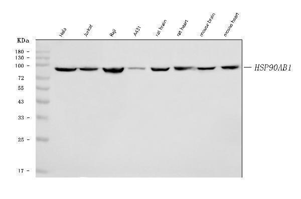
Western blot analysis of Hsp90 beta/HSP90AB1 using anti-Hsp90 beta/HSP90AB1 antibody (M01692-4).
Electrophoresis was performed on a 5-20% SDS-PAGE gel at 70V (Stacking gel) / 90V (Resolving gel) for 2-3 hours. The sample well of each lane was loaded with 30 ug of sample under reducing conditions.
Lane 1: human Hela whole cell lysates,
Lane 2: human Jurkat whole cell lysates,
Lane 3: human Raji whole cell lysates,
Lane 4: human A431 whole cell lysates,
Lane 5: rat brain tissue lysates,
Lane 6: rat heart tissue lysates,
Lane 7: mouse brain tissue lysates,
Lane 8: mouse heart tissue lysates.
After electrophoresis, proteins were transferred to a nitrocellulose membrane at 150 mA for 50-90 minutes. Blocked the membrane with 5% non-fat milk/TBS for 1.5 hour at RT. The membrane was incubated with mouse anti-Hsp90 beta/HSP90AB1 antigen affinity purified monoclonal antibody (Catalog # M01692-4) at 0.5 μg/mL overnight at 4°C, then washed with TBS-0.1%Tween 3 times with 5 minutes each and probed with a goat anti-mouse IgG-HRP secondary antibody at a dilution of 1:10000 for 1.5 hour at RT. The signal is developed using an Enhanced Chemiluminescent detection (ECL) kit (Catalog # EK1001) with Tanon 5200 system. A specific band was detected for Hsp90 beta/HSP90AB1 at approximately 90 kDa. The expected band size for Hsp90 beta/HSP90AB1 is at 84 kDa.

Click image to see more details
IHC analysis of Hsp90 beta/HSP90AB1 using anti-Hsp90 beta/HSP90AB1 antibody (M01692-4).
Hsp90 beta/HSP90AB1 was detected in a paraffin-embedded section of human thyroid cancer tissue. Heat mediated antigen retrieval was performed in EDTA buffer (pH 8.0, epitope retrieval solution). The tissue section was blocked with 10% goat serum. The tissue section was then incubated with 2 μg/ml mouse anti-Hsp90 beta/HSP90AB1 Antibody (M01692-4) overnight at 4°C. Peroxidase Conjugated Goat Anti-mouse IgG was used as secondary antibody and incubated for 30 minutes at 37°C. The tissue section was developed using HRP Conjugated Mouse IgG Super Vision Assay Kit (Catalog # SV0001) with DAB as the chromogen.

Click image to see more details
IF analysis of Hsp90 beta/HSP90AB1 using anti-Hsp90 beta/HSP90AB1 antibody (M01692-4).
Hsp90 beta/HSP90AB1 was detected in an immunocytochemical section of MCF-7 cells. Enzyme antigen retrieval was performed using IHC enzyme antigen retrieval reagent (AR0022) for 15 mins. The cells were blocked with 10% goat serum. And then incubated with 5 μg/mL mouse anti-Hsp90 beta/HSP90AB1 Antibody (M01692-4) overnight at 4°C. DyLight®488 Conjugated Goat Anti-Mouse IgG (BA1126) was used as secondary antibody at 1:100 dilution and incubated for 30 minutes at 37°C. The section was counterstained with DAPI. Visualize using a fluorescence microscope and filter sets appropriate for the label used.

Click image to see more details
IHC analysis of Hsp90 beta/HSP90AB1 using anti-Hsp90 beta/HSP90AB1 antibody (M01692-4).
Hsp90 beta/HSP90AB1 was detected in a paraffin-embedded section of human placenta tissue. Heat mediated antigen retrieval was performed in EDTA buffer (pH 8.0, epitope retrieval solution). The tissue section was blocked with 10% goat serum. The tissue section was then incubated with 2 μg/ml mouse anti-Hsp90 beta/HSP90AB1 Antibody (M01692-4) overnight at 4°C. Peroxidase Conjugated Goat Anti-mouse IgG was used as secondary antibody and incubated for 30 minutes at 37°C. The tissue section was developed using HRP Conjugated Mouse IgG Super Vision Assay Kit (Catalog # SV0001) with DAB as the chromogen.

Click image to see more details
IHC analysis of Hsp90 beta/HSP90AB1 using anti-Hsp90 beta/HSP90AB1 antibody (M01692-4).
Hsp90 beta/HSP90AB1 was detected in a paraffin-embedded section of human laryngeal squamous cell carcinoma tissue. Heat mediated antigen retrieval was performed in EDTA buffer (pH 8.0, epitope retrieval solution). The tissue section was blocked with 10% goat serum. The tissue section was then incubated with 2 μg/ml mouse anti-Hsp90 beta/HSP90AB1 Antibody (M01692-4) overnight at 4°C. Peroxidase Conjugated Goat Anti-mouse IgG was used as secondary antibody and incubated for 30 minutes at 37°C. The tissue section was developed using HRP Conjugated Mouse IgG Super Vision Assay Kit (Catalog # SV0001) with DAB as the chromogen.

Click image to see more details
IHC analysis of Hsp90 beta/HSP90AB1 using anti-Hsp90 beta/HSP90AB1 antibody (M01692-4).
Hsp90 beta/HSP90AB1 was detected in a paraffin-embedded section of mouse brain tissue. Heat mediated antigen retrieval was performed in EDTA buffer (pH 8.0, epitope retrieval solution). The tissue section was blocked with 10% goat serum. The tissue section was then incubated with 2 μg/ml mouse anti-Hsp90 beta/HSP90AB1 Antibody (M01692-4) overnight at 4°C. Peroxidase Conjugated Goat Anti-mouse IgG was used as secondary antibody and incubated for 30 minutes at 37°C. The tissue section was developed using HRP Conjugated Mouse IgG Super Vision Assay Kit (Catalog # SV0001) with DAB as the chromogen.

Click image to see more details
IHC analysis of Hsp90 beta/HSP90AB1 using anti-Hsp90 beta/HSP90AB1 antibody (M01692-4).
Hsp90 beta/HSP90AB1 was detected in a paraffin-embedded section of rat brain tissue. Heat mediated antigen retrieval was performed in EDTA buffer (pH 8.0, epitope retrieval solution). The tissue section was blocked with 10% goat serum. The tissue section was then incubated with 2 μg/ml mouse anti-Hsp90 beta/HSP90AB1 Antibody (M01692-4) overnight at 4°C. Peroxidase Conjugated Goat Anti-mouse IgG was used as secondary antibody and incubated for 30 minutes at 37°C. The tissue section was developed using HRP Conjugated Mouse IgG Super Vision Assay Kit (Catalog # SV0001) with DAB as the chromogen.

Click image to see more details
Flow Cytometry analysis of CACO-2 cells using anti-Hsp90 beta/HSP90AB1 antibody (M01692-4).
Overlay histogram showing CACO-2 cells stained with M01692-4 (Blue line). To facilitate intracellular staining, cells were fixed with 4% paraformaldehyde and permeabilized with permeabilization buffer. The cells were blocked with 10% normal goat serum. And then incubated with mouse anti-Hsp90 beta/HSP90AB1 Antibody (M01692-4, 1 μg/1x106 cells) for 30 min at 20°C. DyLight®488 conjugated goat anti-mouse IgG (BA1126, 5-10 μg/1x106 cells) was used as secondary antibody for 30 minutes at 20°C. Isotype control antibody (Green line) was mouse IgG (1 μg/1x106) used under the same conditions. Unlabelled sample without incubation with primary antibody and secondary antibody (Red line) was used as a blank control.
Specific Publications For Anti-Hsp90 beta/HSP90AB1 Antibody Picoband® (monoclonal, 7B7F5) (M01692-4)
Loading publications
Recommended Resources
Here are featured tools and databases that you might find useful.
- Boster's Pathways Library
- Protein Databases
- Bioscience Research Protocol Resources
- Data Processing & Analysis Software
- Photo Editing Software
- Scientific Literature Resources
- Research Paper Management Tools
- Molecular Biology Software
- Primer Design Tools
- Bioinformatics Tools
- Phylogenetic Tree Analysis
Customer Reviews
Have you used Anti-Hsp90 beta/HSP90AB1 Antibody Picoband® (monoclonal, 7B7F5)?
Share your experimental results or join a short interview to earn up to $1,000 in product credits or other rewards.
0 Reviews For Anti-Hsp90 beta/HSP90AB1 Antibody Picoband® (monoclonal, 7B7F5)
Customer Q&As
Have a question?
Find answers in Q&As, reviews.
Can't find your answer?
Submit your question
