Click image to see more details
-

-
-
-
-
+10
Product Info Summary
| SKU: | A00171 |
|---|---|
| Size: | 100 μg/vial |
| Reactive Species: | Human, Mouse, Rat |
| Host: | Rabbit |
| Application: | ELISA, Flow Cytometry, IF, IHC, ICC, WB |
Customers Who Bought This Also Bought

Product info
Product Name
Anti-ICAM1 Antibody Picoband®
SKU/Catalog Number
A00171
Size
100 μg/vial
Form
Lyophilized
Description
ICAM1 (CD54) is an immunoglobulin superfamily adhesion molecule induced on endothelial/immune cells and binds β2 integrins (e.g., LFA-1), mediating leukocyte adhesion and transendothelial migration during inflammation; it is also described as a rhinovirus receptor (context-dependent). Assay context: antibody generated against recombinant human ICAM1 (reported immunogen region Q28–R268) and validated across ELISA/flow/IF/IHC/WB, enabling combined soluble and tissue-localization readouts with orthogonal confirmation via Western blotting as needed. Often profiled alongside chemokine/cytokine inflammation panels including IL-18 and secreted glycoproteins such as LGALS3BP to link endothelial activation with immune signaling.
Storage & Handling
Store at -20˚C for one year from date of receipt. After reconstitution, at 4˚C for one month. It can also be aliquotted and stored frozen at -20˚C for six months. Avoid repeated freeze-thaw cycles.
Cite This Product
Anti-ICAM1 Antibody Picoband® (Boster Biological Technology, Pleasanton CA, USA, Catalog # A00171)
Host
Rabbit
Contents
Each vial contains 4mg Trehalose, 0.9mg NaCl, 0.2mg Na2HPO4, 0.05mg NaN3.
Clonality
Polyclonal
Isotype
Rabbit IgG
Immunogen
E. coli-derived human ICAM1 recombinant protein (Position: Q28-R268).
Cross-reactivity
No cross-reactivity with other proteins.
Reactive Species
A00171 is reactive to ICAM1 in Human, Mouse, Rat
Observed Molecular Weight
90 kDa
Calculated molecular weight
57.8 kDa
Background of ICAM1
CD54, also known as ICAM-1. Intercellular adhesion molecule-1 (ICAM1) is a ligand for lymphocyte function-associated (LFA) antigens. ICAM-1 is an integral membrane protein, a member of the immunoglobulin superfamily, and a ligand for LFA-1, a beta 2 leukocyte integrin. This protein is the major human rhinovirus receptor. The ICAM1 gene is mapped to human chromosome 19. In humans, lymphocyte adhesion to cells is mediated by the protein heterodimer CD11a/CD18 (Leu-CAMa, LFA-1) and its ligand CD54 (ICAM-1).
Antibody Validation
Boster validates all antibodies on WB, IHC, ICC, Immunofluorescence, and ELISA with known positive control and negative samples to ensure specificity and high affinity, including thorough antibody incubations.
Application & Images
Applications
A00171 is guaranteed for ELISA, Flow Cytometry, IF, IHC, ICC, WB Boster Guarantee
Recommend Dilution
| Application | Dilution | Species |
|---|---|---|
| Western blot | 0.1-0.5μg/ml | |
| Immunohistochemistry (Paraffin-embedded Section) | 0.5-1μg/ml | |
| Immunocytochemistry/Immunofluorescence | 2μg/ml | |
| Flow Cytometry (Fixed) | 1-3μg/1x106 cells | |
| ELISA | 0.1-0.5μg/ml |
Tested application
Suggested blocking solution with 5% non-fat milk or BSA; (*)Recommended protein loading: 20-40 µg per lane
Use TE buffer pH 9.0 for antigen retrieval; (*) citrate buffer pH 6.0 is an alternative.
Validation Images & Assay Conditions
Click image to see more details
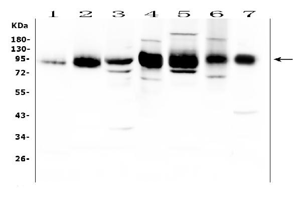
Western blot analysis of ICAM1 using anti-ICAM1 antibody (A00171).
Electrophoresis was performed on a 5-20% SDS-PAGE gel at 70V (Stacking gel) / 90V (Resolving gel) for 2-3 hours. The sample well of each lane was loaded with 30 ug of sample under reducing conditions.
Lane 1: rat spleen tissue lysate,
Lane 2: rat thymus tissue lysate,
Lane 3: rat RH35 cell lysate,
Lane 4: mouse spleen tissue lysate,
Lane 5: mouse thymus tissue lysate,
Lane 6: mouse HEPA1-6 cell lysate,
Lane 7: mouse heart tissue lysate.
After Electrophoresis, proteins were transferred to a Nitrocellulose membrane at 150mA for 50-90 minutes. Blocked the membrane with 5% Non-fat Milk/ TBS for 1.5 hour at RT. The membrane was incubated with rabbit anti-ICAM1 antigen affinity purified polyclonal antibody (Catalog # A00171) at 0.5 μg/mL overnight at 4°C, then washed with TBS-0.1%Tween 3 times with 5 minutes each and probed with a goat anti-rabbit IgG-HRP secondary antibody at a dilution of 1:10000 for 1.5 hour at RT. The signal is developed using an Enhanced Chemiluminescent detection (ECL) kit (Catalog # EK1002) with Tanon 5200 system. A specific band was detected for ICAM1 at approximately 90 kDa. The expected band size for ICAM1 is at 58 kDa.

Click image to see more details
STAT3 inhibitor attenuates L-AKI and restores kidney function. A Schematic diagram of the animal model. B BUN and serum creatinine were measured to assess kidney function. Groups: Control, LPS, LPS + Stattic (5 mg/kg) and LPS + Stattic (10 mg/kg) ( n = 6 in each group). C Flow cytometric comparison of pSTAT3 levels was conducted between two populations (kidney macrophages, CD11b high F4/80 low and CD11b low F4/80 high ) in LPS-induced kidneys. D Western blotting representative image (left) and quantification (right) of pSTAT3, STAT3 and ICAM-1. Each band shows a typical group, as indicated. E Changes in the proportion of two macrophage subpopulations between each group were observed with FACS analysis, and the frequency and ratio of individual populations were quantified using GraphPad Prism. F Upregulated expression of ICAM-1, NGAL, and pSTAT3 by LPS induction in IHC analysis ( n = 6 in each group) decreased with Stattic treatment. Scale bars, 100 μm (100X) and 50 μm (400X). All experiments were independently replicated at least three times, and the data are presented as mean ± SEM. * P < 0.05, ** P < 0.01, *** P < 0.001
Index in PubMed under a CC BY license. PMID: 39367511

Click image to see more details
Single-cell transcriptional profiling of intracranial fusiform aneurysmal cells ( A - K ) and multi-color immunofluorescence (mIF) of smooth muscle cells (SMCs) markers and inflammatory markers between intracranial fusiform aneurysms (IFAs) and normal cerebral arteries (NCAs) ( L - M ). T-SNE visualization of intracranial fusiform aneurysmal cells type. Colored according to cell type ( A ). Visualization of specific gene expression patterns related to cell subsets identified in ( A ) using a bubble plot ( B ). T-SNE visualization of VSMC cell clusters. Colored according to clusters ( C ). Visualization of structural protein and inflammation-related genes expression patterns within the subsets of smooth muscle cells identified in ( C ) using a bubble plot ( D ). The violin plots show the expression differences of structural protein genes and inflammatory genes between the contraction subgroup (VSMC5) and the inflammatory subgroup (VSMC6) ( E ). The volcano plot specifically shows the gene expression differences between the two cell groups ( F ). The petal plot displays the GO enrichment analysis results of differential genes between the VSMC5 and VSMC6 ( G ). The bar graph shows the KEGG enrichment analysis results of differential genes between these two cell subsets ( H ). The GSEA enrichment analysis results of differential genes between the two groups. The enrichment results of differential genes related to signaling pathways ( I ). The enrichment results of differential genes related to structural protein genes ( J ). The enrichment results of differential genes related to the process of inflammatory factor secretion ( K ). α-SMA (green, SMCs marker), MMP-9 (blue, inflammatory marker) and ICAM-1 (red, inflammatory marker) in FIAs and NCAs are detected using mIF. Scale bar, 100 μm ( L ). Statistical analysis of mean fluorescence intensity about SMCs marker (α-SMA) and inflammatory markers (ICAM-1 and MMP-9) between NCAs ( n = 4) and FIAs ( n = 5). 'n' represented the number of samples. Three random fields were selected for statistical analysis in each sample, and the average value represented the detection value of this marker in this sample. The Student's t-test is utilized to examine the statistical differences among each marker. ns, no significant; ** p < 0.01, *** p < 0.01 (M). n values indicate number of independent experiments performed
Index in PubMed under a CC BY license. PMID: 38741091

Click image to see more details
PDGFRB somatic mutation induce phenotypic modulation in SMCs. Immunostaining reveals the expression levels of smooth muscle markers (a-SMA and SM22a) and inflammatory markers (VCAM1, ICAM1, MMP1 and MMP9) in HBVSMCs transfected with different viruses (Control: vector; Wild Type: PDGFRB; Y562D: PDGFRB Y562D ) ( a ). The relative density of immunoblot bands about markers shown in ( A ) were display ( B ) (normalized to those in cells transfected with vector viruses). RT-qPCR ( C ) of SMCs markers (α-SMA and SM22α) and inflammatory markers (VCAM-1, ICAM1, MMP-9 and MMP-1) in HBVSMCs underwent different treatments. Student's t-test and Benjamini–Hochberg correction are employed to assess the statistical significance. Edu assay exhibit the proliferation ability of HBVSMCs under different treatment conditions ( D ). Statistical analysis of the proportion of Edu-positive cells in the different groups from 5 different fields of each group at × 200 magnification. Tukey's multiple comparisons test is used for statistical differences ( E ). Scratch assay displays migratory ability of HBVSMCs underwent different treatment ( F ). Statistical analysis of the rate of wound healing (reduced area at 4H /area at 0H) in the different groups from 9 different fields of each group at × 200 magnification. Tukey's multiple comparisons test is utilized to evaluate the statistical significance. * p adj < 0.05, ** p adj < 0.01, *** p adj < 0.001, **** p adj < 0.0001 ( G ). The above experiments are all repeated three times
Index in PubMed under a CC BY license. PMID: 38741091

Click image to see more details
Protein (A) (immunohistochemistry) and gene (B) expression of ICAM-1. Sham, false-operated rats; BD, rats submitted to brain death; MP, rats treated with methylprednisolone (MP) after 3h of confirmation of BD and MP/E2, rats treated with 17β-estradiol (E2) and methylprednisolone after 3h of confirmation of BD. Data expressed as mean ± SEM from 5-8 animals per group (A) . Data expressed as median and 95th percentile from 6-8 animals (B) . 1 section per animal and 10 areas per section were analyzed. The photomicrographs (x20) are representative of protein expression on each group. (A) p (Kruskal Wallis) =0.6009; (B) p (Kruskal Wallis) =0.7960.
Index in PubMed under a CC BY license. PMID: 38765005

Click image to see more details
mRNA expression of ICAM-1, VCAM-1 and PBEF and protein expression of ICAM-1 and VCAM-1 in lung . A ) Real-time PCR showed that the expression levels of ICAM-1, VCAM-1 and PBEF mRNA decreased significantly in the EP and GL groups compared to the control group. B ) Western-blot showed that the expression levels of ICAM-1 and VCAM-1 protein decreased significantly in the EP and GL groups compared to the control group. C ) Quantitative assessment of protein relative to β-actin showed that the expression levels of ICAM-1 and VCAM-1 protein decreased significantly in the EP and GL groups compared to the control group. * P < 0.05 versus the control group. EP, ethyl pyruvate; GL, glycyrrhizin; ICAM-1, intercellular adhesion molecule-1; PBEF, pre-B-cell colony-enhancing factor; VCAM-1, vascular cell adhesion molecule 1.
Index in PubMed under a CC BY license. PMID: 23497622

Click image to see more details
Western blot analysis of ICAM1 using anti-ICAM1 antibody (A00171).
Electrophoresis was performed on a 5-20% SDS-PAGE gel at 70V (Stacking gel) / 90V (Resolving gel) for 2-3 hours. The sample well of each lane was loaded with 30 ug of sample under reducing conditions.
Lane 1: human Raji whole cell lysates,
Lane 2: human K562 whole cell lysates,
Lane 3: human HepG2 whole cell lysates,
Lane 4: human 293T whole cell lysates.
After electrophoresis, proteins were transferred to a nitrocellulose membrane at 150 mA for 50-90 minutes. Blocked the membrane with 5% non-fat milk/TBS for 1.5 hour at RT. The membrane was incubated with rabbit anti-ICAM1 antigen affinity purified polyclonal antibody (Catalog # A00171) at 0.5 μg/mL overnight at 4°C, then washed with TBS-0.1%Tween 3 times with 5 minutes each and probed with a goat anti-rabbit IgG-HRP secondary antibody at a dilution of 1:5000 for 1.5 hour at RT. The signal is developed using an Enhanced Chemiluminescent detection (ECL) kit (Catalog # EK1002) with Tanon 5200 system. A specific band was detected for ICAM1 at approximately 90 kDa. The expected band size for ICAM1 is at 58 kDa.

Click image to see more details
IHC analysis of ICAM1 using anti-ICAM1 antibody (A00171).
ICAM1 was detected in paraffin-embedded section of human lung cancer tissue. Heat mediated antigen retrieval was performed in EDTA buffer (pH8.0, epitope retrieval solution). The tissue section was blocked with 10% goat serum. The tissue section was then incubated with 1μg/ml rabbit anti-ICAM1 Antibody (A00171) overnight at 4°C. Peroxidase Conjugated Goat Anti-rabbit IgG was used as secondary antibody and incubated for 30 minutes at 37°C. The tissue section was developed using HRP Conjugated Rabbit IgG Super Vision Assay Kit (Catalog # SV0002) with DAB as the chromogen.

Click image to see more details
Flow Cytometry analysis of PC-3 cells using anti-ICAM1 antibody (A00171).

Click image to see more details
IHC analysis of ICAM1 using anti-ICAM1 antibody (A00171).
ICAM1 was detected in paraffin-embedded section of human bladder urothelial carcinoma tissue. Heat mediated antigen retrieval was performed in EDTA buffer (pH8.0, epitope retrieval solution). The tissue section was blocked with 10% goat serum. The tissue section was then incubated with 1μg/ml rabbit anti-ICAM1 Antibody (A00171) overnight at 4°C. Peroxidase Conjugated Goat Anti-rabbit IgG was used as secondary antibody and incubated for 30 minutes at 37°C. The tissue section was developed using HRP Conjugated Rabbit IgG Super Vision Assay Kit (Catalog # SV0002) with DAB as the chromogen.

Click image to see more details
IHC analysis of ICAM1 using anti-ICAM1 antibody (A00171).
ICAM1 was detected in paraffin-embedded section of human colorectal adenocarcinoma tissue. Heat mediated antigen retrieval was performed in EDTA buffer (pH8.0, epitope retrieval solution). The tissue section was blocked with 10% goat serum. The tissue section was then incubated with 1μg/ml rabbit anti-ICAM1 Antibody (A00171) overnight at 4°C. Peroxidase Conjugated Goat Anti-rabbit IgG was used as secondary antibody and incubated for 30 minutes at 37°C. The tissue section was developed using HRP Conjugated Rabbit IgG Super Vision Assay Kit (Catalog # SV0002) with DAB as the chromogen.

Click image to see more details
IHC analysis of ICAM1 using anti-ICAM1 antibody (A00171).
ICAM1 was detected in paraffin-embedded section of human placenta tissue. Heat mediated antigen retrieval was performed in EDTA buffer (pH8.0, epitope retrieval solution). The tissue section was blocked with 10% goat serum. The tissue section was then incubated with 1μg/ml rabbit anti-ICAM1 Antibody (A00171) overnight at 4°C. Peroxidase Conjugated Goat Anti-rabbit IgG was used as secondary antibody and incubated for 30 minutes at 37°C. The tissue section was developed using HRP Conjugated Rabbit IgG Super Vision Assay Kit (Catalog # SV0002) with DAB as the chromogen.

Click image to see more details
IHC analysis of ICAM1 using anti-ICAM1 antibody (A00171).
ICAM1 was detected in paraffin-embedded section of human spleen tissue. Heat mediated antigen retrieval was performed in EDTA buffer (pH8.0, epitope retrieval solution). The tissue section was blocked with 10% goat serum. The tissue section was then incubated with 1μg/ml rabbit anti-ICAM1 Antibody (A00171) overnight at 4°C. Peroxidase Conjugated Goat Anti-rabbit IgG was used as secondary antibody and incubated for 30 minutes at 37°C. The tissue section was developed using HRP Conjugated Rabbit IgG Super Vision Assay Kit (Catalog # SV0002) with DAB as the chromogen.

Click image to see more details
IF analysis of ICAM1 using anti-ICAM1 antibody (A00171).
ICAM1 was detected in immunocytochemical section of A431 cells. Enzyme antigen retrieval was performed using IHC enzyme antigen retrieval reagent (AR0022) for 15 mins. The cells were blocked with 10% goat serum. And then incubated with 2μg/mL rabbit anti-ICAM1 Antibody (A00171) overnight at 4°C. DyLight®488 Conjugated Goat Anti-Rabbit IgG (BA1127) was used as secondary antibody at 1:100 dilution and incubated for 30 minutes at 37°C. The section was counterstained with DAPI. Visualize using a fluorescence microscope and filter sets appropriate for the label used.
Specific Publications For Anti-ICAM1 Antibody Picoband® (A00171)
Loading publications
Recommended Resources
Here are featured tools and databases that you might find useful.
- Boster's Pathways Library
- Protein Databases
- Bioscience Research Protocol Resources
- Data Processing & Analysis Software
- Photo Editing Software
- Scientific Literature Resources
- Research Paper Management Tools
- Molecular Biology Software
- Primer Design Tools
- Bioinformatics Tools
- Phylogenetic Tree Analysis
Customer Reviews
Have you used Anti-ICAM1 Antibody Picoband®?
Share your experimental results or join a short interview to earn up to $1,000 in product credits or other rewards.
0 Reviews For Anti-ICAM1 Antibody Picoband®
Customer Q&As
Have a question?
Find answers in Q&As, reviews.
Can't find your answer?
Submit your question
16 Customer Q&As for Anti-ICAM1 Antibody Picoband®
Question
Would anti-ICAM1 antibody A00171 work for WB with cervix carcinoma?
Verified Customer
Verified customer
Asked: 2020-03-25
Answer
According to the expression profile of cervix carcinoma, ICAM1 is highly expressed in cervix carcinoma. So, it is likely that anti-ICAM1 antibody A00171 will work for WB with cervix carcinoma.
Boster Scientific Support
Answered: 2020-03-25
Question
Do you have a BSA free version of anti-ICAM1 antibody A00171 available?
Verified Customer
Verified customer
Asked: 2020-02-28
Answer
I appreciate your recent telephone inquiry. I can confirm that some lots of this anti-ICAM1 antibody A00171 are BSA free. For now, these lots are available and we can make a BSA free formula for you free of charge. It will take 3 extra days to prepare. If you require this antibody BSA free again in future, please do not hesitate to contact me and I will be pleased to check which lots we have in stock that are BSA free.
Boster Scientific Support
Answered: 2020-02-28
Question
We are currently using anti-ICAM1 antibody A00171 for human tissue, and we are content with the WB results. The species of reactivity given in the datasheet says human, mouse, rat. Is it true that the antibody can work on horse tissues as well?
Verified Customer
Verified customer
Asked: 2019-11-14
Answer
The anti-ICAM1 antibody (A00171) has not been tested for cross reactivity specifically with horse tissues, but there is a good chance of cross reactivity. We have an innovator award program that if you test this antibody and show it works in horse you can get your next antibody for free. Please contact me if I can help you with anything.
Boster Scientific Support
Answered: 2019-11-14
Question
Thanks for helping with my inquiry over the phone. Here are the WB image, lot number and protocol we used for cervix carcinoma using anti-ICAM1 antibody A00171. Let me know if you need anything else.
L. Jackson
Verified customer
Asked: 2019-09-06
Answer
Thank you for the data. You have provided everything we needed. Our lab team are working to resolve your inquiry as quickly as possible, and we appreciate your patience and understanding! Please let me know if there is anything you need in the meantime.
Boster Scientific Support
Answered: 2019-09-06
Question
I see that the anti-ICAM1 antibody A00171 works with WB, what is the protocol used to produce the result images on the product page?
Verified Customer
Verified customer
Asked: 2019-08-06
Answer
You can find protocols for WB on the "support/technical resources" section of our navigation menu. If you have any further questions, please send an email to support@bosterbio.com
Boster Scientific Support
Answered: 2019-08-06
Question
I was wanting to use to test anti-ICAM1 antibody A00171 on mouse cervix carcinoma for research purposes, then I may be interested in using anti-ICAM1 antibody A00171 for diagnostic purposes as well. Is the antibody suitable for diagnostic purposes?
S. Rodriguez
Verified customer
Asked: 2019-07-23
Answer
The products we sell, including anti-ICAM1 antibody A00171, are only intended for research use. They would not be suitable for use in diagnostic work. If you have the means to develop a product into diagnostic use, and are interested in collaborating with us and develop our product into an IVD product, please contact us for more discussions.
Boster Scientific Support
Answered: 2019-07-23
Question
Is this A00171 anti-ICAM1 antibody reactive to the isotypes of ICAM1?
Verified Customer
Verified customer
Asked: 2019-07-11
Answer
The immunogen of A00171 anti-ICAM1 antibody is E. coli-derived human ICAM1 recombinant protein (Position: Q28-R268). Could you tell me which isotype you are interested in so I can help see if the immunogen is part of this isotype?
Boster Scientific Support
Answered: 2019-07-11
Question
We need using your anti-ICAM1 antibody for regulation of ruffle assembly studies. Has this antibody been tested with western blotting on mouse thymus tissue? We would like to see some validation images before ordering.
Verified Customer
Verified customer
Asked: 2019-07-09
Answer
Thanks for your inquiry. This A00171 anti-ICAM1 antibody is tested on rat thymus tissue, spleen tissue, tissue lysate, mouse thymus tissue, heart tissue. It is guaranteed to work for ELISA, WB in human, mouse, rat. Our Boster guarantee will cover your intended experiment even if the sample type has not been be directly tested.
Boster Scientific Support
Answered: 2019-07-09
Question
I was wanting to use your anti-ICAM1 antibody for WB for mouse cervix carcinoma on frozen tissues, but I want to know if it has been tested for this particular application. Has this antibody been tested and is this antibody a good choice for mouse cervix carcinoma identification?
Verified Customer
Verified customer
Asked: 2019-05-22
Answer
As indicated on the product datasheet, A00171 anti-ICAM1 antibody has been tested for ELISA, WB on human, mouse, rat tissues. We have an innovator award program that if you test this antibody and show it works in mouse cervix carcinoma in IHC-frozen, you can get your next antibody for free.
Boster Scientific Support
Answered: 2019-05-22
Question
My question regarding product A00171, anti-ICAM1 antibody. I was wondering if it would be possible to conjugate this antibody with biotin. I would need it to be without BSA or sodium azide. I am planning on using a buffer exchange of sodium azide with PBS only. Would there be problems for me to conjugate the antibody and store it in -20 degrees in small aliquots?
Verified Customer
Verified customer
Asked: 2018-11-16
Answer
We suggest not storing this antibody with PBS buffer only in -20 degrees. If you want to store it in -20 degrees it is best to add some cryoprotectant like glycerol. If you want carrier free A00171 anti-ICAM1 antibody, we can provide it to you in a special formula with trehalose and/or glycerol. These molecules will not interfere with conjugation chemistry and provide a good level of protection for the antibody from degradation. Please be sure to specify this in your purchase order.
Boster Scientific Support
Answered: 2018-11-16
Question
Our lab used your anti-ICAM1 antibody for ELISA on plasma in the past. I am using human, and We intend to use the antibody for WB next. I am interested in examining plasma as well as cervix carcinoma in our next experiment. Do you have any suggestion on which antibody would work the best for WB?
Verified Customer
Verified customer
Asked: 2018-05-16
Answer
I have checked the website and datasheets of our anti-ICAM1 antibody and it seems that A00171 has been validated on human in both ELISA and WB. Thus A00171 should work for your application. Our Boster satisfaction guarantee will cover this product for WB in human even if the specific tissue type has not been validated. We do have a comprehensive range of products for WB detection and you can check out our website bosterbio.com to find out more information about them.
Boster Scientific Support
Answered: 2018-05-16
Question
We have been able to see staining in mouse cervix carcinoma. Do you have any suggestions? Is anti-ICAM1 antibody supposed to stain cervix carcinoma positively?
Verified Customer
Verified customer
Asked: 2017-07-18
Answer
According to literature cervix carcinoma does express ICAM1. According to Uniprot.org, ICAM1 is expressed in lung, kidney, blood, plasma, cervix carcinoma, liver, leukemic t-cell, erythroleukemia, among other tissues. Regarding which tissues have ICAM1 expression, here are a few articles citing expression in various tissues:
Blood, Pubmed ID: 15572059
Cervix carcinoma, Pubmed ID: 18669648, 20068231
Erythroleukemia, Pubmed ID: 23186163
Kidney, Pubmed ID: 15489334
Leukemic T-cell, Pubmed ID: 19349973
Liver, Pubmed ID: 19159218, 24275569
Lung, Pubmed ID: 14702039
Plasma, Pubmed ID: 16335952
Boster Scientific Support
Answered: 2017-07-18
Question
Our team were content with the WB result of your anti-ICAM1 antibody. However we have observed positive staining in kidney membrane using this antibody. Is that expected? Could you tell me where is ICAM1 supposed to be expressed?
W. Johnson
Verified customer
Asked: 2016-09-23
Answer
From literature, kidney does express ICAM1. Generally ICAM1 expresses in membrane. Regarding which tissues have ICAM1 expression, here are a few articles citing expression in various tissues:
Blood, Pubmed ID: 15572059
Cervix carcinoma, Pubmed ID: 18669648, 20068231
Erythroleukemia, Pubmed ID: 23186163
Kidney, Pubmed ID: 15489334
Leukemic T-cell, Pubmed ID: 19349973
Liver, Pubmed ID: 19159218, 24275569
Lung, Pubmed ID: 14702039
Plasma, Pubmed ID: 16335952
Boster Scientific Support
Answered: 2016-09-23
Question
I have attached the WB image, lot number and protocol we used for cervix carcinoma using anti-ICAM1 antibody A00171. Please let me know if you require anything else.
G. Huang
Verified customer
Asked: 2016-09-21
Answer
Thank you very much for the data. Our lab team are working to resolve this as quickly as possible, and we appreciate your patience and understanding! You have provided everything we needed. Please let me know if there is anything you need in the meantime.
Boster Scientific Support
Answered: 2016-09-21
Question
Is a blocking peptide available for product anti-ICAM1 antibody (A00171)?
D. Carter
Verified customer
Asked: 2016-08-12
Answer
We do provide the blocking peptide for product anti-ICAM1 antibody (A00171). If you would like to place an order for it please contact support@bosterbio.com and make a special request.
Boster Scientific Support
Answered: 2016-08-12
Question
Will A00171 anti-ICAM1 antibody work on parafin embedded sections? If so, which fixation method do you recommend we use (PFA, paraformaldehyde, other)?
E. Gonzalez
Verified customer
Asked: 2014-05-05
Answer
As indicated on the product datasheet, A00171 anti-ICAM1 antibody as been tested on WB. It is best to use PFA for fixation because it has better tissue penetration ability. PFA needs to be prepared fresh before use. Long term stored PFA turns into formalin, as the PFA molecules congregate and become formalin.
Boster Scientific Support
Answered: 2014-05-05
