Click image to see more details
-

-
-
-
-
+1
Product Info Summary
| SKU: | A00070 |
|---|---|
| Size: | 100 μg/vial |
| Reactive Species: | Mouse, Rat |
| Host: | Rabbit |
| Application: | ELISA, IHC, WB |
Customers Who Bought This Also Bought

Product info
Product Name
Anti-IGF1 Receptor/Igf1r Antibody Picoband®
SKU/Catalog Number
A00070
Size
100 μg/vial
Form
Lyophilized
Description
Boster Bio Anti-IGF1 Receptor/Igf1r Antibody Picoband® catalog # A00070. Tested in ELISA, IHC, WB applications. This antibody reacts with Mouse, Rat. The brand Picoband indicates this is a premium antibody that guarantees superior quality, high affinity, and strong signals with minimal background in Western blot applications. Only our best-performing antibodies are designated as Picoband, ensuring unmatched performance.
Storage & Handling
Store at -20˚C for one year from date of receipt. After reconstitution, at 4˚C for one month. It can also be aliquotted and stored frozen at -20˚C for six months. Avoid repeated freeze-thaw cycles.
Cite This Product
Anti-IGF1 Receptor/Igf1r Antibody Picoband® (Boster Biological Technology, Pleasanton CA, USA, Catalog # A00070)
Host
Rabbit
Contents
Each vial contains 4mg Trehalose, 0.9mg NaCl, 0.2mg Na2HPO4, 0.05mg NaN3.
Clonality
Polyclonal
Isotype
Rabbit IgG
Immunogen
E. coli-derived mouse IGF1 Receptor recombinant protein (Position: E31-K257).
Cross-reactivity
No cross-reactivity with other proteins.
Reactive Species
A00070 is reactive to Igf1r in Mouse, Rat
Observed Molecular Weight
155 kDa
Calculated molecular weight
155.8 kDa
Background of Igf1r
IGF1R (Insulin-like Growth Factor 1 (IGF-1) Receptor) is a protein found on the surface of human cells. It is a transmembrane receptor that is activated by a hormone called Insulin-like growth factor 1 (IGF-1) and by a related hormone called IGF-2. It belongs to the large class of tyrosine kinase receptors. The IGF1R gene is mapped on 15q26.3. IGF-1 plays an important role in growth and continues to have anabolic effects in adults - meaning that it can induce hypertrophy of skeletal muscle and other target tissues. Using a yeast 2-hybrid system, it was identified a regulatory subunit of phosphatidylinositol (PI) 3-kinase, PIK3R3, as a binding partner of IGF1R. Functional interaction between BRCA1 and SP1 in the regulation of the IGF1R gene was studied in Schneider cells, a Drosophila cell line which lacks endogenous SP1. In these cells, BRCA1 suppressed 45% of the SP1-induced trans-activation of the IGF1R promoter. Overexpression of the Grb10-binding fragment of Gigyf1 resulted in a significant increase in Igf1-stimulated Igf1r tyrosine phosphorylation. Like the insulin receptor, the IGF-1 receptor is a receptor tyrosine kinase - meaning it signals by causing the addition of a phosphate molecule on particular tyrosines. IGF-1 activates the Insulin receptor at approximately 0.1x the potency of insulin. Part of this signaling may be via IGF1R-InsulinReceptor heterodimers.
Antibody Validation
Boster validates all antibodies on WB, IHC, ICC, Immunofluorescence, and ELISA with known positive control and negative samples to ensure specificity and high affinity, including thorough antibody incubations.
Application & Images
Applications
A00070 is guaranteed for ELISA, IHC, WB Boster Guarantee
Recommend Dilution
| Application | Dilution | Species |
|---|---|---|
| Western blot | 0.1-0.5μg/ml | |
| Immunohistochemistry (Paraffin-embedded Section) | 0.5-1μg/ml | |
| ELISA | 0.1-0.5μg/ml |
Tested application
Suggested blocking solution with 5% non-fat milk or BSA; (*)Recommended protein loading: 20-40 µg per lane
Use TE buffer pH 9.0 for antigen retrieval; (*) citrate buffer pH 6.0 is an alternative.
Validation Images & Assay Conditions
Click image to see more details
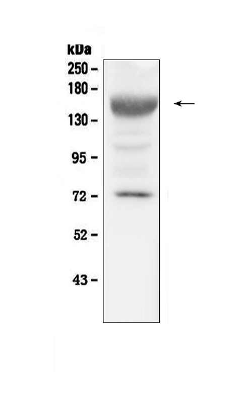
Western blot analysis of IGF1 Receptor using anti-IGF1 Receptor antibody (A00070).
Electrophoresis was performed on a 5-20% SDS-PAGE gel at 70V (Stacking gel) / 90V (Resolving gel) for 2-3 hours. The sample well of each lane was loaded with 50ug of sample under reducing conditions.
Lane 1: mouse liver tissue lysates.
After Electrophoresis, proteins were transferred to a Nitrocellulose membrane at 150mA for 50-90 minutes. Blocked the membrane with 5% Non-fat Milk/ TBS for 1.5 hour at RT. The membrane was incubated with rabbit anti-IGF1 Receptor antigen affinity purified polyclonal antibody (Catalog # A00070) at 0.5 ug/mL overnight at 4 then washed with TBS-0.1%Tween 3 times with 5 minutes each and probed with a goat anti-rabbit IgG-HRP secondary antibody at a dilution of 1:10000 for 1.5 hour at RT. The signal is developed using an Enhanced Chemiluminescent detection (ECL) kit (Catalog # EK1002) with Tanon 5200 system. A specific band was detected for IGF1 Receptor at approximately 155KD. The expected band size for IGF1 Receptor is at 155KD.

Click image to see more details
Overexpression of decorin activated the IGF1R-AKT-AP-1 pathway. ( A , B ) The phosphorylation of AKT was decreased by HG treatment in a time-dependent manner. ( C – E ) The expression of IGF1R, Bcl2, and Bax and the phosphorylation of AKT. ( F , G ) The phosphorylation of AP-1. All data are presented as the mean ± SEM. *p < 0.05; **p < 0.01. # p < 0.05, compared to Con. & p < 0.05, && p < 0.01, compared to HG + GFP.
Index in PubMed under a CC BY license. PMID: 28290552

Click image to see more details
The IGF1R antibody (αIGF1R) blocked the effects induced by overexpression of decorin. ( A , B ) The tube formation test; the photographs were taken at a magnification of 100×. ( C , D ) The cell wound healing test; the photographs were taken at a magnification of 100×. ( E , F ) The apoptosis assay. ( G ) CCK8 assessment. ( H , I ) The expression of VEGF, Bcl2, and Bax and the phosphorylation of AKT and AP 1. All data are presented as the mean ± SEM. *p < 0.05; **p < 0.01. # p < 0.05, ## p < 0.01, compared to Con. & p < 0.05, && p < 0.01, compared to HG + GFP. $ p < 0.05, $$ p < 0.01, compared to HG + DCN.
Index in PubMed under a CC BY license. PMID: 28290552

Click image to see more details
IHC analysis of IGF1 Receptor using anti-IGF1 Receptor antibody (A00070).
IGF1 Receptor was detected in paraffin-embedded section of mouse small intestine tissue. Heat mediated antigen retrieval was performed in citrate buffer (pH6, epitope retrieval solution) for 20 mins. The tissue section was blocked with 10% goat serum. The tissue section was then incubated with 1ug/ml rabbit anti-IGF1 Receptor Antibody (A00070) overnight at 4 Biotinylated goat anti-rabbit IgG was used as secondary antibody and incubated for 30 minutes at 37 The tissue section was developed using Strepavidin-Biotin-Complex (SABC)(Catalog # SA1022) with DAB as the chromogen.

Click image to see more details
IHC analysis of IGF1 Receptor using anti-IGF1 Receptor antibody (A00070).
IGF1 Receptor was detected in paraffin-embedded section of mouse kidney tissue. Heat mediated antigen retrieval was performed in citrate buffer (pH6, epitope retrieval solution) for 20 mins. The tissue section was blocked with 10% goat serum. The tissue section was then incubated with 1ug/ml rabbit anti-IGF1 Receptor Antibody (A00070) overnight at 4 Biotinylated goat anti-rabbit IgG was used as secondary antibody and incubated for 30 minutes at 37 The tissue section was developed using Strepavidin-Biotin-Complex (SABC)(Catalog # SA1022) with DAB as the chromogen.
Specific Publications For Anti-IGF1 Receptor/Igf1r Antibody Picoband® (A00070)
Loading publications
Recommended Resources
Here are featured tools and databases that you might find useful.
- Boster's Pathways Library
- Protein Databases
- Bioscience Research Protocol Resources
- Data Processing & Analysis Software
- Photo Editing Software
- Scientific Literature Resources
- Research Paper Management Tools
- Molecular Biology Software
- Primer Design Tools
- Bioinformatics Tools
- Phylogenetic Tree Analysis
Customer Reviews
Have you used Anti-IGF1 Receptor/Igf1r Antibody Picoband®?
Share your experimental results or join a short interview to earn up to $1,000 in product credits or other rewards.
0 Reviews For Anti-IGF1 Receptor/Igf1r Antibody Picoband®
Customer Q&As
Have a question?
Find answers in Q&As, reviews.
Can't find your answer?
Submit your question
4 Customer Q&As for Anti-IGF1 Receptor/Igf1r Antibody Picoband®
Question
We have observed staining in mouse caput epididymis. Are there any suggestions? Is anti-IGF1 Receptor/Igf1r antibody supposed to stain caput epididymis positively?
Verified Customer
Verified customer
Asked: 2019-08-22
Answer
From literature caput epididymis does express IGF1R. From Uniprot.org, IGF1R is expressed in caput epididymis, placenta, fetal brain, melanocyte, cervix carcinoma, among other tissues. Regarding which tissues have IGF1R expression, here are a few articles citing expression in various tissues:
Cervix carcinoma, Pubmed ID: 18691976, 20068231
Fetal brain, Pubmed ID: 16572171
Melanocyte, Pubmed ID: 8247543, 1846292
Placenta, Pubmed ID: 2877871, 8257688
Boster Scientific Support
Answered: 2019-08-22
Question
We purchased anti-IGF1 Receptor/Igf1r antibody for ELISA on placenta in the past. I am using rat, and We want to use the antibody for WB next. Our lab want to know about examining placenta as well as cervix carcinoma in our next experiment. Could you please give me some suggestion on which antibody would work the best for WB?
Verified Customer
Verified customer
Asked: 2019-06-14
Answer
I viewed the website and datasheets of our anti-IGF1 Receptor/Igf1r antibody and I see that A00070 has been validated on rat in both ELISA and WB. Thus A00070 should work for your application. Our Boster satisfaction guarantee will cover this product for WB in rat even if the specific tissue type has not been validated. We do have a comprehensive range of products for WB detection and you can check out our website bosterbio.com to find out more information about them.
Boster Scientific Support
Answered: 2019-06-14
Question
We are currently using anti-IGF1 Receptor/Igf1r antibody A00070 for rat tissue, and we are well pleased with the WB results. The species of reactivity given in the datasheet says mouse, rat. Is it true that the antibody can work on goat tissues as well?
Verified Customer
Verified customer
Asked: 2019-05-31
Answer
The anti-IGF1 Receptor/Igf1r antibody (A00070) has not been validated for cross reactivity specifically with goat tissues, but there is a good chance of cross reactivity. We have an innovator award program that if you test this antibody and show it works in goat you can get your next antibody for free. Please contact me if I can help you with anything.
Boster Scientific Support
Answered: 2019-05-31
Question
Our lab were well pleased with the WB result of your anti-IGF1 Receptor/Igf1r antibody. However we have observed positive staining in fetal brain cell membrane using this antibody. Is that expected? Could you tell me where is IGF1R supposed to be expressed?
R. Li
Verified customer
Asked: 2013-01-28
Answer
According to literature, fetal brain does express IGF1R. Generally IGF1R expresses in cell membrane. Regarding which tissues have IGF1R expression, here are a few articles citing expression in various tissues:
Cervix carcinoma, Pubmed ID: 18691976, 20068231
Fetal brain, Pubmed ID: 16572171
Melanocyte, Pubmed ID: 8247543, 1846292
Placenta, Pubmed ID: 2877871, 8257688
Boster Scientific Support
Answered: 2013-01-28
