Click image to see more details
Product Info Summary
| SKU: | A01635 |
|---|---|
| Size: | 100 μg/vial |
| Reactive Species: | Mouse |
| Host: | Rabbit |
| Application: | ELISA, IHC, WB |
Customers Who Bought This Also Bought

Product info
Product Name
Anti-IL-16/Il16 Antibody Picoband®
SKU/Catalog Number
A01635
Size
100 μg/vial
Form
Lyophilized
Description
Boster Bio Anti-IL-16/Il16 Antibody Picoband® catalog # A01635. Tested in ELISA, IHC, WB applications. This antibody reacts with Mouse. The brand Picoband indicates this is a premium antibody that guarantees superior quality, high affinity, and strong signals with minimal background in Western blot applications. Only our best-performing antibodies are designated as Picoband, ensuring unmatched performance.
Storage & Handling
Store at -20˚C for one year from date of receipt. After reconstitution, at 4˚C for one month. It can also be aliquotted and stored frozen at -20˚C for six months. Avoid repeated freeze-thaw cycles.
Cite This Product
Anti-IL-16/Il16 Antibody Picoband® (Boster Biological Technology, Pleasanton CA, USA, Catalog # A01635)
Host
Rabbit
Contents
Each vial contains 4mg Trehalose, 0.9mg NaCl, 0.2mg Na2HPO4, 0.05mg NaN3.
Clonality
Polyclonal
Isotype
Rabbit IgG
Immunogen
E. coli-derived mouse IL-16 recombinant protein (Position: S1205-S1322). Mouse IL-16 shares 86.7% amino acid (aa) sequence identity with human IL-16.
Cross-reactivity
No cross-reactivity with other proteins.
Reactive Species
A01635 is reactive to Il16 in Mouse
Observed Molecular Weight
18 kDa
Calculated molecular weight
141.4 kDa
Background of Il16
Interleukin 16 (IL-16) is a cytokine that released by a variety of cells (including lymphocytes and some epithelial cells) that has been characterized as a chemoattractant for certain immune cells expressing the cell surface molecule CD4. It is mapped to 15q25.1. IL-16 was originally described as a factor that could attract activated T cells in humans. It was previously called lymphocyte chemoattractant factor (LCF), and the augmentation of IL16 stimulation by CCR5 plays a role in regulation of Th1 cell recruitment and activation at sites of inflammation.
Antibody Validation
Boster validates all antibodies on WB, IHC, ICC, Immunofluorescence, and ELISA with known positive control and negative samples to ensure specificity and high affinity, including thorough antibody incubations.
Application & Images
Applications
A01635 is guaranteed for ELISA, IHC, WB Boster Guarantee
Recommend Dilution
| Application | Dilution | Species |
|---|---|---|
| Western blot | 0.1-0.5μg/ml | Mouse, |
| Immunohistochemistry (Paraffin-embedded Section) | 0.5-1μg/ml | Mouse, Rat ELISA, 0.1-0.5μg/ml, - |
Tested application
Use TE buffer pH 9.0 for antigen retrieval; (*) citrate buffer pH 6.0 is an alternative.
Validation Images & Assay Conditions
Click image to see more details
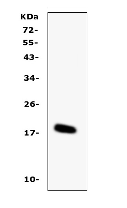
Western blot analysis of IL-16 using anti-IL-16 antibody (A01635).
Electrophoresis was performed on a 5-20% SDS-PAGE gel at 70V (Stacking gel) / 90V (Resolving gel) for 2-3 hours.
Lane 1: recombinant mouse IL-16 protein 1ng.
After Electrophoresis, proteins were transferred to a Nitrocellulose membrane at 150mA for 50-90 minutes. Blocked the membrane with 5% Non-fat Milk/ TBS for 1.5 hour at RT. The membrane was incubated with rabbit anti-IL-16 antigen affinity purified polyclonal antibody (Catalog # A01635) at 0.5 μg/mL overnight at 4°C, then washed with TBS-0.1%Tween 3 times with 5 minutes each and probed with a goat anti-rabbit IgG-HRP secondary antibody at a dilution of 1:10000 for 1.5 hour at RT. The signal is developed using an Enhanced Chemiluminescent detection (ECL) kit (Catalog # EK1002) with Tanon 5200 system. A specific band was detected for IL-16 at approximately 18KD. The expected band size for IL-16 is at 13KD.

Click image to see more details
IHC analysis of IL-16 using anti-IL-16 antibody (A01635). IL-16 was detected in paraffin-embedded section of mouse brain tissues. Heat mediated antigen retrieval was performed in citrate buffer (pH6, epitope retrieval solution) for 20 mins. The tissue section was blocked with 10% goat serum. The tissue section was then incubated with 1μg/ml rabbit anti-IL-16 Antibody (A01635) overnight at 4°C. Biotinylated goat anti-rabbit IgG was used as secondary antibody and incubated for 30 minutes at 37°C. The tissue section was developed using Strepavidin-Biotin-Complex (SABC)(Catalog # SA1022) with DAB as the chromogen.

Click image to see more details
IHC analysis of IL-16 using anti-IL-16 antibody (A01635). IL-16 was detected in paraffin-embedded section of rat brain tissues. Heat mediated antigen retrieval was performed in citrate buffer (pH6, epitope retrieval solution) for 20 mins. The tissue section was blocked with 10% goat serum. The tissue section was then incubated with 1μg/ml rabbit anti-IL-16 Antibody (A01635) overnight at 4°C. Biotinylated goat anti-rabbit IgG was used as secondary antibody and incubated for 30 minutes at 37°C. The tissue section was developed using Strepavidin-Biotin-Complex (SABC)(Catalog # SA1022) with DAB as the chromogen.

Click image to see more details
IHC analysis of IL-16 using anti-IL-16 antibody (A01635). IL-16 was detected in paraffin-embedded section of rat spleen tissues. Heat mediated antigen retrieval was performed in citrate buffer (pH6, epitope retrieval solution) for 20 mins. The tissue section was blocked with 10% goat serum. The tissue section was then incubated with 1μg/ml rabbit anti-IL-16 Antibody (A01635) overnight at 4°C. Biotinylated goat anti-rabbit IgG was used as secondary antibody and incubated for 30 minutes at 37°C. The tissue section was developed using Strepavidin-Biotin-Complex (SABC)(Catalog # SA1022) with DAB as the chromogen.
Specific Publications For Anti-IL-16/Il16 Antibody Picoband® (A01635)
Loading publications
Recommended Resources
Here are featured tools and databases that you might find useful.
- Boster's Pathways Library
- Protein Databases
- Bioscience Research Protocol Resources
- Data Processing & Analysis Software
- Photo Editing Software
- Scientific Literature Resources
- Research Paper Management Tools
- Molecular Biology Software
- Primer Design Tools
- Bioinformatics Tools
- Phylogenetic Tree Analysis
Customer Reviews
Have you used Anti-IL-16/Il16 Antibody Picoband®?
Share your experimental results or join a short interview to earn up to $1,000 in product credits or other rewards.
0 Reviews For Anti-IL-16/Il16 Antibody Picoband®
Customer Q&As
Have a question?
Find answers in Q&As, reviews.
Can't find your answer?
Submit your question
5 Customer Q&As for Anti-IL-16/Il16 Antibody Picoband®
Question
My question regarding product A01635, anti-IL-16/Il16 antibody. I was wondering if it would be possible to conjugate this antibody with biotin. I would need it to be without BSA or sodium azide. I am planning on using a buffer exchange of sodium azide with PBS only. Would there be problems for me to conjugate the antibody and store it in -20 degrees in small aliquots?
Verified Customer
Verified customer
Asked: 2020-03-25
Answer
We do not recommend storing this antibody with PBS buffer only in -20 degrees. If you want to store it in -20 degrees it is best to add some cryoprotectant like glycerol. If you want carrier free A01635 anti-IL-16/Il16 antibody, we can provide it to you in a special formula with trehalose and/or glycerol. These molecules will not interfere with conjugation chemistry and provide a good level of protection for the antibody from degradation. Please be sure to specify this in your purchase order.
Boster Scientific Support
Answered: 2020-03-25
Question
I am interested in to test anti-IL-16/Il16 antibody A01635 on mouse leukocyte for research purposes, then I may be interested in using anti-IL-16/Il16 antibody A01635 for diagnostic purposes as well. Is the antibody suitable for diagnostic purposes?
Verified Customer
Verified customer
Asked: 2019-11-21
Answer
The products we sell, including anti-IL-16/Il16 antibody A01635, are only intended for research use. They would not be suitable for use in diagnostic work. If you have the means to develop a product into diagnostic use, and are interested in collaborating with us and develop our product into an IVD product, please contact us for more discussions.
Boster Scientific Support
Answered: 2019-11-21
Question
Would A01635 anti-IL-16/Il16 antibody work on parafin embedded sections? If so, which fixation method do you recommend we use (PFA, paraformaldehyde, other)?
Verified Customer
Verified customer
Asked: 2019-07-23
Answer
It shows on the product datasheet, A01635 anti-IL-16/Il16 antibody as been validated on IHC. It is best to use PFA for fixation because it has better tissue penetration ability. PFA needs to be prepared fresh before use. Long term stored PFA turns into formalin, as the PFA molecules congregate and become formalin.
Boster Scientific Support
Answered: 2019-07-23
Question
Is a blocking peptide available for product anti-IL-16/Il16 antibody (A01635)?
Verified Customer
Verified customer
Asked: 2019-06-26
Answer
We do provide the blocking peptide for product anti-IL-16/Il16 antibody (A01635). If you would like to place an order for it please contact support@bosterbio.com and make a special request.
Boster Scientific Support
Answered: 2019-06-26
Question
Will anti-IL-16/Il16 antibody A01635 work on zebrafish WB with liver?
W. Dhar
Verified customer
Asked: 2018-10-18
Answer
Our lab technicians have not validated anti-IL-16/Il16 antibody A01635 on zebrafish. You can run a BLAST between zebrafish and the immunogen sequence of anti-IL-16/Il16 antibody A01635 to see if they may cross-react. If the sequence homology is close, then you can perform a pilot test. Keep in mind that since we have not validated zebrafish samples, this use of the antibody is not covered by our guarantee. However we have an innovator award program that if you test this antibody and show it works in zebrafish liver in WB, you can get your next antibody for free.
Boster Scientific Support
Answered: 2018-10-18
