Click image to see more details
-

-
-
-
-
+2
Product Info Summary
| SKU: | A02902-1 |
|---|---|
| Size: | 100 μg/vial |
| Reactive Species: | Human |
| Host: | Rabbit |
| Application: | ELISA, Flow Cytometry, IF, IHC, ICC, WB |
Customers Who Bought This Also Bought

Product info
Product Name
Anti-Integrin alpha 3/ITGA3 Antibody Picoband®
SKU/Catalog Number
A02902-1
Size
100 μg/vial
Form
Lyophilized
Description
Boster Bio Anti-Integrin alpha 3/ITGA3 Antibody Picoband® catalog # A02902-1. Tested in ELISA, Flow Cytometry, IF, IHC, ICC, WB applications. This antibody reacts with Human. The brand Picoband indicates this is a premium antibody that guarantees superior quality, high affinity, and strong signals with minimal background in Western blot applications. Only our best-performing antibodies are designated as Picoband, ensuring unmatched performance.
Storage & Handling
At -20°C for one year from date of receipt. After reconstitution, at 4°C for one month. It can also be aliquotted and stored frozen at -20°C for six months. Avoid repeated freezing and thawing.
Cite This Product
Anti-Integrin alpha 3/ITGA3 Antibody Picoband® (Boster Biological Technology, Pleasanton CA, USA, Catalog # A02902-1)
Host
Rabbit
Contents
Each vial contains 4 mg Trehalose, 0.9 mg NaCl, 0.2 mg Na2HPO4.
Clonality
Polyclonal
Isotype
Rabbit IgG
Immunogen
E.coli-derived human Integrin alpha 3/ITGA3 recombinant protein (Position: L96-L324).
Cross-reactivity
No cross-reactivity with other proteins.
Reactive Species
A02902-1 is reactive to ITGA3 in Human
Observed Molecular Weight
130 kDa
Calculated molecular weight
116.6 kDa
Background of ITGA3
ITGA3(INTEGRIN, ALPHA-3), also called CD49C, VLA3 or GAPB3, is a protein that in humans is encoded by the ITGA3 gene. It is an integrin alpha subunit which is also a member of the family of cell surface adhesion molecules. This gene is mapped to chromosome 17 and its exact cytogenetic location is 17q21.33. ITGA3 makes up half of the α3β1 integrin duplex that plays a role in neural migration and corticogenesis together with beta-1 subunit.A functional link between DAB1 phosphorylation and ITGA3 signaling drives the timely detachment of migrating neurons from radial glial fibers. Expression of human ITGA3 increased the infectivity of virus for Chinese hamster ovary cells. ITGA3 also contains 13 potential N-glycosylation sites, 2 potential cleavage sites, and the 7 N-terminal repeating units characteristic of ITGAs. Recombinant ITGA3 is expressed as a 150-kD protein as the same size as the native protein by the western blot analysis.
Antibody Validation
Boster validates all antibodies on WB, IHC, ICC, Immunofluorescence, and ELISA with known positive control and negative samples to ensure specificity and high affinity, including thorough antibody incubations.
Application & Images
Applications
A02902-1 is guaranteed for ELISA, Flow Cytometry, IF, IHC, ICC, WB Boster Guarantee
Recommend Dilution
| Application | Dilution | Species |
|---|---|---|
| Western blot | 0.25-0.5 μg/ml | Human |
| Immunohistochemistry(Paraffin-embedded Section) | 2-5 μg/ml | Human |
| Immunocytochemistry/Immunofluorescence | 5 μg/ml | Human |
| Flow Cytometry(Fixed) | 1-3 μg/1x106 cells | Human |
| ELISA | 0.1-0.5 μg/ml | - |
Tested application
Suggested blocking solution with 5% non-fat milk or BSA; (*)Recommended protein loading: 20-40 µg per lane
Use TE buffer pH 9.0 for antigen retrieval; (*) citrate buffer pH 6.0 is an alternative.
Validation Images & Assay Conditions
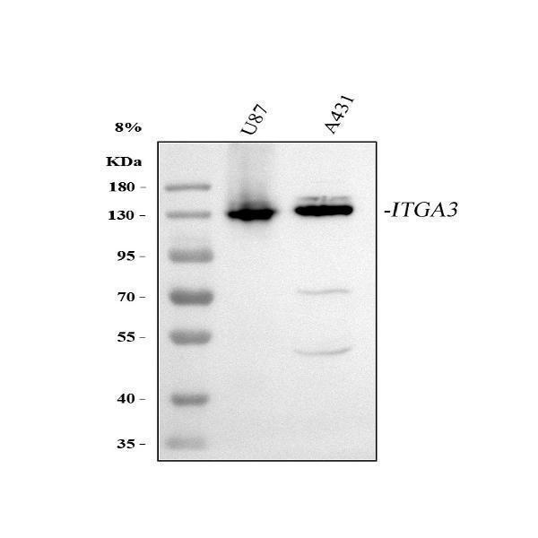
Click image to see more details
Western blot analysis of Integrin Alpha 3/ITGA3 using anti-Integrin Alpha 3/ITGA3 antibody (A02902-1).
Electrophoresis was performed on a 5-20% SDS-PAGE gel at 70V (Stacking gel) / 90V (Resolving gel) for 2-3 hours. The sample well of each lane was loaded with 30 ug of sample under reducing conditions.
Lane 1: human U87 whole cell lysates,
Lane 2: human A431 whole cell lysates.
After electrophoresis, proteins were transferred to a nitrocellulose membrane at 150 mA for 50-90 minutes. Blocked the membrane with 5% non-fat milk/TBS for 1.5 hour at RT. The membrane was incubated with rabbit anti-Integrin Alpha 3/ITGA3 antigen affinity purified polyclonal antibody (Catalog # A02902-1) at 0.5 μg/mL overnight at 4°C, then washed with TBS-0.1%Tween 3 times with 5 minutes each and probed with a goat anti-rabbit IgG-HRP secondary antibody at a dilution of 1:5000 for 1.5 hour at RT. The signal is developed using an Enhanced Chemiluminescent detection (ECL) kit (Catalog # EK1002) with Tanon 5200 system. A specific band was detected for Integrin Alpha 3/ITGA3 at approximately 130 kDa. The expected band size for Integrin Alpha 3/ITGA3 is at 130 kDa.

Click image to see more details
IHC analysis of Integrin alpha 3/ITGA3 using anti-Integrin alpha 3/ITGA3 antibody (A02902-1).
Integrin alpha 3/ITGA3 was detected in a paraffin-embedded section of human colorectal adenocarcinoma tissue. Heat mediated antigen retrieval was performed in EDTA buffer (pH 8.0, epitope retrieval solution). The tissue section was blocked with 10% goat serum. The tissue section was then incubated with 2 μg/ml rabbit anti-Integrin alpha 3/ITGA3 Antibody (A02902-1) overnight at 4°C. Peroxidase Conjugated Goat Anti-rabbit IgG was used as secondary antibody and incubated for 30 minutes at 37°C. The tissue section was developed using HRP Conjugated Rabbit IgG Super Vision Assay Kit (Catalog # SV0002) with DAB as the chromogen.

Click image to see more details
IHC analysis of Integrin alpha 3/ITGA3 using anti-Integrin alpha 3/ITGA3 antibody (A02902-1).
Integrin alpha 3/ITGA3 was detected in a paraffin-embedded section of human esophageal squamous carcinoma tissue. Heat mediated antigen retrieval was performed in EDTA buffer (pH 8.0, epitope retrieval solution). The tissue section was blocked with 10% goat serum. The tissue section was then incubated with 2 μg/ml rabbit anti-Integrin alpha 3/ITGA3 Antibody (A02902-1) overnight at 4°C. Peroxidase Conjugated Goat Anti-rabbit IgG was used as secondary antibody and incubated for 30 minutes at 37°C. The tissue section was developed using HRP Conjugated Rabbit IgG Super Vision Assay Kit (Catalog # SV0002) with DAB as the chromogen.

Click image to see more details
IHC analysis of Integrin alpha 3/ITGA3 using anti-Integrin alpha 3/ITGA3 antibody (A02902-1).
Integrin alpha 3/ITGA3 was detected in a paraffin-embedded section of human lung adenocarcinoma tissue. Heat mediated antigen retrieval was performed in EDTA buffer (pH 8.0, epitope retrieval solution). The tissue section was blocked with 10% goat serum. The tissue section was then incubated with 2 μg/ml rabbit anti-Integrin alpha 3/ITGA3 Antibody (A02902-1) overnight at 4°C. Peroxidase Conjugated Goat Anti-rabbit IgG was used as secondary antibody and incubated for 30 minutes at 37°C. The tissue section was developed using HRP Conjugated Rabbit IgG Super Vision Assay Kit (Catalog # SV0002) with DAB as the chromogen.

Click image to see more details
IF analysis of Integrin alpha 3/ITGA3 using anti-Integrin alpha 3/ITGA3 antibody (A02902-1).
Integrin alpha 3/ITGA3 was detected in an immunocytochemical section of A431 cells. Enzyme antigen retrieval was performed using IHC enzyme antigen retrieval reagent (AR0022) for 15 mins. The cells were blocked with 10% goat serum. And then incubated with 5 μg/mL rabbit anti-Integrin alpha 3/ITGA3 Antibody (A02902-1) overnight at 4°C. DyLight®488 Conjugated Goat Anti-Rabbit IgG (BA1127) was used as secondary antibody at 1:500 dilution and incubated for 30 minutes at 37°C. The section was counterstained with DAPI. Visualize using a fluorescence microscope and filter sets appropriate for the label used.

Click image to see more details
Flow Cytometry analysis of U87 cells using anti-Integrin alpha 3/ITGA3 antibody (A02902-1).
Overlay histogram showing U87 cells stained with A02902-1 (Blue line). The cells were fixed with 4% paraformaldehyde and blocked with 10% normal goat serum. And then incubated with rabbit anti-Integrin alpha 3/ITGA3 Antibody (A02902-1, 1 μg/1x106 cells) for 30 min at 20°C. DyLight®488 conjugated goat anti-rabbit IgG (BA1127, 5-10 μg/1x106 cells) was used as secondary antibody for 30 minutes at 20°C. Isotype control antibody (Green line) was rabbit IgG (1 μg/1x106) used under the same conditions. Unlabelled sample (Red line) was also used as a control.
Specific Publications For Anti-Integrin alpha 3/ITGA3 Antibody Picoband® (A02902-1)
Loading publications
Recommended Resources
Here are featured tools and databases that you might find useful.
- Boster's Pathways Library
- Protein Databases
- Bioscience Research Protocol Resources
- Data Processing & Analysis Software
- Photo Editing Software
- Scientific Literature Resources
- Research Paper Management Tools
- Molecular Biology Software
- Primer Design Tools
- Bioinformatics Tools
- Phylogenetic Tree Analysis
Customer Reviews
Have you used Anti-Integrin alpha 3/ITGA3 Antibody Picoband®?
Share your experimental results or join a short interview to earn up to $1,000 in product credits or other rewards.
0 Reviews For Anti-Integrin alpha 3/ITGA3 Antibody Picoband®
Customer Q&As
Have a question?
Find answers in Q&As, reviews.
Can't find your answer?
Submit your question
