Click image to see more details
Product Info Summary
| SKU: | PA1709 |
|---|---|
| Size: | 100 μg/vial |
| Reactive Species: | Mouse, Rat |
| Host: | Rabbit |
| Application: | IHC, WB |
Customers Who Bought This Also Bought

Product info
Product Name
Anti-Kallikrein 1/KLK1 Antibody Picoband®
SKU/Catalog Number
PA1709
BA3829 is an alternative SKU for this antibody, used in previous lots.
Size
100 μg/vial
Form
Lyophilized
Description
Boster Bio Anti-Kallikrein 1/KLK1 Antibody catalog # PA1709. Tested in IHC, WB applications. This antibody reacts with Mouse, Rat. The brand Picoband indicates this is a premium antibody that guarantees superior quality, high affinity, and strong signals with minimal background in Western blot applications. Only our best-performing antibodies are designated as Picoband, ensuring unmatched performance.
Storage & Handling
Store at -20˚C for one year from date of receipt. After reconstitution, at 4˚C for one month. It can also be aliquotted and stored frozen at -20˚C for six months. Avoid repeated freeze-thaw cycles.
Cite This Product
Anti-Kallikrein 1/KLK1 Antibody Picoband® (Boster Biological Technology, Pleasanton CA, USA, Catalog # PA1709)
Host
Rabbit
Contents
Each vial contains antibody formulated with stabilizing components, 0.9mg NaCl, 0.2mg Na2HPO4, 0.05mg Thimerosal, 0.05mg NaN3.
*This antibody is supplied in a stabilized formulation.
Compatibility with conjugation reactions depends on the chemistry of the conjugation method used.
For conjugation methods that are not compatible with the stabilizing components present in this formulation, a carrier-free antibody format is required.
Clonality
Polyclonal
Isotype
Rabbit IgG
Immunogen
A synthetic peptide corresponding to a sequence at the C-terminus of mouse Kallikrein 1.
Cross-reactivity
No cross-reactivity with other proteins
Reactive Species
PA1709 is reactive to Klk1 in Mouse, Rat
Observed Molecular Weight
29 kDa
Calculated molecular weight
28.8 kDa
Background of Klk1
KLK1 (KALLIKREIN 1), also called KLKR, is a protein that in humans is encoded by the KLK1 gene. KLK1 is a member of the peptidase S1 family. KLK1 is a serine protease that generates Lys-bradykinin by specific proteolysis of kininogen-1. The KLK1 gene is one of the fifteen kallikrein subfamily members located in a cluster on chromosome 19 and its exact cytogenetic location is 19q13.33. The KLK1 gene contains 5 coding exons. And KLK1 is the most centromeric gene in the cluster. Mice lacking tissue kallikrein are unable to generate significant levels of kinins in most tissues and develop cardiovascular abnormalities early in adulthood despite normal blood pressure. The protein is functionally conserved in its capacity to release the vasoactive peptide, Lys-bradykinin, from low molecular weight kininogen.
Antibody Validation
Boster validates all antibodies on WB, IHC, ICC, Immunofluorescence, and ELISA with known positive control and negative samples to ensure specificity and high affinity, including thorough antibody incubations.
Application & Images
Applications
PA1709 is guaranteed for IHC, WB Boster Guarantee
Recommend Dilution
| Application | Dilution | Species |
|---|---|---|
| Immunohistochemistry (Paraffin-embedded Section) | 0.5-1μg/ml | Mouse |
| Western blot | 0.1-0.5μg/ml | Mouse, Rat |
Tested application
Suggested blocking solution with 5% non-fat milk or BSA; (*)Recommended protein loading: 20-40 µg per lane
Use TE buffer pH 9.0 for antigen retrieval; (*) citrate buffer pH 6.0 is an alternative.
Validation Images & Assay Conditions
Click image to see more details
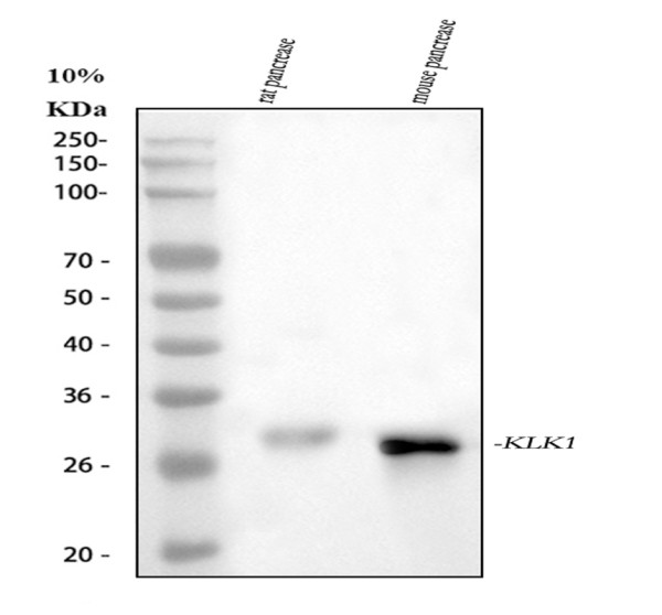
Western blot analysis of Kallikrein 1/KLK1 using anti-Kallikrein 1/KLK1 antibody (PA1709).
Electrophoresis was performed on a 10% SDS-PAGE gel at 80V (StacKallikrein 1/KLK1g gel) / 120V (Resolving gel) for 2 hours. The sample well of each lane was loaded with 30 ug of sample under reducing conditions.
Lane 1: rat pancrease tissue lysates,
Lane 2: mouse pancrease tissue lysates.
After electrophoresis, proteins were transferred to a nitrocellulose membrane at 150 mA for 50-90 minutes. Blocked the membrane with 5% non-fat milk/TBS for 1.5 hour at RT. The membrane was incubated with rabbit anti-Kallikrein 1/KLK1 antigen affinity purified polyclonal antibody (PA1709) at 0.5 μg/mL overnight at 4°C, then washed with TBS-0.1%Tween 3 times with 5 minutes each and probed with a goat anti-rabbit IgG-HRP secondary antibody at a dilution of 1:5000 for 1.5 hour at RT. The signal is developed using an ECL Plus Western Blotting Substrate (Catalog # AR1196-200) with Tanon 5200 system. A specific band was detected for Kallikrein 1/KLK1 at approximately 29 kDa. The expected band size for Kallikrein 1/KLK1 is at 29 kDa.

Click image to see more details
Anti-Kallikrein 1 antibody, PA1709, IHC(P)
IHC(P): Mouse Kidney Tissue

Click image to see more details
Acinar cell apoptosis is delayed in post-ligated SMGs of ATG5-deficient mice. ( a ) ATG5 status impinges upon duct ligation-triggered acinar apoptosis. Apoptosis by ApopTag Peroxidase (brown nuclear staining) was visualized using an In situ Apoptosis Detection Kit ( left panel ). Peak apoptosis was detected in L1 and L3 SMGs of Atg5 WT mice, whereas the strongest ApopTag signals were detected in L3 and L7 SMGs of Atg5 KO mice. Magnification: × 100, and enlarged view ( inset ): × 400. Bar: 100 μ m. Percent ApopTag-positive cells quantified by dividing the total ApopTag-positive cells by total number of cells examined from 10 randomly chosen fields are shown ( right panel ). Student’s t -test was employed to determine statistically significant differences in percentage of ApopTag-positive cells between Atg5 WT and Atg5 KO groups. Results are shown as mean±S.D.; * P <0.05. ( b ) ATG5 deficiency delays caspase-3 activation. Equal amounts of whole SMG lysates from Ctrl, L1, L3 and L7 SMGs of Atg5 WT and Atg5 KO mice were analyzed on western blots using indicated antibodies. The cleavage of caspase-3 represents caspase-3 activation. ( c ) Gene expression analyses of Prol1/Muc-10 and Klk1 , markers for acinar and duct cells, respectively. SMG duct ligation was performed as described in . Expression of the indicated messages in ligated and control SMGs was analyzed by quantitative RT-PCR analyses. Relative gene expression was calculated where respective mean value for control SMG set to 1 for each amplification ( N ≥4). Non-parametric Mann–Whitney test was performed to compare expression levels between respective ligated and control SMGs ( *; below bars), and between same-day ligated SMGs and control SMGs from Atg5 WT and Atg5 KO ( *; above bars). Results are shown as mean±S.D.; * P <0.05; ** P <0.01. ( d ) Autophagy inhibition correlates with resistance to H 2 O 2 -induced cell death. Cell viability was measured in m5-7, Atg7 -KO MEF, WT MEF and salivary Pa-4 cells treated with indicated concentrations of H 2 O 2 in combination with chloroquine (CQ; 20 μ M) or baflomycin A1 (BafA1; 10 nM) for 48 h and compared with that of the control cells. Data were analyzed with ANOVA followed by Bonferroni t -test to determine the statistical differences between treatment and control group ( left panels ). Representative western blots ( right panels ), in which total lysates from cells treated for 6 h with vehicle, CQ (20 μ M) or BafA1 (10 nM) were stained with indicated antibodies, are shown. Results are shown as mean±S.E.M.; N =3; * P <0.05; ** P <0.01; *** P <0.001
Index in PubMed under a CC BY license. PMID: 25341032
Specific Publications For Anti-Kallikrein 1/KLK1 Antibody Picoband® (PA1709)
Loading publications
Recommended Resources
Here are featured tools and databases that you might find useful.
- Boster's Pathways Library
- Protein Databases
- Bioscience Research Protocol Resources
- Data Processing & Analysis Software
- Photo Editing Software
- Scientific Literature Resources
- Research Paper Management Tools
- Molecular Biology Software
- Primer Design Tools
- Bioinformatics Tools
- Phylogenetic Tree Analysis
Customer Reviews
Have you used Anti-Kallikrein 1/KLK1 Antibody Picoband®?
Share your experimental results or join a short interview to earn up to $1,000 in product credits or other rewards.
0 Reviews For Anti-Kallikrein 1/KLK1 Antibody Picoband®
Customer Q&As
Have a question?
Find answers in Q&As, reviews.
Can't find your answer?
Submit your question
