Click image to see more details
-

-
-
-
-
+1
Product Info Summary
| SKU: | M03267 |
|---|---|
| Size: | 80 µl |
| Reactive Species: | Human |
| Host: | Rabbit |
| Application: | Flow Cytometry, IF, IHC-P, WB |
Customers Who Bought This Also Bought

Product info
Product Name
Anti-ITGA7 Antibody (N-term)
SKU/Catalog Number
M03267
Size
80 µl
Description
Boster Bio Anti-ITGA7 Antibody (N-term) (Catalog # M03267). Tested in WB, Flow Cytometry, IHC-P, IF application(s). This antibody reacts with Human.
Storage & Handling
Maintain refrigerated at 2-8°C for up to 2 weeks. For long-term storage, store at -20°C in small aliquots to prevent freeze-thaw cycles.
Cite This Product
Anti-ITGA7 Antibody (N-term) (Boster Biological Technology, Pleasanton CA, USA, Catalog # M03267)
Host
Rabbit
Contents
Purified polyclonal antibody supplied in PBS with 0.09% (W/V) sodium azide.
Clonality
Polyclonal
Isotype
Rabbit IgG
Immunogen
This ITGA7 antibody is generated from a rabbit immunized with a KLH conjugated synthetic peptide between 247-279 amino acids from the N-terminal region of human ITGA7.
Reactive Species
M03267 is reactive to ITGA7 in Human
Calculated molecular weight
128.9 kDa
Background of ITGA7
Integrin alpha-7/beta-1 is the primary laminin receptor on skeletal myoblasts and adult myofibers. During myogenic differentiation, it may induce changes in the shape and mobility of myoblasts, and facilitate their localization at laminin-rich sites of secondary fiber formation. It is involved in the maintenance of the myofibers cytoarchitecture as well as for their anchorage, viability and functional integrity. Isoform Alpha-7X2B and isoform Alpha-7X1B promote myoblast migration on laminin 1 and laminin 2/4, but isoform Alpha-7X1B is less active on laminin 1 (In vitro). Acts as Schwann cell receptor for laminin-2. Acts as a receptor of COMP and mediates its effect on vascular smooth muscle cells (VSMCs) maturation (By similarity). Required to promote contractile phenotype acquisition in differentiated airway smooth muscle (ASM) cells.
Antibody Validation
Boster validates all antibodies on WB, IHC, ICC, Immunofluorescence, and ELISA with known positive control and negative samples to ensure specificity and high affinity, including thorough antibody incubations.
Application & Images
Applications
M03267 is guaranteed for Flow Cytometry, IF, IHC-P, WB Boster Guarantee
Recommend Dilution
IF: 1:25
WB: 1:4000
IHC-P: 1:25
FC: 1:25
Validation Images & Assay Conditions
Click image to see more details
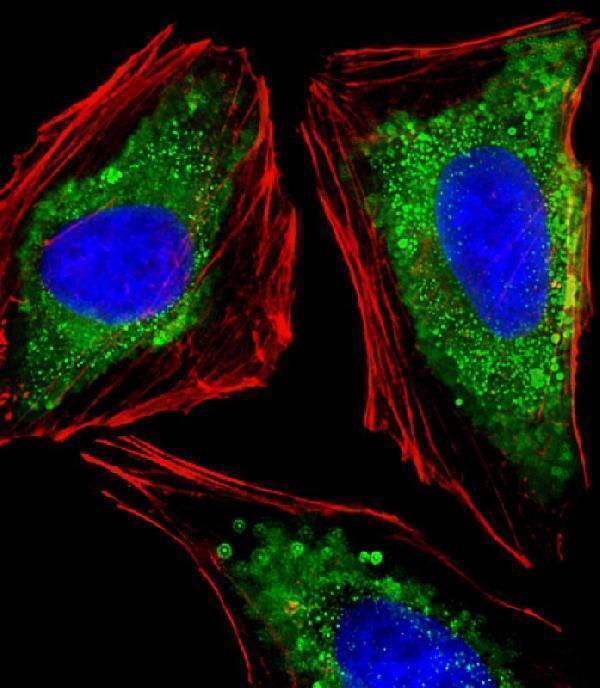
Immunofluorescent analysis of 4% paraformaldehyde-fixed, 0.1% Triton X-100 permeabilized HeLa (human cervical epithelial adenocarcinoma cell line) cells labeling ITGA7 with M03267 at 1/25 dilution, followed by Dylight® 488-conjugated goat anti-rabbit IgG secondary antibody at 1/200 dilution (green). Immunofluorescence image showing vesicles,cytoplasm and weakly nucleus staining on HeLa cell line. Cytoplasmic actin is detected with Dylight® 554 Phalloidin at 1/100 dilution (red).The nuclear counter stain is DAPI (blue).

Click image to see more details
All lanes : Anti-ITGA7 Antibody (N-term) at 1:2000 dilution
Lane 1: Hela whole cell lysates
Lane 2: A431 whole cell lysates
Lane 3: Jurkat whole cell lysates
Lysates/proteins at 20 µg per lane.
Secondary
Goat Anti-Rabbit IgG, (H+L), Peroxidase conjugated at 1/10000 dilution
Predicted band size : 129 kDa
Blocking/Dilution buffer: 5% NFDM/TBST.

Click image to see more details
Anti-ITGA7 Antibody (N-term)at 1:4000 dilution + A431 whole cell lysates
Lysates/proteins at 20 µg per lane.
Secondary
Goat Anti-Rabbit IgG, (H+L), Peroxidase conjugated at 1/10000 dilution
Predicted band size : 129 kDa
Blocking/Dilution buffer: 5% NFDM/TBST.

Click image to see more details
M03267 staining ITGA7 in human skeletal muscle sections by Immunohistochemistry (IHC-P -paraformaldehyde-fixed, paraffin-embedded sections). Tissue was fixed with formaldehyde and blocked with 3% BSA for 0. 5 hour at room temperature; antigen retrieval was by heat mediation with a citrate buffer (pH6). Samples were incubated with primary antibody (1/25) for 1 hours at 37°C. A undiluted biotinylated goat polyvalent antibody was used as the secondary antibody.

Click image to see more details
Overlay histogram showing U-2 OS cells stained with M03267 (green line). The cells were fixed with 2% paraformaldehyde (10 min) and then permeabilized with 90% methanol for 10 min. The cells were then icubated in 2% bovine serum albumin to block non-specific protein-protein interactions followed by the antibody (M03267, 1:25 dilution) for 60 min at 37ºC. The secondary antibody used was Goat-Anti-Rabbit IgG, DyLight® 488 Conjugated Highly Cross-Adsorbed at 1/400 dilution for 40 min at 37ºC. Isotype control antibody (blue line) was rabbit IgG (1g/1x10^6 cells) used under the same conditions. Acquisition of >10, 000 events was performed.
Specific Publications For Anti-ITGA7 Antibody (N-term) (M03267)
Loading publications
Recommended Resources
Here are featured tools and databases that you might find useful.
- Boster's Pathways Library
- Protein Databases
- Bioscience Research Protocol Resources
- Data Processing & Analysis Software
- Photo Editing Software
- Scientific Literature Resources
- Research Paper Management Tools
- Molecular Biology Software
- Primer Design Tools
- Bioinformatics Tools
- Phylogenetic Tree Analysis
Customer Reviews
Have you used Anti-ITGA7 Antibody (N-term)?
Share your experimental results or join a short interview to earn up to $1,000 in product credits or other rewards.
0 Reviews For Anti-ITGA7 Antibody (N-term)
Customer Q&As
Have a question?
Find answers in Q&As, reviews.
Can't find your answer?
Submit your question
