Click image to see more details
-

-
-
-
-
+2
Product Info Summary
| SKU: | A00514-3 |
|---|---|
| Size: | 100 μg/vial |
| Reactive Species: | Human, Mouse, Rat |
| Host: | Rabbit |
| Application: | ELISA, Flow Cytometry, IF, ICC, WB |
Customers Who Bought This Also Bought

Product info
Product Name
Anti-KEAP1 Antibody Picoband®
SKU/Catalog Number
A00514-3
Size
100 μg/vial
Form
Lyophilized
Description
Boster Bio Anti-KEAP1 Antibody Picoband® catalog # A00514-3. Tested in ELISA, Flow Cytometry, IF, ICC, WB applications. This antibody reacts with Human, Mouse, Rat. The brand Picoband indicates this is a premium antibody that guarantees superior quality, high affinity, and strong signals with minimal background in Western blot applications. Only our best-performing antibodies are designated as Picoband, ensuring unmatched performance.
Storage & Handling
At -20°C for one year from date of receipt. After reconstitution, at 4°C for one month. It can also be aliquotted and stored frozen at -20°C for six months. Avoid repeated freezing and thawing.
Cite This Product
Anti-KEAP1 Antibody Picoband® (Boster Biological Technology, Pleasanton CA, USA, Catalog # A00514-3)
Host
Rabbit
Contents
Each vial contains 4 mg Trehalose, 0.9 mg NaCl, 0.2 mg Na2HPO4.
Clonality
Polyclonal
Isotype
Rabbit IgG
Immunogen
E.coli-derived human KEAP1 recombinant protein (Position: K84-K312).
Cross-reactivity
No cross-reactivity with other proteins.
Reactive Species
A00514-3 is reactive to KEAP1 in Human, Mouse, Rat
Observed Molecular Weight
66-72 kDa
Calculated molecular weight
69.7 kDa
Background of KEAP1
KEAP1 (KELCH-LIKE ECH-ASSOCIATED PROTEIN 1), is a protein that in humans is encoded by the Keap1 gene. The KIAA0132 gene is mapped on 19p13.2. Keap1 contains a central BTB/POZ domain and a C-terminal double glycine repeat (DGR), or Kelch, module. Keap1 has been shown to interact with Nrf2, a master regulator of the antioxidant response, which is important for the amelioration of oxidative stress. In the presence of the electrophilic agent diethylmalate, Nrf2 activity is released from Keap1 and Nrf2 translocate to the nucleus. Under quiescent conditions, Nrf2 is anchored in the cytoplasm through binding to Keap1, which, in turn, facilitates the ubiquitination and subsequent proteolysis of Nrf2. Because Nrf2 activation leads to a coordinated antioxidant and anti-inflammatory response, and Keap1 represses Nrf2 activation, Keap1 has become a very attractive drug target.
Antibody Validation
Boster validates all antibodies on WB, IHC, ICC, Immunofluorescence, and ELISA with known positive control and negative samples to ensure specificity and high affinity, including thorough antibody incubations.
Application & Images
Applications
A00514-3 is guaranteed for ELISA, Flow Cytometry, IF, ICC, WB Boster Guarantee
Recommend Dilution
| Application | Dilution | Species |
|---|---|---|
| Western blot | 0.25-0.5 μg/ml | Human, Mouse, Rat |
| Immunocytochemistry/Immunofluorescence | 5 μg/ml | Human |
| Flow Cytometry (Fixed) | 1-3 μg/1x106 cells | Human |
| ELISA | 0.1-0.5 μg/ml | - |
Tested application
Suggested blocking solution with 5% non-fat milk or BSA; (*)Recommended protein loading: 20-40 µg per lane
Validation Images & Assay Conditions
Click image to see more details
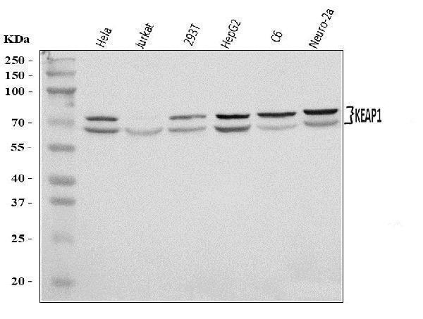
Western blot analysis of KEAP1 using anti-KEAP1 antibody (A00514-3).
Electrophoresis was performed on a 5-20% SDS-PAGE gel at 70V (Stacking gel) / 90V (Resolving gel) for 2-3 hours. The sample well of each lane was loaded with 30 ug of sample under reducing conditions.
Lane 1: human Hela whole cell lysates,
Lane 2: human Jurkat whole cell lysates,
Lane 3: human 293T whole cell lysates,
Lane 4: human HepG2 whole cell lysates,
Lane 5: rat C6 whole cell lysates,
Lane 6: mouse Neuro-2a whole cell lysates.
After electrophoresis, proteins were transferred to a nitrocellulose membrane at 150 mA for 50-90 minutes. Blocked the membrane with 5% non-fat milk/TBS for 1.5 hour at RT. The membrane was incubated with rabbit anti-KEAP1 antigen affinity purified polyclonal antibody (Catalog # A00514-3) at 0.5 μg/mL overnight at 4°C, then washed with TBS-0.1%Tween 3 times with 5 minutes each and probed with a goat anti-rabbit IgG-HRP secondary antibody at a dilution of 1:5000 for 1.5 hour at RT. The signal is developed using an Enhanced Chemiluminescent detection (ECL) kit (Catalog # EK1002) with Tanon 5200 system. A specific band was detected for KEAP1 at approximately 66-72 kDa. The expected band size for KEAP1 is at 70 kDa.

Click image to see more details
Exo-miR-1290 promoted CAFs activation by upregulating Nrf2 expression in fibroblasts. A GO enrichment analysis showed that the antioxidant activities were involved in the COX-2 exosomes-mediated CAFs activation. B Western blotting assays of NIH-3T3 cells detected with anti-Nrf2 antibody after treatments with exosomes from A549-Con or A549-COX-2 cells. C Western blotting assays of fibroblasts detected with anti-α-SMA, FN1, and FAP-1 antibodies after treatments with Nrf2 overexpression. D , E Western blotting assays of fibroblasts detected with anti-FN1, and FAP-1 antibodies after treatments with siNrf2 and/or miR-1290 mimic transfection. (D. NIH-3T3, E. MRC-5). F Western blotting assays of fibroblasts detected with anti-FN1, α-SMA, and FAP-1 antibodies after treatments with siNrf2 and exosomes. β-actin was used as an internal reference. Data were presented as the mean ± SEM from three independent experiments. * P < 0.05, ** P < 0.01 compared with corresponding control; # P < 0.05, ## P < 0.01 compared with miR-1290 mimic or COX-2-exo groups
Index in PubMed under a CC BY license. PMID: 37723559

Click image to see more details
The effects of TSP on the Nrf2/HO-1 signaling pathway. (A) The representative images of western blotting results. (B–D) Quantification of the protein expression of Keap1, Nrf2, and HO-1. All data were expressed as mean ± SEM ( n = 3). # P < 0.05, ## P < 0.01 vs. Control group; * P < 0.05, ** P < 0.01 vs. Model group.
Index in PubMed under a CC BY license. PMID: 35719151

Click image to see more details
CUL3 was involved in exo-miR-1290-mediated Nrf2 upregulation and CAFs activation. A qRT-PCR assays of Nrf2 mRNA expression in NIH-3T3 cells treated with miR-1290 mimic or A549-COX-2 exosomes. B Western blotting assays of NIH-3T3 cells were detected with anti-Nrf2 antibody after treatments with MG132 and exosomes from A549-Con or A549-COX-2 cells. C Western blotting assays of NIH-3T3 cells detected with anti-CUL3 and KEAP1 antibodies after treatments with A549-con or A549-COX-2 exosomes. E Western blotting assays of fibroblasts detected with anti-CUL3 antibody after treatments with miR-1290 mimic or inhibitor. F The miR-1290 binding site of 3’UTR of CUL3 mRNA predicted by RNAhybrid 2.2 and miRWalk. Firefly luciferase reporter was used to analyze the activity of the miR-1290 binding site of the reporter with the wild-type CUL3 3’UTR (CUL3) or with the mutational CUL3 3’UTR (CUL3-M). G Western blotting assays of fibroblasts detected with anti-CUL3 antibody after CUL3 overexpression or siCUL3 treatments. H Western blotting assays of fibroblasts detected with anti- FN1, α-SMA, and FAP-1 antibodies after treatments with CUL3 overexpression and miR-1290 mimic transfection, or exosome. I Western blotting assays of fibroblasts were detected with anti- FN1, α-SMA, and FAP-1 antibodies after treatments with siCUL3. β-actin was used as an internal reference. Data were presented as the means ± SEM from three independent experiments. * P < 0.05, ** P < 0.01 compared with corresponding control; # P < 0.05, ## P < 0.01 compared with miR-1290 mimic or COX-2-exo groups
Index in PubMed under a CC BY license. PMID: 37723559

Click image to see more details
IF analysis of KEAP1 using anti-KEAP1 antibody (A00514-3).
KEAP1 was detected in an immunocytochemical section of A431 cells. Enzyme antigen retrieval was performed using IHC enzyme antigen retrieval reagent (AR0022) for 15 mins. The cells were blocked with 10% goat serum. And then incubated with 5 μg/mL rabbit anti-KEAP1 Antibody (A00514-3) overnight at 4°C. DyLight®488 Conjugated Goat Anti-Rabbit IgG (BA1127) was used as secondary antibody at 1:100 dilution and incubated for 30 minutes at 37°C. The section was counterstained with DAPI. Visualize using a fluorescence microscope and filter sets appropriate for the label used.

Click image to see more details
Flow Cytometry analysis of U251 cells using anti-KEAP1 antibody (A00514-3).
Overlay histogram showing U251 cells stained with A00514-3 (Blue line). To facilitate intracellular staining, cells were fixed with 4% paraformaldehyde and permeabilized with permeabilization buffer. The cells were blocked with 10% normal goat serum. And then incubated with rabbit anti-KEAP1 Antibody (A00514-3, 1 μg/1x106 cells) for 30 min at 20°C. DyLight®488 conjugated goat anti-rabbit IgG (BA1127, 5-10 μg/1x106 cells) was used as secondary antibody for 30 minutes at 20°C. Isotype control antibody (Green line) was rabbit IgG (1 μg/1x106) used under the same conditions. Unlabelled sample without incubation with primary antibody and secondary antibody (Red line) was used as a blank control.
Specific Publications For Anti-KEAP1 Antibody Picoband® (A00514-3)
Loading publications
Recommended Resources
Here are featured tools and databases that you might find useful.
- Boster's Pathways Library
- Protein Databases
- Bioscience Research Protocol Resources
- Data Processing & Analysis Software
- Photo Editing Software
- Scientific Literature Resources
- Research Paper Management Tools
- Molecular Biology Software
- Primer Design Tools
- Bioinformatics Tools
- Phylogenetic Tree Analysis
Customer Reviews
Have you used Anti-KEAP1 Antibody Picoband®?
Share your experimental results or join a short interview to earn up to $1,000 in product credits or other rewards.
0 Reviews For Anti-KEAP1 Antibody Picoband®
Customer Q&As
Have a question?
Find answers in Q&As, reviews.
Can't find your answer?
Submit your question
