Click image to see more details
Product Info Summary
| SKU: | PB9814 |
|---|---|
| Size: | 100 μg/vial |
| Reactive Species: | Human, Mouse, Rat |
| Host: | Rabbit |
| Application: | Flow Cytometry, IP, WB |
Customers Who Bought This Also Bought

Product info
Product Name
Anti-PRC1 Antibody Picoband®
SKU/Catalog Number
PB9814
PB0858 is an alternative SKU for this antibody, used in previous lots.
Size
100 μg/vial
Form
Lyophilized
Description
Boster Bio Anti-PRC1 Antibody Picoband® catalog # PB9814. Tested in Flow Cytometry, IP, WB applications. This antibody reacts with Human, Mouse, Rat. The brand Picoband indicates this is a premium antibody that guarantees superior quality, high affinity, and strong signals with minimal background in Western blot applications. Only our best-performing antibodies are designated as Picoband, ensuring unmatched performance.
Storage & Handling
Store at -20˚C for one year from date of receipt. After reconstitution, at 4˚C for one month. It can also be aliquotted and stored frozen at -20˚C for six months. Avoid repeated freeze-thaw cycles.
Cite This Product
Anti-PRC1 Antibody Picoband® (Boster Biological Technology, Pleasanton CA, USA, Catalog # PB9814)
Host
Rabbit
Contents
Each vial contains 4 mg Trehalose, 0.9 mg NaCl and 0.2 mg Na2HPO4.
Clonality
Polyclonal
Isotype
Rabbit IgG
Immunogen
E.coli-derived human PRC1 recombinant protein (Position: M1-E233). Human PRC1 shares 85% amino acid (aa) sequence identity with mouse PRC1.
Cross-reactivity
No cross-reactivity with other proteins.
Reactive Species
PB9814 is reactive to PRC1 in Human, Mouse, Rat
Observed Molecular Weight
72 kDa
Calculated molecular weight
71.6 kDa
Background of PRC1
Protein Regulator of cytokinesis 1 (PRC1) is a protein that in humans is encoded by the PRC1 gene and is involved in cytokinesis. This gene is mapped to chromosome 15q26.1 based on an alignment of the PRC1 sequence with the genomic sequence. PRC1 protein is expressed at relatively high levels during S and G2/M phases of the cell cycle before dropping dramatically after mitotic exit and entrance into G1 phase. It is located in the nucleus during interphase, becomes associated with the mitotic spindle in a highly dynamic manner during anaphase, and localizes to the cell midbody during cytokinesis. And this protein has been shown to be a substrate of several cyclin-dependent kinases (CDKs). It is necessary for polarizing parallel microtubules and concentrating the factors responsible for contractile ring assembly. Alternative splicing results in multiple transcript variants.
Antibody Validation
Boster validates all antibodies on WB, IHC, ICC, Immunofluorescence, and ELISA with known positive control and negative samples to ensure specificity and high affinity, including thorough antibody incubations.
Application & Images
Applications
PB9814 is guaranteed for Flow Cytometry, IP, WB Boster Guarantee
Recommend Dilution
| Application | Dilution | Species |
|---|---|---|
| Western blot | 0.1-0.5μg/ml | Human, Mouse, Rat |
| Immunoprecipitation | 0.5-2 μg/ml | Human |
| Flow Cytometry(Fixed) | 1-3 μg/1x106 cells | Human |
Tested application
Suggested blocking solution with 5% non-fat milk or BSA; (*)Recommended protein loading: 20-40 µg per lane
Validation Images & Assay Conditions
Click image to see more details
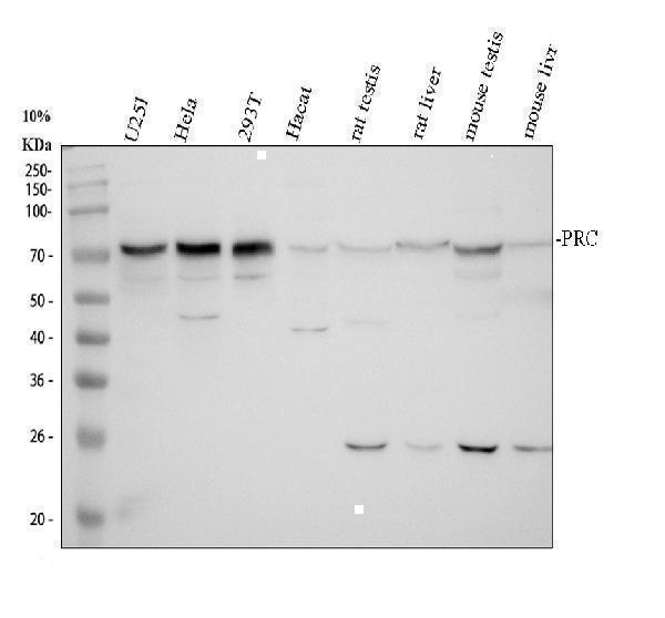
Western blot analysis of PRC1 using anti-PRC1 antibody (PB9814).
Electrophoresis was performed on a 10% SDS-PAGE gel at 80V (Stacking gel) / 120V (Resolving gel) for 2 hours. The sample well of each lane was loaded with 30 ug of sample under reducing conditions.
Lane 1: human U251 whole cell lysates,
Lane 2: human Hela whole cell lysates,
Lane 3: human 293T whole cell lysates,
Lane 4: human Hacat whole cell lysates,
Lane 5: rat testis tissue lysates,
Lane 6: rat liver tissue lysates,
Lane 7: mouse testis tissue lysates,
Lane 8: mouse liver tissue lysates.
After electrophoresis, proteins were transferred to a nitrocellulose membrane at 150 mA for 50-90 minutes. Blocked the membrane with 5% non-fat milk/TBS for 1.5 hour at RT. The membrane was incubated with rabbit anti-PRC1 antigen affinity purified polyclonal antibody (PB9814) at 0.5 μg/mL overnight at 4°C, then washed with TBS-0.1%Tween 3 times with 5 minutes each and probed with a goat anti-rabbit IgG-HRP secondary antibody (Catalog # BA1054) at a dilution of 1:5000 for 1.5 hour at RT. The signal is developed using an ECL Plus Western Blotting Substrate (Catalog # AR1196-200) with Tanon 5200 system. A specific band was detected for PRC1 at approximately 72 kDa. The expected band size for PRC1 is at 72 kDa.

Click image to see more details
Immunoprecipitating (IP) PRC1 in 293T whole cell lysate.
Western blot analysis of PRC1 using anti-PRC1 antibody (PB9814);
Lane 1: 293T whole cell lysates (30ug);
Lane 2: Rabbit control IgG instead of anti-PRC1 antibody in 293T whole cell lysate;
Lane 3: anti-PRC1 antibody (2μg) + 293T whole cell lysate (500μg).
After electrophoresis, proteins were transferred to a membrane. Then the membrane was incubated with rabbit anti-PRC1 antigen affinity purified polyclonal antibody (PB9814) at a dilution of 0.5 μg/mL and probed with a goat anti-rabbit IgG-HRP secondary antibody (Catalog # BA1054). The signal is developed using ECL Plus Western Blotting Substrate (Catalog # AR1196-200). A specific band was detected for PRC1 at approximately 72 kDa. The expected band size for PRC1 is at 72 kDa.

Click image to see more details
Flow Cytometry analysis of 293T cells using anti-PRC1 antibody (PB9814).
Overlay histogram showing 293T cells stained with PB9814 (Blue line). To facilitate intracellular staining, cells were fixed with 4% paraformaldehyde and permeabilized with permeabilization buffer. The cells were blocked with 10% normal goat serum. And then incubated with rabbit anti-PRC1 Antibody (PB9814, 1 μg/1x106 cells) for 30 min at 20°C. DyLight®488 conjugated goat anti-rabbit IgG (BA1127, 5-10 μg/1x106 cells) was used as secondary antibody for 30 minutes at 20°C. Isotype control antibody (Green line) was rabbit IgG (1 μg/1x106) used under the same conditions. Unlabelled sample without incubation with primary antibody and secondary antibody (Red line) was used as a blank control.
Specific Publications For Anti-PRC1 Antibody Picoband® (PB9814)
Loading publications
Recommended Resources
Here are featured tools and databases that you might find useful.
- Boster's Pathways Library
- Protein Databases
- Bioscience Research Protocol Resources
- Data Processing & Analysis Software
- Photo Editing Software
- Scientific Literature Resources
- Research Paper Management Tools
- Molecular Biology Software
- Primer Design Tools
- Bioinformatics Tools
- Phylogenetic Tree Analysis
Customer Reviews
Have you used Anti-PRC1 Antibody Picoband®?
Share your experimental results or join a short interview to earn up to $1,000 in product credits or other rewards.
0 Reviews For Anti-PRC1 Antibody Picoband®
Customer Q&As
Have a question?
Find answers in Q&As, reviews.
Can't find your answer?
Submit your question
4 Customer Q&As for Anti-PRC1 Antibody Picoband®
Question
My colleagues were well pleased with the WB result of your anti-PRC1 antibody. However we have been able to see positive staining in cervix carcinoma nucleus. using this antibody. Is that expected? Could you tell me where is PRC1 supposed to be expressed?
Verified Customer
Verified customer
Asked: 2020-05-05
Answer
From what I have seen in literature, cervix carcinoma does express PRC1. Generally PRC1 expresses in nucleus. Regarding which tissues have PRC1 expression, here are a few articles citing expression in various tissues:
Cervix carcinoma, Pubmed ID: 18669648, 18691976, 20068231
Cervix carcinoma, and Erythroleukemia, Pubmed ID: 23186163
Kidney, and Placenta, Pubmed ID: 15489334
Boster Scientific Support
Answered: 2020-05-05
Question
We are currently using anti-PRC1 antibody PB9814 for human tissue, and we are happy with the WB results. The species of reactivity given in the datasheet says human. Is it possible that the antibody can work on horse tissues as well?
Verified Customer
Verified customer
Asked: 2020-04-29
Answer
The anti-PRC1 antibody (PB9814) has not been validated for cross reactivity specifically with horse tissues, but there is a good chance of cross reactivity. We have an innovator award program that if you test this antibody and show it works in horse you can get your next antibody for free. Please contact me if I can help you with anything.
Boster Scientific Support
Answered: 2020-04-29
Question
We have observed staining in human cervix carcinoma erythroleukemia. Are there any suggestions? Is anti-PRC1 antibody supposed to stain cervix carcinoma erythroleukemia positively?
Verified Customer
Verified customer
Asked: 2019-11-26
Answer
From what I have seen in literature cervix carcinoma erythroleukemia does express PRC1. From what I have seen in Uniprot.org, PRC1 is expressed in testis, kidney eco:0000312, embl:aah03138.1
rc placenta eco:0000312, embl:aah05140.1, cervix carcinoma, cervix carcinoma erythroleukemia, among other tissues. Regarding which tissues have PRC1 expression, here are a few articles citing expression in various tissues:
Cervix carcinoma, Pubmed ID: 18669648, 18691976, 20068231
Cervix carcinoma, and Erythroleukemia, Pubmed ID: 23186163
Kidney, and Placenta, Pubmed ID: 15489334
Boster Scientific Support
Answered: 2019-11-26
Question
you antibody using your anti-PRC1 antibody for mitotic spindle elongation studies. Has this antibody been tested with western blotting on sgc whole cell lysate? We would like to see some validation images before ordering.
Verified Customer
Verified customer
Asked: 2019-01-09
Answer
Thank you for your inquiry. This PB9814 anti-PRC1 antibody is tested on sgc whole cell lysate, hela whole cell lysate. It is guaranteed to work for WB in human. Our Boster guarantee will cover your intended experiment even if the sample type has not been be directly tested.
Boster Scientific Support
Answered: 2019-01-09
