This website uses cookies to ensure you get the best experience on our website.
- Table of Contents
62 Citations 3 Q&As
23 Citations 7 Q&As
1 Citations 7 Q&As
18 Citations
1 Citations
9 Citations
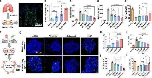
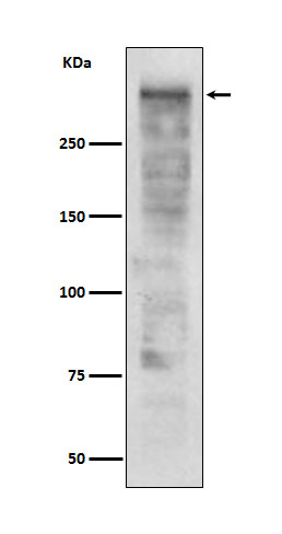
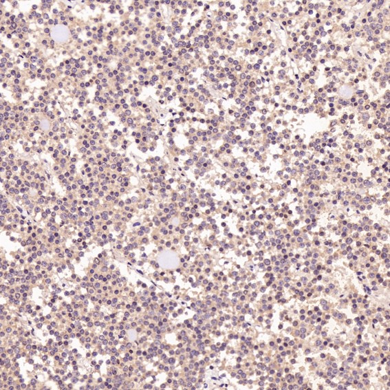
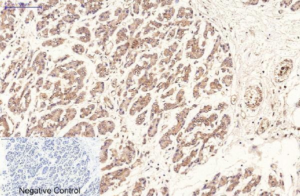
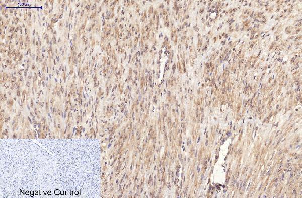
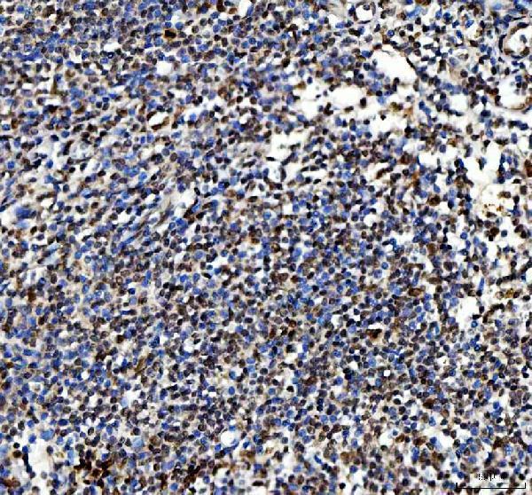
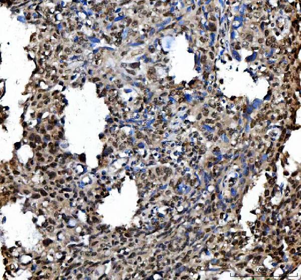
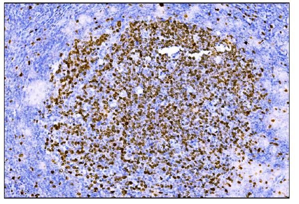
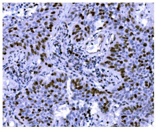
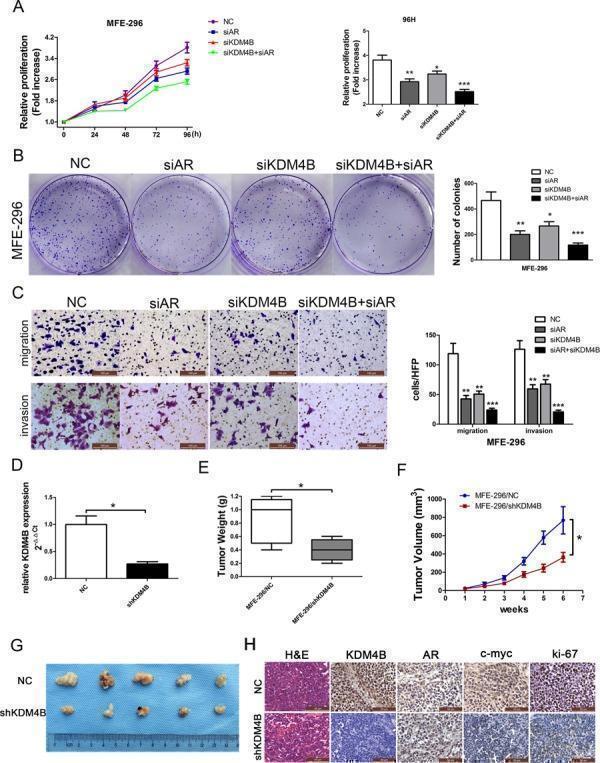
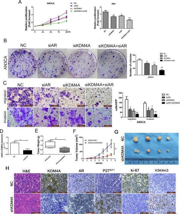
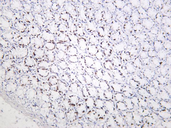
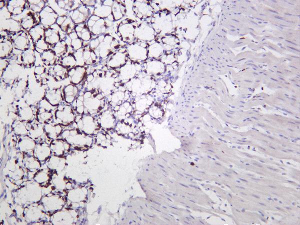
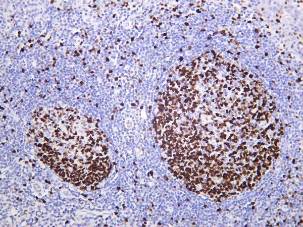
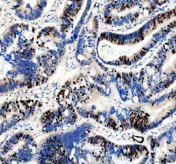
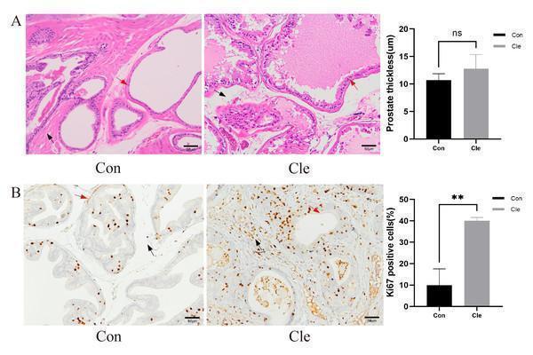
Facts about Proliferation marker protein Ki-67.
Prevents chromosomes from collapsing to a single chromatin mass by forming a steric and electrostatic charge barrier: the protein has a high net electrical charge and acts as a surfactant, dispersing chromosomes and enabling independent chromosome motility (PubMed:27362226). Binds DNA, with a preference for supercoiled DNA and AT-rich DNA (PubMed:10878551).
Human | |
---|---|
Gene Name: | MKI67 |
Uniprot: | P46013 |
Entrez: | 4288 |
Belongs to: |
---|
No superfamily |
antigen identified by monoclonal Ki-67; Antigen Ki67; antigen KI-67; antiKi67; Ki67 ihc; Ki-67 ihc; Ki67 mouse; Ki-67 mouse; Ki67 western blot; Ki-67 western blot; Ki67; Ki-67; KIA; Marker Of Proliferation Ki-67; MIB-; MIB-1; MKI67; PPP1R105; Proliferation Marker Protein Ki-67; proliferation-related Ki-67 antigen; Protein Phosphatase 1; Regulatory Subunit 105; TSG126
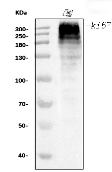
Mass (kDA):
358.694 kDA
Human | |
---|---|
Location: | 10q26.2 |
Sequence: | 10; NC_000010.11 (128096659..128126423, complement) |
Chromosome. Nucleus. Nucleus, nucleolus. Associates with the surface of the mitotic chromosome, the perichromosomal layer, and covers a substantial fraction of the mitotic chromosome surface (PubMed:27362226). Associates with satellite DNA in G1 phase (PubMed:9510506). Binds tightly to chromatin in interphase, chromatin-binding decreases in mitosis when it associates with the surface of the condensed chromosomes (PubMed:15896774, PubMed:22002106). Predominantly localized in the G1 phase in the perinucleolar region, in the later phases it is also detected throughout the nuclear interior, being
In this article, we'll review the advantages of the MKI67 Marker Kits for ELISA, and also discuss the characteristics and advantages of primary antibodies and IHC optimized polyclonal antibodies. You will learn about the advantages of each of the three products, and also learn what they can do to aid you in achieving the most effective results. Boster Bio can provide additional details. We are looking forward to hearing from you.
The MKI67 ELISA Kits by Boster Bio are available for use in immunological studies. These kits are designed to identify biomarkers of inflammation, cancer neurobiology, developmental biology and other areas of research. These kits are highly sensitive and can determine the concentrations of these proteins down to the picogram level. Boster Bio offers ELISA kits for all of these applications and more. For more information, go to tebu-bio.
The MBS8291369 Human Ki67 ELISA Kit is a single-wash sandwich ELISA that measures Ki67 protein present in cell lysates. The the sensitivity of this kit is 21.2 pg/ml. Additionally the 384-well version this kit is available in case you're looking to get greater sensitivity. The MBS8291369 ELISA kit is compatible with both microplates of 96-well and 384-well formats.
The MKI67 ELISA Kit comes with a reagent solution as well as a standard curve of each antibody. Instructions sheets are included in the kit. The instructions are clear and easy to comprehend. The kit also comes with buffers, sample reagents and other useful items. Additionally, Boster Bio has several other kits for MKI67 and you can be sure that you choose the correct one for your experiment.
The MKI67 ELISA Kits from Boster Bio are a great option for research and development process. The MKI67 Monoclonal Antibody contains a monoclonal antibody that reacts with the Ki67 protein in Human, Mouse, and Rat. These kits are able to generate three kinds of information:
MKI67 is a nuclear protein which is connected to the mitotic chromosomes, and is expressed in a variety of cells. It has recently gained importance as a prognostic biomarker for malignancies of the hematological or solid type. This molecule is bound to the UBF protein and binds to the Hklp2 Hklp2 protein with a 160 kD. It is a length of 3256 amino acids and was originally identified as an antigen in association with a monoclonal antibody formulated against Hodgkin's nuclear lymphoma.
Immunocytochemistry was employed to show Ki67/MKI67 specificity in an immersion-fixed cervical epithelial carcinoma HeLa line. Immunocytochemistry revealed specific staining at 320 kDa in HeLa cells. However, Ki67 knockout mutations did not show any Ki67 staining. To demonstrate the specificity of the MKI67 marker it was used to perform immunocytochemistry using an antibody monoclonal to Ki67/MKI67 at a concentration of 1 ug/mL over 3 hours at ambient temperature. Cells were counter-stained with DAPI and stained using NorthernLights 557 antibody. The specific staining was localized to the nuclei of the cells. A fluorescent ICC staining method was developed to demonstrate the specificity of the antibody against
As a leading antibody manufacturer, Boster Bio develops research antibodies, ELISA kits, and other immunological reagents to aid in biomarker identification in cancer, neurosciences inflammation, and other areas of biology. Boster Bio's antibody products have been fully tested against more than 250 human tissues as well as non-transfected cells. A high-quality technical support service is available to help you get these top-quality antibodies.
The Boster Bio MKI67-optimised polyclonal antibodies are based on the MKI67 protein, a 350-370 kDa nuclear protein that belongs to the mitotic chromosome-associated proteins family. It has been identified as an antigen that was previously associated with monoclonal antibody. It is a cell-proliferation marker since it is expressed in a variety of kinds of cells. MKI67 binds to the Hklp2 protein of 160 kDa and enhances centrosome seperation. It is also a key factor in ribosomal DNA synthesis.
To improve Boster Bio MKI67-optimised, polyclonal antibodies to study cancer, Sanbio conducted a mAb screening to determine the presence of human ACE2 and RBD. Competing mAbs were considered inactive if they produced no additional BLI signal. This was a clear indication of ineffectiveness of the antibody to stop RBD binding to ACE2.
The antigen-targeting ability of OTI T cells TG2P-CD8+ was examined. These cells had a greater capacity to respond to the antigen as well as cytotoxic activity. The mICAM-1 as well as mCD8T antibodies were also used to detect the presence of the apoptosis. In addition to the antigen-specific antibody OTI T cells with TG2P showed a dramatic increase in the production of cytokine.
The MKI67 marker is an amino acid that interacts with DNA and heterochromatin. It is a popular marker for cancers, tumors and other illnesses in humans. The cDNA sequences reveal that the MKI67 protein contains 40 weak and 10 strong PEST sites. These PEST sites are found in proteins that play an important function in the regulation of cell cycles as well as transcription and other functions. The 10 most powerful PEST sites are located in exon 13 around conserved cysteine residues in position 8 of the Ki67 repeats.
The MKI67 marker can also be utilized in clinical trials for lung cancer and prostate cancer. It has been proven to predict the fate of pituitary tumor adenomas. MKI67 also has a correlation with high-grade gastric cancer. Both genders are more likely to develop cancer-related differentiation with high levels of MKI67 gene expression.
The MKI67 gene is located on chromosome 10, at locus 10q26.2. The gene is located in an flanking 5' region with putative Sp1 binding sites. The MKI67 promoter is home to two evolutionary conserved E2F transcription factor binding sites. The MKI67 gene is also present in two distinct forms both the heavy and light. The heavy form has 3256 aminoacids, and 395 kDa. The 7th exon is not present from the light isoform.
Currently, MKI67 expression has been shown to be a significant prognostic marker for survival in patients with node-negative breast cancer. Patients suffering from this type cancer typically receive radiation, surgery, or no adjuvant systemic treatment. MKI67 is also comparable to histological evaluation. Similarly, MKI67 expression is also useful for patients suffering from breast cancer with a BRCA1 mutation.
PMID: 8227122 by Schlueter C., et al. The cell proliferation-associated antigen of antibody Ki-67: a very large, ubiquitous nuclear protein with numerous repeated elements, representing a new kind of cell cycle-maintaining proteins.
PMID: 6339421 by Gerdes J., et al. Production of a mouse monoclonal antibody reactive with a human nuclear antigen associated with cell proliferation.
*Showing only the more recent 20. More publications can be found for each product on its corresponding product page