Click image to see more details
Product Info Summary
| SKU: | PA2238 |
|---|---|
| Size: | 100 μg/vial |
| Reactive Species: | Human, Mouse, Rat |
| Host: | Rabbit |
| Application: | IF, ICC, WB |
Customers Who Bought This Also Bought

Product info
Product Name
Anti-DNA/RNA-binding protein KIN17 KIN Antibody Picoband®
SKU/Catalog Number
PA2238
BA3419 is an alternative SKU for this antibody, used in previous lots.
Size
100 μg/vial
Form
Lyophilized
Description
Boster Bio Anti-DNA/RNA-binding protein KIN17 KIN Antibody catalog # PA2238. Tested in ICC/IF, WB applications. This antibody reacts with Human, Mouse, Rat. The brand Picoband indicates this is a premium antibody that guarantees superior quality, high affinity, and strong signals with minimal background in Western blot applications. Only our best-performing antibodies are designated as Picoband, ensuring unmatched performance.
Storage & Handling
Store at -20˚C for one year from date of receipt. After reconstitution, at 4˚C for one month. It can also be aliquotted and stored frozen at -20˚C for six months. Avoid repeated freeze-thaw cycles.
Cite This Product
Anti-DNA/RNA-binding protein KIN17 KIN Antibody Picoband® (Boster Biological Technology, Pleasanton CA, USA, Catalog # PA2238)
Host
Rabbit
Contents
Each vial contains 4mg Trehalose, 0.9mg NaCl, 0.2mg Na2HPO4, 0.01mg NaN3.
Clonality
Polyclonal
Isotype
Rabbit IgG
Immunogen
A synthetic peptide corresponding to a sequence at the N-terminus of human KIN, identical to the related mouse and rat sequences.
Cross-reactivity
No cross-reactivity with other proteins
Reactive Species
PA2238 is reactive to KIN in Human, Mouse, Rat
Observed Molecular Weight
45 kDa
Calculated molecular weight
45.4 kDa
Background of KIN
DNA/RNA-binding protein KIN17, also known as BTCD or KIN17 is a protein that in humans is encoded by the KIN gene. This gene is mapped to 10p14. The protein encoded by this gene is a nuclear protein that forms intranuclear foci during proliferation and is redistributed in the nucleoplasm during the cell cycle. Short-wave ultraviolet light provokes the relocalization of the protein, suggesting its participation in the cellular response to DNA damage. Originally selected based on protein-binding with RecA antibodies, the mouse protein presents a limited similarity with a functional domain of the bacterial RecA protein, a characteristic shared by this human ortholog. Alternative splicing of this gene results in multiple transcript variants.
Antibody Validation
Boster validates all antibodies on WB, IHC, ICC, Immunofluorescence, and ELISA with known positive control and negative samples to ensure specificity and high affinity, including thorough antibody incubations.
Application & Images
Applications
PA2238 is guaranteed for IF, ICC, WB Boster Guarantee
Recommend Dilution
| Application | Dilution | Species |
|---|---|---|
| Western blot | 0.1-0.5μg/ml | Human, Mouse, Rat |
| Immunocytochemistry/Immunofluorescence | 5 μg/ml | Human |
Tested application
Suggested blocking solution with 5% non-fat milk or BSA; (*)Recommended protein loading: 20-40 µg per lane
Validation Images & Assay Conditions
Click image to see more details
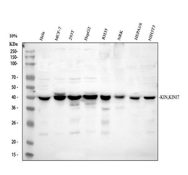
Western blot analysis of KIN using anti-KIN antibody (PA2238).
Electrophoresis was performed on a 10% SDS-PAGE gel at 80V (Stacking gel) / 120V (Resolving gel) for 2 hours. The sample well of each lane was loaded with 30 ug of sample under reducing conditions.
Lane 1: human Hela whole cell lysates,
Lane 2: human MCF-7 whole cell lysates,
Lane 3: human 293T whole cell lysates,
Lane 4: human HepG2 whole cell lysates,
Lane 5: rat RH35 whole cell lysates,
Lane 6: rat NRK whole cell lysates,
Lane 7: mouse HEPA1-6 whole cell lysates,
Lane 8: mouse NIH/3T3 whole cell lysates.
After electrophoresis, proteins were transferred to a nitrocellulose membrane at 150 mA for 50-90 minutes. Blocked the membrane with 5% non-fat milk/TBS for 1.5 hour at RT. The membrane was incubated with rabbit anti-KIN antigen affinity purified polyclonal antibody (PA2238) at 0.5 μg/mL overnight at 4°C, then washed with TBS-0.1%Tween 3 times with 5 minutes each and probed with a goat anti-rabbit IgG-HRP secondary antibody at a dilution of 1:5000 for 1.5 hour at RT. The signal is developed using an ECL Plus Western Blotting Substrate (Catalog # AR1196-200) with Tanon 5200 system. A specific band was detected for KIN at approximately 45 kDa. The expected band size for KIN is at 45 kDa.

Click image to see more details
IF analysis of KIN using anti-KIN antibody (PA2238) and anti-Alpha Tubulin antibody (M03989-3).
KIN was detected in an immunocytochemical section of A549 cells. Enzyme antigen retrieval was performed using IHC enzyme antigen retrieval reagent (AR0022) for 15 mins. The cells were blocked with 10% goat serum. And then incubated with 5 μg/mL rabbit anti-KIN Antibody (PA2238) and mouse anti-Alpha Tubulin antibody (M03989-3) overnight at 4°C. Fluoro594 Conjugated Goat Anti-Rabbit IgG (BA1142) and Fluoro488 Conjugated Goat Anti-Mouse IgG (BA1126) were used as secondary antibody at 1:500 dilution and incubated for 30 minutes at 37°C. Visualize using a fluorescence microscope and filter sets appropriate for the label used.
Specific Publications For Anti-DNA/RNA-binding protein KIN17 KIN Antibody Picoband® (PA2238)
Loading publications
Recommended Resources
Here are featured tools and databases that you might find useful.
- Boster's Pathways Library
- Protein Databases
- Bioscience Research Protocol Resources
- Data Processing & Analysis Software
- Photo Editing Software
- Scientific Literature Resources
- Research Paper Management Tools
- Molecular Biology Software
- Primer Design Tools
- Bioinformatics Tools
- Phylogenetic Tree Analysis
Customer Reviews
Have you used Anti-DNA/RNA-binding protein KIN17 KIN Antibody Picoband®?
Share your experimental results or join a short interview to earn up to $1,000 in product credits or other rewards.
0 Reviews For Anti-DNA/RNA-binding protein KIN17 KIN Antibody Picoband®
Customer Q&As
Have a question?
Find answers in Q&As, reviews.
Can't find your answer?
Submit your question
