Click image to see more details
Product Info Summary
| SKU: | A00760-2 |
|---|---|
| Size: | 100 μg/vial |
| Reactive Species: | Mouse, Rat |
| Host: | Rabbit |
| Application: | ELISA, WB |
Customers Who Bought This Also Bought

Product info
Product Name
Anti-LOX 1/Olr1 Antibody Picoband®
SKU/Catalog Number
A00760-2
Size
100 μg/vial
Form
Lyophilized
Description
Boster Bio Anti-LOX 1/Olr1 Antibody Picoband® catalog # A00760-2. Tested in ELISA, WB applications. This antibody reacts with Mouse, Rat. The brand Picoband indicates this is a premium antibody that guarantees superior quality, high affinity, and strong signals with minimal background in Western blot applications. Only our best-performing antibodies are designated as Picoband, ensuring unmatched performance.
Storage & Handling
Store at -20˚C for one year from date of receipt. After reconstitution, at 4˚C for one month. It can also be aliquotted and stored frozen at -20˚C for six months. Avoid repeated freeze-thaw cycles.
Cite This Product
Anti-LOX 1/Olr1 Antibody Picoband® (Boster Biological Technology, Pleasanton CA, USA, Catalog # A00760-2)
Host
Rabbit
Contents
Each vial contains 4mg Trehalose, 0.9mg NaCl, 0.2mg Na2HPO4, 0.05mg NaN3.
Clonality
Polyclonal
Isotype
Rabbit IgG
Immunogen
E.coli-derived mouse LOX 1/Olr1 recombinant protein (Position: Q55-I363).
Cross-reactivity
No cross-reactivity with other proteins.
Reactive Species
A00760-2 is reactive to Olr1 in Mouse, Rat
Observed Molecular Weight
52 kDa
Calculated molecular weight
41.6 kDa
Background of Olr1
OLR1 (oxidized low density lipoprotein (lectin-like) receptor 1) also called CLEC8A, LOX-1, SCARE1, is a receptor protein which belongs to the C-type lectin superfamily. The OLR1 gene encodes a cell-surface endocytosis receptor for oxidized low density lipoprotein (OxLDL). This gene is mapped on 12p13.2. Incubation of the cells with LDL had no effect on LOX1 expression, but incubation with OxLDL resulted in a dose-dependent increase in LOX1 mRNA and protein expression; however, very high concentrations of OxLDL caused a decrease in OxLDL expression, perhaps indicating toxic effects on endothelial cells. LOX1 was also expressed in macrophages, but not in vascular smooth muscle cells. The findings suggested a role for LOX1 in the pathophysiology of atherosclerotic cardiovascular disease. LOX1 expression was detected in all choroidal neovascular membranes, regardless of structure, whereas there was no evidence of LOX1 within the posterior segments of normal eyes. LOX1 plays an active role in the pathogenesis of choroidal neovascularization, especially in ARMD.
Antibody Validation
Boster validates all antibodies on WB, IHC, ICC, Immunofluorescence, and ELISA with known positive control and negative samples to ensure specificity and high affinity, including thorough antibody incubations.
Application & Images
Applications
A00760-2 is guaranteed for ELISA, WB Boster Guarantee
Recommend Dilution
| Application | Dilution | Species |
|---|---|---|
| Western blot | 0.25-0.5μg/ml | Mouse, Rat |
| ELISA | 0.1-0.5μg/ml | - |
Tested application
Suggested blocking solution with 5% non-fat milk or BSA; (*)Recommended protein loading: 20-40 µg per lane
Validation Images & Assay Conditions
Click image to see more details
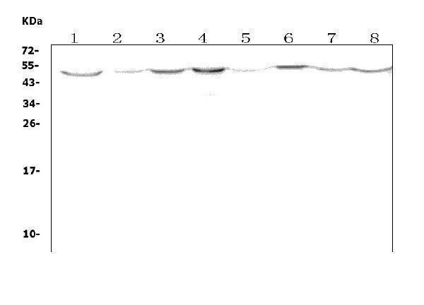
Western blot analysis of Olr1 using anti-Olr1 antibody (A00760-2).
Electrophoresis was performed on a 5-20% SDS-PAGE gel at 70V (Stacking gel) / 90V (Resolving gel) for 2-3 hours. The sample well of each lane was loaded with 50ug of sample under reducing conditions.
Lane 1: mouse brain tissue lysates,
Lane 2: mouse lung tissue lysates,
Lane 3: mouse liver tissue lysates,
Lane 4: mouse kidney tissue lysates,
Lane 5: mouse testicular tissue lysates,
Lane 6: mouse smooth muscle tissue lysates,
Lane 7: mouse SP20 whole cell lysates,
Lane 8: mouse Neuro-2a whole cell lysates.
After Electrophoresis, proteins were transferred to a Nitrocellulose membrane at 150mA for 50-90 minutes. Blocked the membrane with 5% Non-fat Milk/ TBS for 1.5 hour at RT. The membrane was incubated with rabbit anti-Olr1 antigen affinity purified polyclonal antibody (Catalog # A00760-2) at 0.5 μg/mL overnight at 4°C, then washed with TBS-0.1%Tween 3 times with 5 minutes each and probed with a goat anti-rabbit IgG-HRP secondary antibody at a dilution of 1:5000 for 1.5 hour at RT. The signal is developed using an Enhanced Chemiluminescent detection (ECL) kit (Catalog # EK1002) with Tanon 5200 system. A specific band was detected for Olr1 at approximately 52KD. The expected band size for Olr1 is at 31KD.
Specific Publications For Anti-LOX 1/Olr1 Antibody Picoband® (A00760-2)
Loading publications
Recommended Resources
Here are featured tools and databases that you might find useful.
- Boster's Pathways Library
- Protein Databases
- Bioscience Research Protocol Resources
- Data Processing & Analysis Software
- Photo Editing Software
- Scientific Literature Resources
- Research Paper Management Tools
- Molecular Biology Software
- Primer Design Tools
- Bioinformatics Tools
- Phylogenetic Tree Analysis
Customer Reviews
Have you used Anti-LOX 1/Olr1 Antibody Picoband®?
Share your experimental results or join a short interview to earn up to $1,000 in product credits or other rewards.
0 Reviews For Anti-LOX 1/Olr1 Antibody Picoband®
Customer Q&As
Have a question?
Find answers in Q&As, reviews.
Can't find your answer?
Submit your question
