Click image to see more details
-

-
-
-
-
+7
Product Info Summary
| SKU: | M00829 |
|---|---|
| Size: | 100 μl |
| Reactive Species: | Human, Mouse, Rat |
| Host: | Rabbit |
| Application: | Flow Cytometry, IP, IF, IHC, ICC, WB |
Customers Who Bought This Also Bought

Product info
Product Name
Anti-LRP1/Lrp 1 Cluster Ii Rabbit Monoclonal Antibody
SKU/Catalog Number
M00829
BM4098 is an alternative SKU for this antibody, used in previous lots.
Size
100 μl
Form
Liquid
Description
Boster Bio Anti-LRP1/Lrp 1 Cluster Ii Rabbit Monoclonal Antibody catalog # M00829. Tested in WB, IHC, ICC/IF, IP, Flow Cytometry applications. This antibody reacts with Human, Mouse, Rat.
Storage & Handling
Store at -20°C for one year. For short term storage and frequent use, store at 4°C for up to one month. Avoid repeated freeze-thaw cycles.
Cite This Product
Anti-LRP1/Lrp 1 Cluster Ii Rabbit Monoclonal Antibody (Boster Biological Technology, Pleasanton CA, USA, Catalog # M00829)
Host
Rabbit
Contents
Rabbit IgG in stabilizing components, phosphate buffered saline, pH 7.4, 150mM NaCl, 0.02% sodium azide and 50% glycerol.
*This antibody is supplied in a stabilized formulation.
Compatibility with conjugation reactions depends on the chemistry of the conjugation method used.
For conjugation methods that are not compatible with the stabilizing components present in this formulation, a carrier-free antibody format is required.
Clonality
Monoclonal
Clone Number
BDC-12
Isotype
Rabbit IgG
Immunogen
A synthesized peptide derived from human LRP1
Reactive Species
M00829 is reactive to LRP1 in Human, Mouse, Rat
Observed Molecular Weight
85 kDa
Calculated molecular weight
504.6 kDa
Antibody Validation
Boster validates all antibodies on WB, IHC, ICC, Immunofluorescence, and ELISA with known positive control and negative samples to ensure specificity and high affinity, including thorough antibody incubations.
Application & Images
Applications
M00829 is guaranteed for Flow Cytometry, IP, IF, IHC, ICC, WB Boster Guarantee
Recommend Dilution
WB 1:1000-5000
IHC 1:50-200
ICC/IF 1:50-200
IP 1:20
FC 1:20
Tested application
Use TE buffer pH 9.0 for antigen retrieval; (*) citrate buffer pH 6.0 is an alternative.
Validation Images & Assay Conditions
Click image to see more details
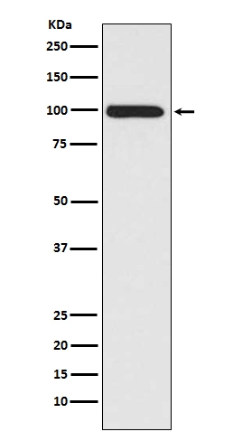
Western blot analysis of LRP1 expression in A549 cell lysate.

Click image to see more details
Western blot analysis of LRP1/Lrp 1 Cluster Ii using anti-LRP1/Lrp 1 Cluster Ii antibody (M00829).
Electrophoresis was performed on a 10% SDS-PAGE gel at 80V (Stacking gel) / 120V (Resolving gel) for 2 hours. The sample well of each lane was loaded with 30 ug of sample under reducing conditions.
Lane 1: human U87 whole cell lysates,
Lane 2: human HepG2 whole cell lysates,
Lane 3: human U20S whole cell lysates,
Lane 4: human Hela whole cell lysates,
Lane 5: rat brain tissue lysates,
Lane 6: rat heart muscle tissue lysates,
Lane 7: mouse brain tissue lysates,
Lane 8: mouse heart tissue lysates.
After electrophoresis, proteins were transferred to a nitrocellulose membrane at 150 mA for 50-90 minutes. Blocked the membrane with 5% non-fat milk/TBS for 1.5 hour at RT. The membrane was incubated with rabbit anti-LRP1/Lrp 1 Cluster Ii antigen affinity purified monoclonal antibody (M00829) at 1:500 overnight at 4°C, then washed with TBS-0.1%Tween 3 times with 5 minutes each and probed with a goat anti-rabbit IgG-HRP secondary antibody at a dilution of 1:1000 for 1.5 hour at RT. The signal is developed using an ECL Plus Western Blotting Substrate (Catalog # AR1196-200) with Tanon 5200 system. A specific band was detected for LRP1/Lrp 1 Cluster Ii at approximately 85 kDa. The expected band size for LRP1/Lrp 1 Cluster Ii is at 85 kDa.

Click image to see more details
Membrane-receptor LRP1 is responsible for the stimulatory effect of CD44s-tPA on lamellipodia formation (A) The distribution patterns of cortactin (red) and F-actin (green) demonstrated by immunofluorescence staining in control and LRP1 inhibitor (RAP, 200 nM), Annexin A2 inhibitor (LCKLSL, 2.5 µM), and EGFR inhibitor (AG1478, 10 µM) pretreated CD44s-overexpressing MCF7 cells. The elongated lamellipodia are highlighted by the arrows. The proportion of cells with lamellipodia in control and RAP, LCKLSL, and AG1478 pretreated groups were calculated from triplicate independent experiments, means ± SD from triplicate experiments were plotted. (B) Representative images and quantitative analysis of migration assay showing the wound closure rate of control and RAP pretreated MCF7 CD44s cells. The means ± SD of wound closure rates from triplicate experiments were plotted. (C) Analysis of LRP1 expression in control and LRP1 knockdown MCF7CD44s cells. (D) Representative images and quantitative analysis of migration assay showing the wound closure rate of control and LRP1 knockdown MCF7 CD44s cells. The means ± SD of wound closure rates from triplicate experiments were plotted. (E) Representative images and quantitative analysis of migration assay showing the wound closure rate of control and LRP1 knockdown MCF7 CD44s cells. The means ± SD of wound closure rates from triplicate experiments were plotted. n. s. Indicates no significant, ** p < 0.01, *** p < 0.001.
Index in PubMed under a CC BY license. PMID: 37842093

Click image to see more details
tPA/LRP1 axis enhances the lamellipodia formation through NFκB signaling pathway. (A) Gene set enrichment analysis (GSEA) enrichment plots of the hallmark of NFκB gene sets in MCF7 CD44s compared with MCF7 vector groups. (B) Analysis of phosphorylation of p65, total p65 protein levels in MCF7 vector , MCF7 CD44s , and LRP1 knockdown MCF7 CD44s cells by western blot. (C) Analysis of phosphorylation of p65, total p65 protein levels in control and RAP pretreated MCF7 CD44s cells by western blot. (D) Analysis of phosphorylation of p65, total p65 protein levels in control and NFκB pathway inhibitor (BAY 11-7082, 10 µM) pretreated MCF7 CD44s cells by western blot. (E) The distribution patterns of cortactin (red) and F-actin (green) demonstrated by immunofluorescence staining in control and BAY 11-7082 pretreated CD44s-overexpressing MCF7 cells. The elongated lamellipodia are highlighted by the arrows. The proportion of cells with lamellipodia in control and BAY 11-7082 pretreated groups were calculated from triplicate independent experiments, means ± SD from triplicate experiments were plotted. (F) Representative images and quantitative analysis of migration assay showing the wound closure rate of control and BAY 11-7082 pretreated MCF7 CD44s cells. The means ± SD of wound closure rates from triplicate experiments were plotted. * p < 0.05.
Index in PubMed under a CC BY license. PMID: 37842093

Click image to see more details
All lanes use the Antibody at 1:3W dilution for 1 hour at room temperature.

Click image to see more details
Immunohistochemical analysis of paraffin-embedded human liver carcinoma, using LRP1 Antibody.

Click image to see more details
Immunohistochemical analysis of paraffin-embedded Human pituitary tumor, using the Antibody at 1:300 dilution.

Click image to see more details
Immunohistochemical analysis of paraffin-embedded Human prostate cancer, using the Antibody at 1:300 dilution.

Click image to see more details
Immunohistochemical analysis of paraffin-embedded Human breast cancer, using the Antibody at 1:300 dilution.

Click image to see more details
Immunofluorescent analysis using the Antibody at 1:50 dilution.

Click image to see more details
Flow cytometric analysis of LRP1 was done on Hela cells. The cells were fixed, permeabilized and stained with the primary antibody (1/50) (blue). After incubation of the primary antibody at room temperature for an hour, the cells were stained with a Alexa Fluor 488-conjugated Goat anti-Rabbit IgG Secondary antibody at 1/1000 dilution for 30 minutes.Unlabelled sample was used as a control (cells without incubation with primary antibody; red).
Specific Publications For Anti-LRP1/Lrp 1 Cluster Ii Rabbit Monoclonal Antibody (M00829)
Loading publications
Recommended Resources
Here are featured tools and databases that you might find useful.
- Boster's Pathways Library
- Protein Databases
- Bioscience Research Protocol Resources
- Data Processing & Analysis Software
- Photo Editing Software
- Scientific Literature Resources
- Research Paper Management Tools
- Molecular Biology Software
- Primer Design Tools
- Bioinformatics Tools
- Phylogenetic Tree Analysis
Customer Reviews
Have you used Anti-LRP1/Lrp 1 Cluster Ii Rabbit Monoclonal Antibody?
Share your experimental results or join a short interview to earn up to $1,000 in product credits or other rewards.
0 Reviews For Anti-LRP1/Lrp 1 Cluster Ii Rabbit Monoclonal Antibody
Customer Q&As
Have a question?
Find answers in Q&As, reviews.
Can't find your answer?
Submit your question
5 Customer Q&As for Anti-LRP1/Lrp 1 Cluster Ii Rabbit Monoclonal Antibody
Question
We are currently using anti-LRP1/Lrp 1 Cluster Ii Rabbit Monoclonal antibody M00829 for rat tissue, and we are satisfied with the WB results. The species of reactivity given in the datasheet says human, mouse, rat. Is it likely that the antibody can work on horse tissues as well?
Verified Customer
Verified customer
Asked: 2020-01-29
Answer
The anti-LRP1/Lrp 1 Cluster Ii Rabbit Monoclonal antibody (M00829) has not been tested for cross reactivity specifically with horse tissues, though there is a good chance of cross reactivity. We have an innovator award program that if you test this antibody and show it works in horse you can get your next antibody for free. Please contact me if I can help you with anything.
Boster Scientific Support
Answered: 2020-01-29
Question
Is a blocking peptide available for product anti-LRP1/Lrp 1 Cluster Ii Rabbit Monoclonal antibody (M00829)?
Verified Customer
Verified customer
Asked: 2019-07-30
Answer
We do provide the blocking peptide for product anti-LRP1/Lrp 1 Cluster Ii Rabbit Monoclonal antibody (M00829). If you would like to place an order for it please contact support@bosterbio.com and make a special request.
Boster Scientific Support
Answered: 2019-07-30
Question
Thank you for helping with my inquiry over the phone. Here are the WB image, lot number and protocol we used for placenta using anti-LRP1/Lrp 1 Cluster Ii Rabbit Monoclonal antibody M00829. Let me know if you need anything else.
Verified Customer
Verified customer
Asked: 2018-08-06
Answer
Thanks for the data. You have provided everything we needed. Our lab team are working to resolve your inquiry as quickly as possible, and we appreciate your patience and understanding! Please let me know if there is anything you need in the meantime.
Boster Scientific Support
Answered: 2018-08-06
Question
Is this M00829 anti-LRP1/Lrp 1 Cluster Ii Rabbit Monoclonal antibody reactive to the isotypes of LRP1?
Verified Customer
Verified customer
Asked: 2017-11-29
Answer
The immunogen of M00829 anti-LRP1/Lrp 1 Cluster Ii Rabbit Monoclonal antibody is A synthesized peptide derived from human LRP1. Could you tell me which isotype you are interested in so I can help see if the immunogen is part of this isotype?
Boster Scientific Support
Answered: 2017-11-29
Question
Does anti-LRP1/Lrp 1 Cluster Ii Rabbit Monoclonal antibody M00829 work on primate IHC with plasma?
J. Anderson
Verified customer
Asked: 2013-01-24
Answer
Our lab technicians have not validated anti-LRP1/Lrp 1 Cluster Ii Rabbit Monoclonal antibody M00829 on primate. You can run a BLAST between primate and the immunogen sequence of anti-LRP1/Lrp 1 Cluster Ii Rabbit Monoclonal antibody M00829 to see if they may cross-react. If the sequence homology is close, then you can perform a pilot test. Keep in mind that since we have not validated primate samples, this use of the antibody is not covered by our guarantee. However we have an innovator award program that if you test this antibody and show it works in primate plasma in IHC, you can get your next antibody for free.
Boster Scientific Support
Answered: 2013-01-24
