Click image to see more details
Product Info Summary
| SKU: | A00261-1 |
|---|---|
| Size: | 100 μg/vial |
| Reactive Species: | Human, Mouse, Rat |
| Host: | Rabbit |
| Application: | ELISA, IHC, WB |
Customers Who Bought This Also Bought

Product info
Product Name
Anti-NAK/TBK1 Antibody Picoband®
SKU/Catalog Number
A00261-1
Size
100 μg/vial
Form
Lyophilized
Description
Boster Bio Anti-NAK/TBK1 Antibody Picoband® catalog # A00261-1. Tested in ELISA, IHC, WB applications. This antibody reacts with Human, Mouse, Rat. The brand Picoband indicates this is a premium antibody that guarantees superior quality, high affinity, and strong signals with minimal background in Western blot applications. Only our best-performing antibodies are designated as Picoband, ensuring unmatched performance.
Storage & Handling
At -20°C for one year from date of receipt. After reconstitution, at 4°C for one month. It can also be aliquotted and stored frozen at -20°C for six months. Avoid repeated freezing and thawing.
Cite This Product
Anti-NAK/TBK1 Antibody Picoband® (Boster Biological Technology, Pleasanton CA, USA, Catalog # A00261-1)
Host
Rabbit
Contents
Each vial contains 4 mg Trehalose, 0.9 mg NaCl, 0.2 mg Na2HPO4.
Clonality
Polyclonal
Isotype
Rabbit IgG
Immunogen
E.coli-derived human NAK/TBK1 recombinant protein (Position: M1-L729).
Cross-reactivity
No cross-reactivity with other proteins.
Reactive Species
A00261-1 is reactive to TBK1 in Human, Mouse, Rat
Observed Molecular Weight
84 kDa
Calculated molecular weight
83.6 kDa
Background of TBK1
Serine/threonine-protein kinase TBK1, also called TANK-binding kinase 1 or NF-kappa-B-activating kinase is an enzyme that in humans is encoded by the TBK1 gene. The gene was assigned to human chromosome 12q14.2. Serine/threonine kinase plays an essential role in regulating inflammatory responses to foreign agents. TBK1 and NF-kappa-B signaling are essential in KRAS mutant tumors, and established a general approach for the rational identification of codependent pathways in cancer.
Antibody Validation
Boster validates all antibodies on WB, IHC, ICC, Immunofluorescence, and ELISA with known positive control and negative samples to ensure specificity and high affinity, including thorough antibody incubations.
Application & Images
Applications
A00261-1 is guaranteed for ELISA, IHC, WB Boster Guarantee
Recommend Dilution
| Application | Dilution | Species |
|---|---|---|
| Western blot | 0.1-0.25 μg/ml | Human, Mouse, Rat |
| Immunohistochemistry(Paraffin-embedded Section) | 2-5 μg/ml | Mouse |
| ELISA | 0.1-0.5 μg/ml | - |
Tested application
Suggested blocking solution with 5% non-fat milk or BSA; (*)Recommended protein loading: 20-40 µg per lane
Use TE buffer pH 9.0 for antigen retrieval; (*) citrate buffer pH 6.0 is an alternative.
Validation Images & Assay Conditions
Click image to see more details
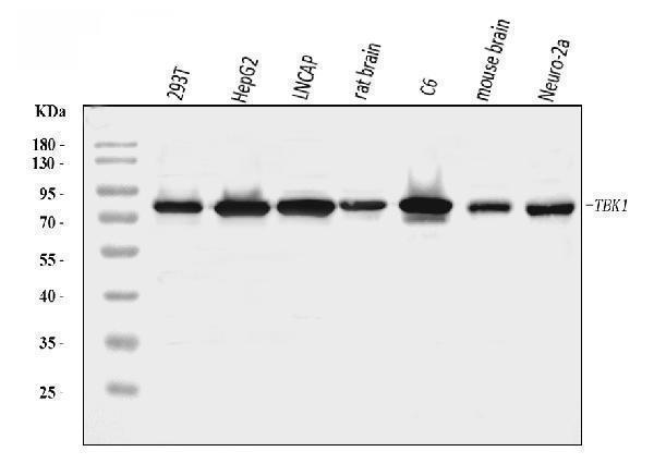
Western blot analysis of NAK/TBK1 using anti-NAK/TBK1 antibody (A00261-1).
Electrophoresis was performed on a 5-20% SDS-PAGE gel at 70V (Stacking gel) / 90V (Resolving gel) for 2-3 hours. The sample well of each lane was loaded with 30 ug of sample under reducing conditions.
Lane 1: human 293T whole cell lysates,
Lane 2: human HepG2 whole cell lysates,
Lane 3: human LNCAP whole cell lysates,
Lane 4: rat brain tissue lysates,
Lane 5: rat C6 whole cell lysates,
Lane 7: mouse brain tissue lysates,
Lane 8: mouse Neuro-2a whole cell lysates.
After electrophoresis, proteins were transferred to a nitrocellulose membrane at 150 mA for 50-90 minutes. Blocked the membrane with 5% non-fat milk/TBS for 1.5 hour at RT. The membrane was incubated with rabbit anti-NAK/TBK1 antigen affinity purified polyclonal antibody (Catalog # A00261-1) at 0.25 μg/mL overnight at 4°C, then washed with TBS-0.1%Tween 3 times with 5 minutes each and probed with a goat anti-rabbit IgG-HRP secondary antibody at a dilution of 1:5000 for 1.5 hour at RT. The signal is developed using an Enhanced Chemiluminescent detection (ECL) kit (Catalog # EK1002) with Tanon 5200 system. A specific band was detected for NAK/TBK1 at approximately 84 kDa. The expected band size for NAK/TBK1 is at 84 kDa.

Click image to see more details
HMGCR inhibition combined with radiotherapy significantly activates the cGAS–STING pathway. a , A volcano plot of differentially expressed genes between the combination group and the radiotherapy group. b GSEA of the TCR signaling pathway (KEGG: MMU04660) and T-cell-mediated immunity (GO: 0002456) between the combination therapy group and the radiotherapy group. c A heatmap of log 2 FC to depict the gene expression associated with type I IFN. d The expression of p-TBK1, p-IRF3, TBK1 and IRF3 protein extracted from CT26 tumors was detected by western blot. e , The relative expression of CCL5, CXCL10 and IFNβ mRNA extracted from CT26 tumors was detected by qPCR. f , Representative IF images of p-TBK1 and p-IRF3. g , Representative IHC images of IFN-β. h , The levels of IFN-β, CCL5 and IFN-γ protein in tumor tissues were measured via ELISA. Scale bar, 50 μm. ** P < 0.01; * P < 0.05.
Index in PubMed under a CC BY license. PMID: 40355720

Click image to see more details
Cholesterol impairs radiotherapy-induced cGAS–STING activation and lovastatin rescues this activation in vitro. a The expression of p-TBK1, p-IRF3, TBK1 and IRF3 protein extracted from CT26 cells with different treatments (ctrl, cholesterol 50 μM, MβCD 2 mM, IR 6 Gy, IR + cholesterol and IR + MβCD) and detected by western blot. b The relative expression of CCL5, CXCL10 and IFNβ mRNA extracted from CT26 cells was detected by qPCR. c The expression of p-TBK1, p-IRF3, TBK1 and IRF3 protein was extracted from HCT116 cells with different treatments (ctrl, cholesterol 50 μM, MβCD 2 mM, IR 6 Gy, IR + cholesterol and IR + MβCD) and detected by western blot. d The relative expression of CCL5, CXCL10 and IFNβ mRNA extracted from HCT116 cells was detected by qPCR. e , f The expression of p-TBK1, p-IRF3, TBK1 and IRF3 protein extracted from CT26 cells and HCT116 cells with different treatments (ctrl, lovastatin 10 μM, IR 6 Gy and IR + lovastatin) was detected by western blot ( e ) and quantitative anslysis ( f ). g , h , Confocal fluorescence microscopy was conducted on CT26 ( g ) and HCT116 ( h ) cells with different treatments. The cells were labeled with DAPI (blue) and p-TBK1 (green) or p-IRF3 (green). i The levels of IFN-β and IFN-γ in the supernatant of co-cultures of MC38-OVA cells and OT-1 mouse spleen cells as well as HCT116 cells and human PBMCs were measured using ELISA. The ratio of immune cells to tumor cells is 10:1. The co-cultures were maintained for 36 h. j LDH release assay was performed using the supernatants from co-cultures of MC38-OVA cells and OT-1 mouse spleen cells as well as HCT116 cells and human PBMCs. The ratio of immune cells to tumor cells is 10:1. The co-cultures were maintained for 36 h. Scale bar, 5 μm. ** P < 0.01; * P < 0.05; ns, not significant.
Index in PubMed under a CC BY license. PMID: 40355720

Click image to see more details
IHC analysis of NAK/TBK1 using anti-NAK/TBK1 antibody (A00261-1).
NAK/TBK1 was detected in a paraffin-embedded section of mouse testis tissue. Heat mediated antigen retrieval was performed in EDTA buffer (pH 8.0, epitope retrieval solution). The tissue section was blocked with 10% goat serum. The tissue section was then incubated with 2 μg/ml rabbit anti-NAK/TBK1 Antibody (A00261-1) overnight at 4°C. Peroxidase Conjugated Goat Anti-rabbit IgG was used as secondary antibody and incubated for 30 minutes at 37°C. The tissue section was developed using HRP Conjugated Rabbit IgG Super Vision Assay Kit (Catalog # SV0002) with DAB as the chromogen.
Specific Publications For Anti-NAK/TBK1 Antibody Picoband® (A00261-1)
Loading publications
Recommended Resources
Here are featured tools and databases that you might find useful.
- Boster's Pathways Library
- Protein Databases
- Bioscience Research Protocol Resources
- Data Processing & Analysis Software
- Photo Editing Software
- Scientific Literature Resources
- Research Paper Management Tools
- Molecular Biology Software
- Primer Design Tools
- Bioinformatics Tools
- Phylogenetic Tree Analysis
Customer Reviews
Have you used Anti-NAK/TBK1 Antibody Picoband®?
Share your experimental results or join a short interview to earn up to $1,000 in product credits or other rewards.
0 Reviews For Anti-NAK/TBK1 Antibody Picoband®
Customer Q&As
Have a question?
Find answers in Q&As, reviews.
Can't find your answer?
Submit your question
