This website uses cookies to ensure you get the best experience on our website.
- Table of Contents
1 Citations 17 Q&As
2 Citations 16 Q&As
8 Citations
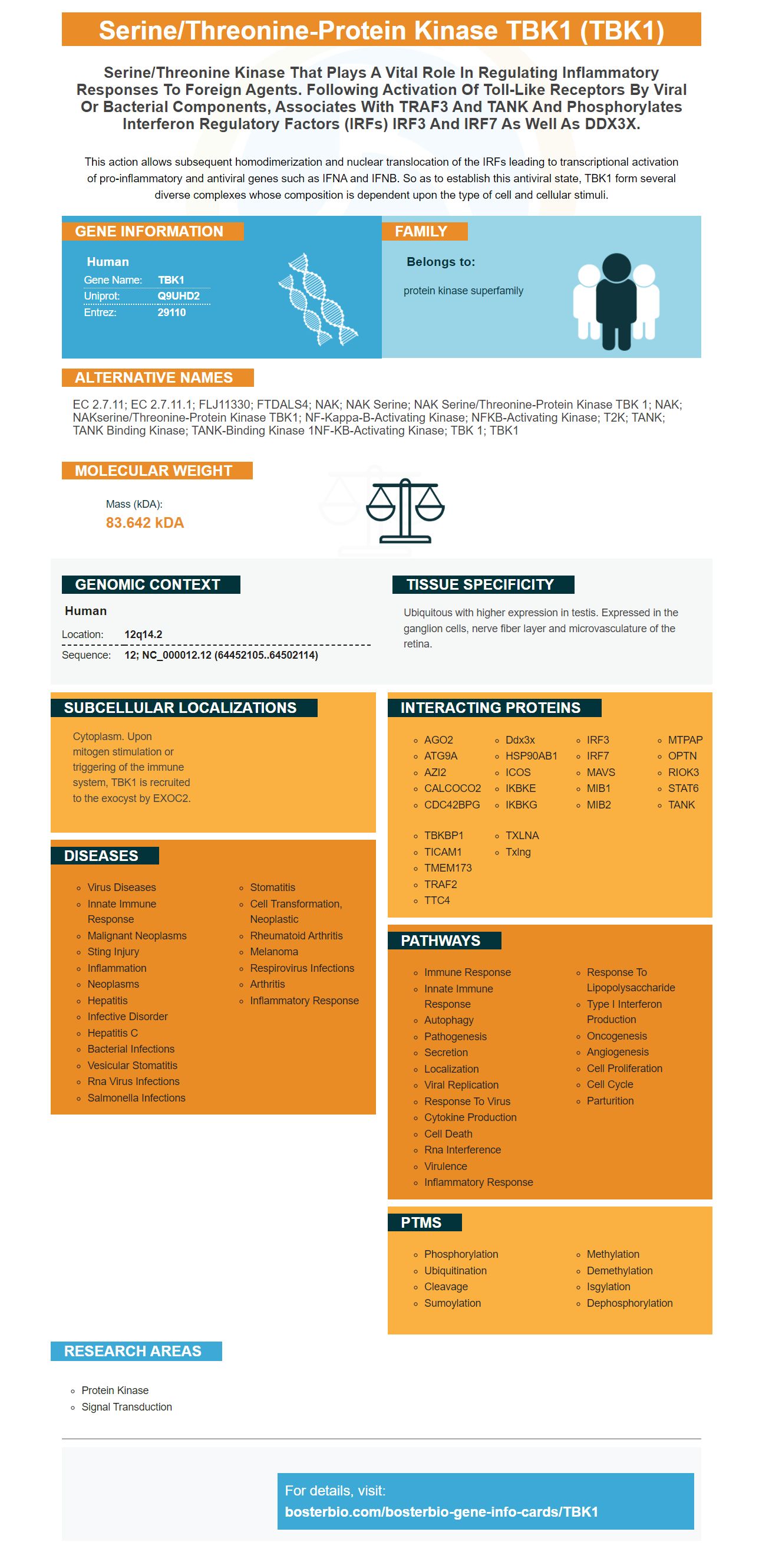
Facts about Serine/threonine-protein kinase TBK1.

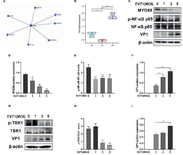
This action allows subsequent homodimerization and nuclear translocation of the IRFs leading to transcriptional activation of pro-inflammatory and antiviral genes such as IFNA and IFNB. So as to establish this antiviral state, TBK1 form several diverse complexes whose composition is dependent upon the type of cell and cellular stimuli.
| Human | |
|---|---|
| Gene Name: | TBK1 |
| Uniprot: | Q9UHD2 |
| Entrez: | 29110 |

| Belongs to: |
|---|
| protein kinase superfamily |

EC 2.7.11; EC 2.7.11.1; FLJ11330; FTDALS4; NAK; NAK serine; NAK serine/threonine-protein kinase TBK 1; NAK; NAKserine/threonine-protein kinase TBK1; NF-kappa-B-activating kinase; NFKB-Activating Kinase; T2K; TANK; TANK binding kinase; TANK-binding kinase 1NF-kB-activating kinase; TBK 1; TBK1
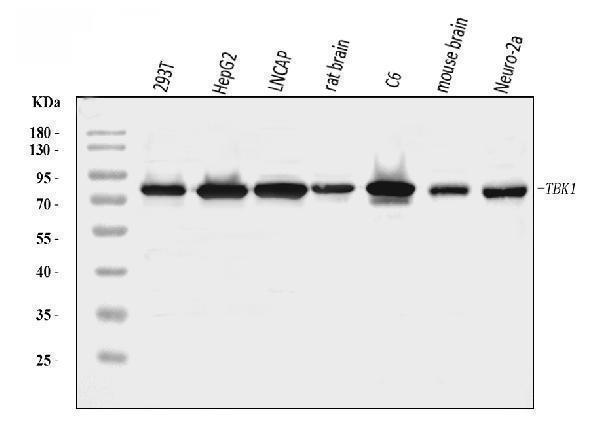
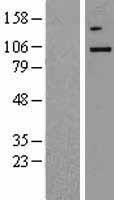
Mass (kDA):
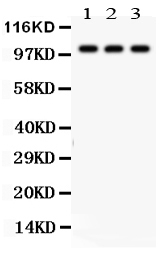
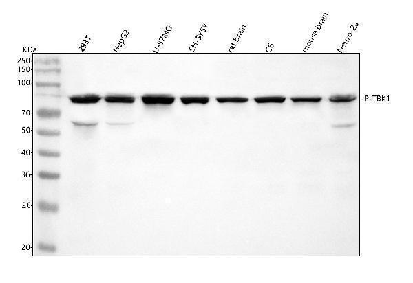
83.642 kDA

| Human | |
|---|---|
| Location: | 12q14.2 |
| Sequence: | 12; NC_000012.12 (64452105..64502114) |
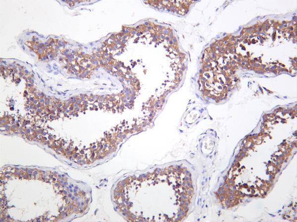
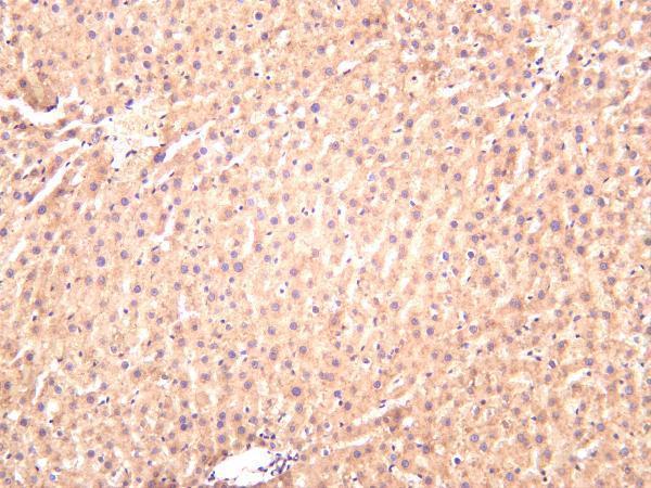
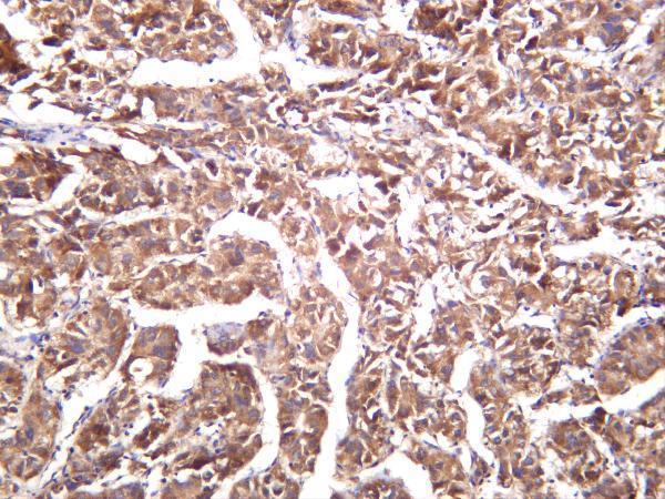
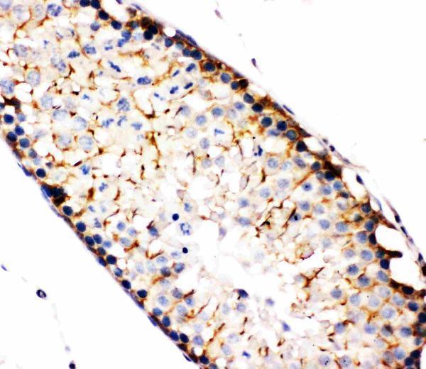
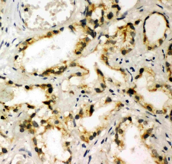
Ubiquitous with higher expression in testis. Expressed in the ganglion cells, nerve fiber layer and microvasculature of the retina.
Cytoplasm. Upon mitogen stimulation or triggering of the immune system, TBK1 is recruited to the exocyst by EXOC2.


PMID: 10581243 by Pomerantz J.L., et al. NF-kB activation by a signaling complex containing TRAF2, TANK, and TBK1, a novel IKK-related kinase.
PMID: 10783893 by Tojima Y., et al. NAK is an IkappaB kinase-activating kinase.