Click image to see more details
Product Info Summary
| SKU: | PA1416 |
|---|---|
| Size: | 100 μg/vial |
| Reactive Species: | Human, Mouse, Rat |
| Host: | Rabbit |
| Application: | IF, ICC, WB |
Customers Who Bought This Also Bought

Product info
Product Name
Anti-Protein NDRG1 NDRG1 Antibody Picoband®
SKU/Catalog Number
PA1416
BA3535 is an alternative SKU for this antibody, used in previous lots.
Size
100 μg/vial
Form
Lyophilized
Description
Boster Bio Anti-Protein NDRG1 NDRG1 Antibody catalog # PA1416. Tested in ICC/IF, WB applications. This antibody reacts with Human, Mouse, Rat. The brand Picoband indicates this is a premium antibody that guarantees superior quality, high affinity, and strong signals with minimal background in Western blot applications. Only our best-performing antibodies are designated as Picoband, ensuring unmatched performance.
Storage & Handling
Store at -20˚C for one year from date of receipt. After reconstitution, at 4˚C for one month. It can also be aliquotted and stored frozen at -20˚C for six months. Avoid repeated freeze-thaw cycles.
Cite This Product
Anti-Protein NDRG1 NDRG1 Antibody Picoband® (Boster Biological Technology, Pleasanton CA, USA, Catalog # PA1416)
Host
Rabbit
Contents
Each vial contains 4 mg Trehalose, 0.9 mg NaCl and 0.2 mg Na2HPO4.
Clonality
Polyclonal
Isotype
Rabbit IgG
Immunogen
A synthetic peptide corresponding to a sequence in the middle region of human NDRG1.
Cross-reactivity
No cross-reactivity with other proteins
Reactive Species
PA1416 is reactive to NDRG1 in Human, Mouse, Rat
Observed Molecular Weight
43 kDa
Calculated molecular weight
42.8 kDa
Background of NDRG1
Protein NDRG1 is a protein that in humans is encoded by the NDRG1 gene. It is a member of the N-myc downregulated gene family which belong to the alpha/beta hydrolase superfamily. The NDRG1 gene is mapped to chromosome 8q24. It has got a deduced 394 amino acid protein with a molecular mass of 43kD. NDRG1 is expressed in the cytoplasm and basolateral membranes of gut lumen surface epithelial cells. The protein appears to play a vital role in growth arrest and cell differentiation.
Antibody Validation
Boster validates all antibodies on WB, IHC, ICC, Immunofluorescence, and ELISA with known positive control and negative samples to ensure specificity and high affinity, including thorough antibody incubations.
Application & Images
Applications
PA1416 is guaranteed for IF, ICC, WB Boster Guarantee
Recommend Dilution
| Application | Dilution | Species |
|---|---|---|
| Western blot | 0.1-0.5μg/ml | Human, Mouse, Rat |
| Immunocytochemistry/Immunofluorescence | 5 μg/ml | Human |
Tested application
Suggested blocking solution with 5% non-fat milk or BSA; (*)Recommended protein loading: 20-40 µg per lane
Validation Images & Assay Conditions
Click image to see more details
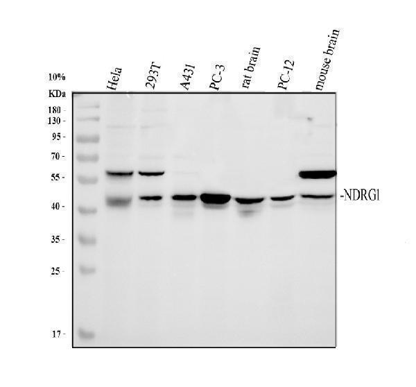
Western blot analysis of NDRG1 using anti-NDRG1 antibody (PA1416).
Electrophoresis was performed on a 10% SDS-PAGE gel at 80V (Stacking gel) / 120V (Resolving gel) for 2 hours. The sample well of each lane was loaded with 30 ug of sample under reducing conditions.
Lane 1: human Hela whole cell lysates,
Lane 2: human 293T whole cell lysates,
Lane 3: human A431 whole cell lysates,
Lane 4: human PC-3 whole cell lysates,
Lane 5: rat brain tissue lysates,
Lane 6: rat PC-12 whole cell lysates,
Lane 7: mouse brain tissue lysates.
After electrophoresis, proteins were transferred to a nitrocellulose membrane at 150 mA for 50-90 minutes. Blocked the membrane with 5% non-fat milk/TBS for 1.5 hour at RT. The membrane was incubated with rabbit anti-NDRG1 antigen affinity purified polyclonal antibody (PA1416) at 0.5 μg/mL overnight at 4°C, then washed with TBS-0.1%Tween 3 times with 5 minutes each and probed with a goat anti-rabbit IgG-HRP secondary antibody (Catalog # BA1054) at a dilution of 1:5000 for 1.5 hour at RT. The signal is developed using an ECL Plus Western Blotting Substrate (Catalog # AR1196-200) with Tanon 5200 system. A specific band was detected for NDRG1 at approximately 43 kDa. The expected band size for NDRG1 is at 43 kDa.

Click image to see more details
IF analysis of NDRG1 using anti-NDRG1 antibody (PA1416).
NDRG1 was detected in an immunocytochemical section of A431 cells. Enzyme antigen retrieval was performed using IHC enzyme antigen retrieval reagent (AR0022) for 15 mins. The cells were blocked with 10% goat serum. And then incubated with 5 μg/mL rabbit anti-NDRG1 Antibody (PA1416) overnight at 4°C. DyLight®488 Conjugated Goat Anti-Rabbit IgG (BA1127) was used as secondary antibody at 1:500 dilution and incubated for 30 minutes at 37°C. The section was counterstained with DAPI. Visualize using a fluorescence microscope and filter sets appropriate for the label used.
Specific Publications For Anti-Protein NDRG1 NDRG1 Antibody Picoband® (PA1416)
Loading publications
Recommended Resources
Here are featured tools and databases that you might find useful.
- Boster's Pathways Library
- Protein Databases
- Bioscience Research Protocol Resources
- Data Processing & Analysis Software
- Photo Editing Software
- Scientific Literature Resources
- Research Paper Management Tools
- Molecular Biology Software
- Primer Design Tools
- Bioinformatics Tools
- Phylogenetic Tree Analysis
Customer Reviews
Have you used Anti-Protein NDRG1 NDRG1 Antibody Picoband®?
Share your experimental results or join a short interview to earn up to $1,000 in product credits or other rewards.
0 Reviews For Anti-Protein NDRG1 NDRG1 Antibody Picoband®
Customer Q&As
Have a question?
Find answers in Q&As, reviews.
Can't find your answer?
Submit your question
1 Customer Q&As for Anti-Protein NDRG1 NDRG1 Antibody Picoband®
Question
We are currently using anti-NDRG1 antibody PA1416 for human tissue, and we are satisfied with the IHC results. The species of reactivity given in the datasheet says human. Is it possible that the antibody can work on primate tissues as well?
Verified Customer
Verified customer
Asked: 2020-04-27
Answer
The anti-NDRG1 antibody (PA1416) has not been validated for cross reactivity specifically with primate tissues, but there is a good chance of cross reactivity. We have an innovator award program that if you test this antibody and show it works in primate you can get your next antibody for free. Please contact me if I can help you with anything.
Boster Scientific Support
Answered: 2020-04-27
