Click image to see more details
Product Info Summary
| SKU: | A01070-2 |
|---|---|
| Size: | 100 μg/vial |
| Reactive Species: | Human, Mouse, Rat |
| Host: | Rabbit |
| Application: | ELISA, Flow Cytometry, WB |
Customers Who Bought This Also Bought

Product info
Product Name
Anti-nNOS (neuronal)/NOS1 Antibody Picoband®
SKU/Catalog Number
A01070-2
Size
100 μg/vial
Form
Lyophilized
Description
Boster Bio Anti-nNOS (neuronal)/NOS1 Antibody Picoband® catalog # A01070-2. Tested in ELISA, Flow Cytometry, WB applications. This antibody reacts with Human, Mouse, Rat. The brand Picoband indicates this is a premium antibody that guarantees superior quality, high affinity, and strong signals with minimal background in Western blot applications. Only our best-performing antibodies are designated as Picoband, ensuring unmatched performance.
Storage & Handling
At -20°C for one year from date of receipt. After reconstitution, at 4°C for one month. It can also be aliquotted and stored frozen at -20°C for six months. Avoid repeated freezing and thawing.
Cite This Product
Anti-nNOS (neuronal)/NOS1 Antibody Picoband® (Boster Biological Technology, Pleasanton CA, USA, Catalog # A01070-2)
Host
Rabbit
Contents
Each vial contains 4 mg Trehalose, 0.9 mg NaCl, 0.2 mg Na2HPO4.
Clonality
Polyclonal
Isotype
Rabbit IgG
Immunogen
E.coli-derived human nNOS (neuronal)/NOS1 recombinant protein (Position: R19-E1320).
Cross-reactivity
No cross-reactivity with other proteins.
Reactive Species
A01070-2 is reactive to NOS1 in Human, Mouse, Rat
Observed Molecular Weight
161 kDa
Calculated molecular weight
161.0 kDa
Background of NOS1
Nitric oxide synthase 1 (neuronal), also known as NOS1, is an enzyme that in humans is encoded by the NOS1 gene. The protein encoded by this gene belongs to the family of nitric oxide synthases, which synthesize nitric oxide from L-arginine. Nitric oxide is a reactive free radical, which acts as a biologic mediator in several processes, including neurotransmission, and antimicrobial and antitumoral activities. In the brain and peripheral nervous system, nitric oxide displays many properties of a neurotransmitter, and has been implicated in neurotoxicity associated with stroke and neurodegenerative diseases, neural regulation of smooth muscle, including peristalsis, and penile erection. This protein is ubiquitously expressed, with high level of expression in skeletal muscle. Multiple transcript variants that differ in the 5' UTR have been described for this gene but the full-length nature of these transcripts is not known. Additionally, alternatively spliced transcript variants encoding different isoforms (some testis-specific) have been found for this gene.
Antibody Validation
Boster validates all antibodies on WB, IHC, ICC, Immunofluorescence, and ELISA with known positive control and negative samples to ensure specificity and high affinity, including thorough antibody incubations.
Application & Images
Applications
A01070-2 is guaranteed for ELISA, Flow Cytometry, WB Boster Guarantee
Assay Dilutions Recommendation
The recommendations below provide a starting point for assay optimization. The actual working concentration varies and should be decided by the user.
Western blot, 0.25-0.5 μg/ml, Mouse, Rat
Flow Cytometry (Fixed), 1-3 μg/1x106 cells, Human
ELISA, 0.1-0.5 μg/ml, -
Positive Control
WB: rat brain tissue, mouse brain tissue
FCM: U87 cell
Validation Images & Assay Conditions
Click image to see more details
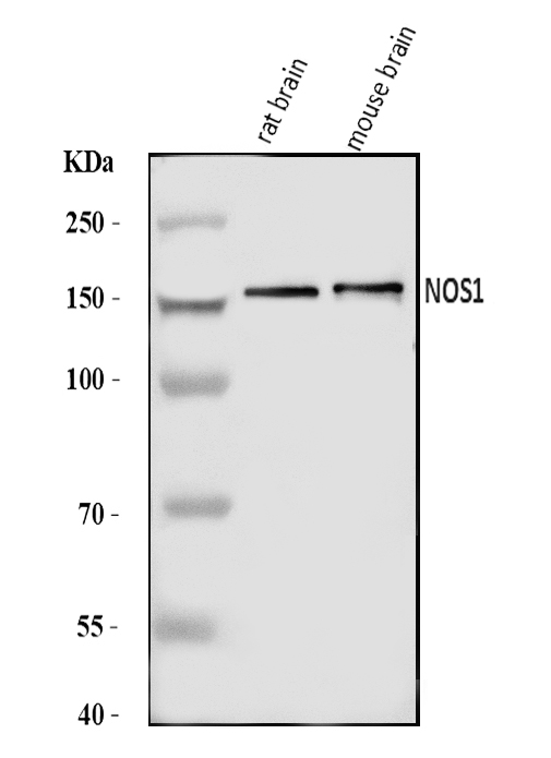
Western blot analysis of nNOS (neuronal)/NOS1 using anti-nNOS (neuronal)/NOS1 antibody (A01070-2).
Electrophoresis was performed on a 5-20% SDS-PAGE gel at 70V (Stacking gel) / 90V (Resolving gel) for 2-3 hours. The sample well of each lane was loaded with 30 ug of sample under reducing conditions.
Lane 1: rat brain tissue lysates,
Lane 2: mouse brain tissue lysates.
After electrophoresis, proteins were transferred to a nitrocellulose membrane at 150 mA for 50-90 minutes. Blocked the membrane with 5% non-fat milk/TBS for 1.5 hour at RT. The membrane was incubated with rabbit anti-nNOS (neuronal)/NOS1 antigen affinity purified polyclonal antibody (Catalog # A01070-2) at 0.5 μg/mL overnight at 4°C, then washed with TBS-0.1%Tween 3 times with 5 minutes each and probed with a goat anti-rabbit IgG-HRP secondary antibody at a dilution of 1:5000 for 1.5 hour at RT. The signal is developed using an Enhanced Chemiluminescent detection (ECL) kit (Catalog # EK1002) with Tanon 5200 system. A specific band was detected for nNOS (neuronal)/NOS1 at approximately 161 kDa. The expected band size for nNOS (neuronal)/NOS1 is at 161 kDa.

Click image to see more details
Effect of exercise on alcohol induced neuronal damage. ( a,b ) Representative western blot analysis showing the levels of neuronal proteins (NeuN and NSC) in different mice groups ( a ). Histogram showing the quantitative estimation of nNOS and NSE proteins after normalization with GAPDH (b). ( c,d ) Representative images showing coronal slices of mice brains stained with cresyl violet (40× magnification) ( c ). Scatter dot plot showing the number of cresyl violet positive cells in different groups of mice ( d ). ( e,f ) Representative images showing Fluoro-Jade C (FJC) staining in brain sections of the different groups of mice (10× magnification). A marked decrease of FJC-stained degenerating neurons (arrows) were observed in CT, EX and AL+EX groups, indicating a lesser degree of neuronal cell death. Brain sections of AL treated mice showing a greater number of FJC-positive neurons (arrows), reflecting increased neuronal cell death ( e ). Scatter dot plot showing the numbers of degenerating neurons in different experimental mice groups ( f ). All the data are represented as mean values ± standard error (SE) in 5 independent experiments. * ,# p < 0.05 considered significant. *p < 0.05 vs. CT and # p < 0.05 vs. AL group. Uncropped blots for a are presented in Supplementary Fig. .
Index in PubMed under a CC BY license. PMID: 29581524

Click image to see more details
Flow Cytometry analysis of U87 cells using anti-nNOS (neuronal)/NOS1 antibody (A01070-2).
Overlay histogram showing U87 cells stained with A01070-2 (Blue line). The cells were fixed with 4% paraformaldehyde and blocked with 10% normal goat serum. And then incubated with rabbit anti-nNOS (neuronal)/NOS1 Antibody (A01070-2, 1 μg/1x106 cells) for 30 min at 20°C. DyLight®488 conjugated goat anti-rabbit IgG (BA1127, 5-10 μg/1x106 cells) was used as secondary antibody for 30 minutes at 20°C. Isotype control antibody (Green line) was rabbit IgG (1 μg/1x106) used under the same conditions. Unlabelled sample without incubation with primary antibody and secondary antibody (Red line) was used as a blank control.
Specific Publications For Anti-nNOS (neuronal)/NOS1 Antibody Picoband® (A01070-2)
Loading publications
Recommended Resources
Here are featured tools and databases that you might find useful.
- Boster's Pathways Library
- Protein Databases
- Bioscience Research Protocol Resources
- Data Processing & Analysis Software
- Photo Editing Software
- Scientific Literature Resources
- Research Paper Management Tools
- Molecular Biology Software
- Primer Design Tools
- Bioinformatics Tools
- Phylogenetic Tree Analysis
Customer Reviews
Have you used Anti-nNOS (neuronal)/NOS1 Antibody Picoband®?
Share your experimental results or join a short interview to earn up to $1,000 in product credits or other rewards.
0 Reviews For Anti-nNOS (neuronal)/NOS1 Antibody Picoband®
Customer Q&As
Have a question?
Find answers in Q&As, reviews.
Can't find your answer?
Submit your question
