Click image to see more details
-

-
-
-
-
+17
Product Info Summary
| SKU: | M03515-1 |
|---|---|
| Size: | 100 µl |
| Reactive Species: | Dog, Human, Monkey, Mouse, Rat |
| Host: | Mouse |
| Application: | Flow Cytometry, IF, IHC, WB |
Customers Who Bought This Also Bought

Product info
Product Name
Anti-NONO Mouse Monoclonal Antibody [Clone ID: OTI4D9]
SKU/Catalog Number
M03515-1
Size
100 µl
Description
Boster Bio NONO mouse monoclonal antibody, clone OTI4D9 (formerly 4D9). Catalog# M03515-1. Tested in FC, IF, IHC, WB. This antibody reacts with Human, Monkey, Mouse, Rat, Dog.
Storage & Handling
Store at -20°C for one year. For short term storage and frequent use, store at 4°C for up to one month. Avoid repeated freeze-thaw cycles.
Cite This Product
Anti-NONO Mouse Monoclonal Antibody [Clone ID: OTI4D9] (Boster Biological Technology, Pleasanton CA, USA, Catalog # M03515-1)
Host
Mouse
Contents
PBS (pH 7.3) containing 1% stabilizing protein, 50% glycerol and 0.02% sodium azide.
This antibody is supplied in a stabilized formulation.
Compatibility with conjugation reactions depends on the chemistry of the conjugation method used.
For conjugation methods that are not compatible with the stabilizing components present in this formulation, a carrier-free antibody format is required.
Clonality
Monoclonal
Clone Number
OTI4D9
Isotype
IgG1
Immunogen
Human recombinant protein fragment correspongding to amino acids 184-385 of human NONO (NP_031389) produced in E.coli.
Reactive Species
M03515-1 is reactive to NONO in Dog, Human, Monkey, Mouse, Rat
Calculated molecular weight
54.2 kDa
Antibody Validation
Boster validates all antibodies on WB, IHC, ICC, Immunofluorescence, and ELISA with known positive control and negative samples to ensure specificity and high affinity, including thorough antibody incubations.
Application & Images
Applications
M03515-1 is guaranteed for Flow Cytometry, IF, IHC, WB Boster Guarantee
Recommend Dilution
WB 1:500~2000
IHC 1:150
IF 1:100
Flow Cytometry 1:100
Validation Images & Assay Conditions
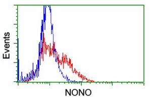
Click image to see more details
HEK293T cells transfected with either NONO (Myc-DDK-tagged) overexpress plasmid (Red) or empty vector control plasmid (Blue) were immunostained by anti-NONO antibody (M03515-1)

Click image to see more details
Anti-NONO mouse monoclonal antibody (M03515-1) immunofluorescent staining of COS7 cells transiently transfected by pCMV6-ENTRY NONO.

Click image to see more details
Immunohistochemical staining of paraffin-embedded Carcinoma of Human thyroid tissue using anti-NONO mouse monoclonal antibody. (Heat-induced epitope retrieval by 10mM citric buffer

Click image to see more details
Immunohistochemical staining of paraffin-embedded Adenocarcinoma of Human endometrium tissue using anti-NONO mouse monoclonal antibody. (Heat-induced epitope retrieval by 10mM citric buffer

Click image to see more details
Immunohistochemical staining of paraffin-embedded Human prostate tissue within the normal limits using anti-NONO mouse monoclonal antibody. (Heat-induced epitope retrieval by 10mM citric buffer

Click image to see more details
Immunohistochemical staining of paraffin-embedded Carcinoma of Human prostate tissue using anti-NONO mouse monoclonal antibody. (Heat-induced epitope retrieval by 10mM citric buffer

Click image to see more details
Immunohistochemical staining of paraffin-embedded Carcinoma of Human bladder tissue using anti-NONO mouse monoclonal antibody. (Heat-induced epitope retrieval by 10mM citric buffer

Click image to see more details
Immunohistochemical staining of paraffin-embedded Human lymphoma tissue using anti-NONO mouse monoclonal antibody. (Heat-induced epitope retrieval by 10mM citric buffer

Click image to see more details
Immunohistochemical staining of paraffin-embedded Human Kidney tissue within the normal limits using anti-NONO mouse monoclonal antibody. (Heat-induced epitope retrieval by 10mM citric buffer

Click image to see more details
Immunohistochemical staining of paraffin-embedded Carcinoma of Human kidney tissue using anti-NONO mouse monoclonal antibody. (Heat-induced epitope retrieval by 10mM citric buffer

Click image to see more details
Immunohistochemical staining of paraffin-embedded Human liver tissue within the normal limits using anti-NONO mouse monoclonal antibody. (Heat-induced epitope retrieval by 10mM citric buffer

Click image to see more details
Immunohistochemical staining of paraffin-embedded Carcinoma of Human liver tissue using anti-NONO mouse monoclonal antibody. (Heat-induced epitope retrieval by 10mM citric buffer

Click image to see more details
Immunohistochemical staining of paraffin-embedded Carcinoma of Human lung tissue using anti-NONO mouse monoclonal antibody. (Heat-induced epitope retrieval by 10mM citric buffer

Click image to see more details
Immunohistochemical staining of paraffin-embedded Human Ovary tissue within the normal limits using anti-NONO mouse monoclonal antibody. (Heat-induced epitope retrieval by 10mM citric buffer

Click image to see more details
Immunohistochemical staining of paraffin-embedded Adenocarcinoma of Human ovary tissue using anti-NONO mouse monoclonal antibody. (Heat-induced epitope retrieval by 10mM citric buffer

Click image to see more details
Immunohistochemical staining of paraffin-embedded Human pancreas tissue within the normal limits using anti-NONO mouse monoclonal antibody. (Heat-induced epitope retrieval by 10mM citric buffer

Click image to see more details
HEK293T cells were transfected with the pCMV6-ENTRY control (Left lane) or pCMV6-ENTRY NONO (Right lane) cDNA for 48 hrs and lysed. Equivalent amounts of cell lysates (5 ug per lane) were separated by SDS-PAGE and immunoblotted with anti-NONO.

Click image to see more details
Western blot analysis of extracts (10ug) from 9 Human tissue by using anti-NONO monoclonal antibody at 1:200 (1: Testis; 2: Omentum; 3: Uterus; 4: Breast; 5: Brain; 6: Liver; 7: Ovary; 8: Thyroid gland; 9: Colon).

Click image to see more details
Western blot analysis of extracts (35ug) from 9 different cell lines by usin g anti-NONO monoclonal antibody (HepG2: human; HeLa: human; SVT2: mouse; A549: human; COS7: monkey; Jurkat: human; MDCK: canine; PC12: rat; MCF7: human).

Click image to see more details
Equivalent amounts of cell lysates (10 ug per lane) of wild-type HEK293T cells (WT) and NONO-Knockout HEK293T cells (KO) were separated by SDS-PAGE and immunoblotted with anti-NONO monoclonal antibody M03515-1 (1:500). Then the blotted membrane was stripped and reprobed with anti-PCNA antibody as a loading control.

Click image to see more details
Western blot analysis of extracts (10ug) from a mouse cell line and 3 different mouse tissues by using anti-NONO monoclonal antibody (1:200).
Specific Publications For Anti-NONO Mouse Monoclonal Antibody [Clone ID: OTI4D9] (M03515-1)
Loading publications
Recommended Resources
Here are featured tools and databases that you might find useful.
- Boster's Pathways Library
- Protein Databases
- Bioscience Research Protocol Resources
- Data Processing & Analysis Software
- Photo Editing Software
- Scientific Literature Resources
- Research Paper Management Tools
- Molecular Biology Software
- Primer Design Tools
- Bioinformatics Tools
- Phylogenetic Tree Analysis
Customer Reviews
Have you used Anti-NONO Mouse Monoclonal Antibody [Clone ID: OTI4D9]?
Share your experimental results or join a short interview to earn up to $1,000 in product credits or other rewards.
0 Reviews For Anti-NONO Mouse Monoclonal Antibody [Clone ID: OTI4D9]
Customer Q&As
Have a question?
Find answers in Q&As, reviews.
Can't find your answer?
Submit your question
