Click image to see more details
-

-
-
-
-
+15
Product Info Summary
| SKU: | PB9309 |
|---|---|
| Size: | 100 μg/vial |
| Reactive Species: | Human, Mouse, Rat |
| Host: | Rabbit |
| Application: | Flow Cytometry, IF, IHC, ICC, WB |
Customers Who Bought This Also Bought

Product info
Product Name
Anti-PARP/PARP1 Antibody Picoband®
SKU/Catalog Number
PB9309
Size
100 μg/vial
Form
Lyophilized
Description
Boster Bio Anti-PARP/PARP1 Antibody Picoband® catalog # PB9309. Tested in Flow Cytometry, IF, IHC, ICC, WB applications. This antibody reacts with Human, Mouse, Rat. The brand Picoband indicates this is a premium antibody that guarantees superior quality, high affinity, and strong signals with minimal background in Western blot applications. Only our best-performing antibodies are designated as Picoband, ensuring unmatched performance.
Storage & Handling
Store at -20˚C for one year from date of receipt. After reconstitution, at 4˚C for one month. It can also be aliquotted and stored frozen at -20˚C for six months. Avoid repeated freeze-thaw cycles.
Cite This Product
Anti-PARP/PARP1 Antibody Picoband® (Boster Biological Technology, Pleasanton CA, USA, Catalog # PB9309)
Host
Rabbit
Contents
Each vial contains antibody formulated with stabilizing components, 0.9 mg NaCl, 0.2 mg Na2HPO4, and 0.05 mg NaN3.
*This antibody is supplied in a stabilized formulation.
Compatibility with conjugation reactions depends on the chemistry of the conjugation method used.
For conjugation methods that are not compatible with the stabilizing components present in this formulation, a carrier-free antibody format is required.
Clonality
Polyclonal
Isotype
Rabbit IgG
Immunogen
E.coli-derived human PARP recombinant protein (Position: Q670-R858). Human PARP shares 94% and 95% amino acid (aa) sequence identity with mouse and rat PARP, respectively.
Cross-reactivity
No cross-reactivity with other proteins
Reactive Species
PB9309 is reactive to PARP1 in Human, Mouse, Rat
Observed Molecular Weight
120 kDa
Calculated molecular weight
113.1 kDa
Background of PARP1
Poly [ADP-ribose] polymerase 1 (PARP1), also known as ADPRT or PPOL is an enzyme that in humans is encoded by the PARP1 gene. PARP1 gene is mapped to 1q42.12. This gene encodes a chromatin-associated enzyme, poly (ADP-ribosyl)transferase, which modifies various nuclear proteins by poly (ADP-ribosyl)ation. The modification is dependent on DNA and is involved in the regulation of various important cellular processes such as differentiation, proliferation, and tumor transformation and also in the regulation of the molecular events involved in the recovery of cell from DNA damage. In addition, this enzyme may be the site of mutation in Fanconi anemia, and may participate in the pathophysiology of type I diabetes.
Antibody Validation
Boster validates all antibodies on WB, IHC, ICC, Immunofluorescence, and ELISA with known positive control and negative samples to ensure specificity and high affinity, including thorough antibody incubations.
Application & Images
Applications
PB9309 is guaranteed for Flow Cytometry, IF, IHC, ICC, WB Boster Guarantee
Recommend Dilution
| Application | Dilution | Species |
|---|---|---|
| Western blot | 0.1-0.5μg/ml | Human, Mouse, Rat |
| Immunohistochemistry (Paraffin-embedded Section) | 0.5-1μg/ml | Human, Mouse, Rat |
| Immunocytochemistry | 2μg/ml | Human, Rat |
| Immunofluorescence | 2μg/ml | Human |
| Flow Cytometry (Fixed) | 1-3μg/1x106 cells | Human |
Tested application
Suggested blocking solution with 5% non-fat milk or BSA; (*)Recommended protein loading: 20-40 µg per lane
Use TE buffer pH 9.0 for antigen retrieval; (*) citrate buffer pH 6.0 is an alternative.
Validation Images & Assay Conditions
Click image to see more details
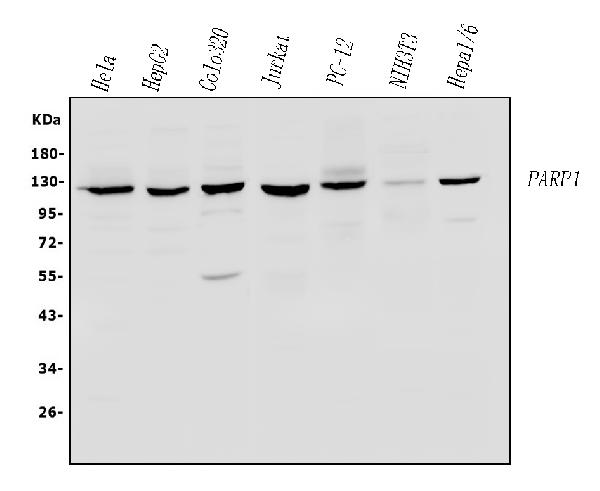
Western blot analysis of PARP using anti-PARP antibody (PB9309).
Electrophoresis was performed on a 5-20% SDS-PAGE gel at 70V (Stacking gel) / 90V (Resolving gel) for 2-3 hours. The sample well of each lane was loaded with 50ug of sample under reducing conditions.
Lane 1: human Hela whole cell lysates,
Lane 2: human HepG2 whole cell lysates,
Lane 3: human COLO-320 whole cell lysates,
Lane 4: human Jurkat whole cell lysates,
Lane 5: rat PC-12 whole cell lysates,
Lane 6: mouse NIH3T3 whole cell lysates,
Lane 7: mouse HEPA1-6 whole cell lysates.
After Electrophoresis, proteins were transferred to a Nitrocellulose membrane at 150mA for 50-90 minutes. Blocked the membrane with 5% Non-fat Milk/ TBS for 1.5 hour at RT. The membrane was incubated with rabbit anti-PARP antigen affinity purified polyclonal antibody (Catalog # PB9309) at 0.5 μg/mL overnight at 4°C, then washed with TBS-0.1%Tween 3 times with 5 minutes each and probed with a goat anti-rabbit IgG-HRP secondary antibody at a dilution of 1:10000 for 1.5 hour at RT. The signal is developed using an Enhanced Chemiluminescent detection (ECL) kit (Catalog # EK1002) with Tanon 5200 system. A specific band was detected for PARP at approximately 120KD. The expected band size for PARP is at 113KD.

Click image to see more details
Western blot analysis of PARP1 using anti-PARP1 antibody (PB9309).
Electrophoresis was performed on a 8% SDS-PAGE gel at 80V (Stacking gel) / 120V (Resolving gel) for 2 hours. The sample well of each lane was loaded with 30 ug of sample under reducing conditions.
Lane 1: human Hela- WT whole cell lysates,
Lane 2: human Hela-GPX4 KO whole cell lysates.
After electrophoresis, proteins were transferred to a nitrocellulose membrane at 150 mA for 50-90 minutes. Blocked the membrane with 5% non-fat milk/TBS for 1.5 hour at RT. Then the membrane was incubated with rabbit anti-PARP1 antigen affinity purified polyclonal antibody (PB9309) at 0.5 μg/mL overnight at 4°C, then washed with TBS-0.1%Tween 3 times with 5 minutes each and probed with a goat anti-rabbit IgG-HRP secondary antibody (Catalog # BA1054) at a dilution of 1:5000 for 1.5 hour at RT. The signal is developed using an ECL Plus Western Blotting Substrate (Catalog # AR1196-200) with Tanon 5200 system. A specific band was detected for PARP1 at approximately 118 kDa. The expected band size for PARP1 is at 118 kDa.

Click image to see more details
IHC analysis of PARP using anti-PARP antibody (PB9309).
PARP was detected in paraffin-embedded section of Mouse Intestine Tissue. Heat mediated antigen retrieval was performed in citrate buffer (pH6, epitope retrieval solution) for 20 mins. The tissue section was blocked with 10% goat serum. The tissue section was then incubated with 1μg/ml rabbit anti-PARP Antibody (PB9309) overnight at 4°C. Biotinylated goat anti-rabbit IgG was used as secondary antibody and incubated for 30 minutes at 37°C. The tissue section was developed using Strepavidin-Biotin-Complex (SABC)(Catalog # SA1022) with DAB as the chromogen.

Click image to see more details
IHC analysis of PARP using anti-PARP antibody (PB9309).
PARP was detected in paraffin-embedded section of Human Placenta Tissue. Heat mediated antigen retrieval was performed in citrate buffer (pH6, epitope retrieval solution) for 20 mins. The tissue section was blocked with 10% goat serum. The tissue section was then incubated with 1μg/ml rabbit anti-PARP Antibody (PB9309) overnight at 4°C. Biotinylated goat anti-rabbit IgG was used as secondary antibody and incubated for 30 minutes at 37°C. The tissue section was developed using Strepavidin-Biotin-Complex (SABC)(Catalog # SA1022) with DAB as the chromogen.

Click image to see more details
IHC analysis of PARP using anti-PARP antibody (PB9309).
PARP was detected in immunocytochemical section of A549 Cell. Enzyme antigen retrieval was performed using IHC enzyme antigen retrieval reagent (AR0022) for 15 mins. The cells were blocked with 10% goat serum. And then incubated with 1μg/ml rabbit anti-PARP Antibody (PB9309) overnight at 4°C. Biotinylated goat anti-rabbit IgG was used as secondary antibody and incubated for 30 minutes at 37°C. The section was developed using Strepavidin-Biotin-Complex (SABC)(Catalog # SA1022) with DAB as the chromogen.

Click image to see more details
IHC analysis of PAPR using anti-PAPR antibody (PB9309).
PAPR was detected in a paraffin-embedded section of mouse liver tissue. Heat mediated antigen retrieval was performed in EDTA buffer (pH 8.0, epitope retrieval solution). The tissue section was blocked with 10% goat serum. The tissue section was then incubated with 5 μg/ml rabbit anti-PAPR Antibody (PB9309) overnight at 4°C. HRP-AffiniPure Goat Anti-Rabbit IgG was used as secondary antibody and incubated for 30 minutes at 37°C. The tissue section was developed using HRP Conjugated Rabbit IgG Super Vision Assay Kit (Catalog # SV0002) with DAB as the chromogen.

Click image to see more details
IHC analysis of PAPR using anti-PAPR antibody (PB9309).
PAPR was detected in a paraffin-embedded section of rat kidney tissue. Heat mediated antigen retrieval was performed in EDTA buffer (pH 8.0, epitope retrieval solution). The tissue section was blocked with 10% goat serum. The tissue section was then incubated with 5 μg/ml rabbit anti-PAPR Antibody (PB9309) overnight at 4°C. HRP-AffiniPure Goat Anti-Rabbit IgG was used as secondary antibody and incubated for 30 minutes at 37°C. The tissue section was developed using HRP Conjugated Rabbit IgG Super Vision Assay Kit (Catalog # SV0002) with DAB as the chromogen.

Click image to see more details
IHC analysis of PAPR using anti-PAPR antibody (PB9309).
PAPR was detected in a paraffin-embedded section of rat spleen tissue. Heat mediated antigen retrieval was performed in EDTA buffer (pH 8.0, epitope retrieval solution). The tissue section was blocked with 10% goat serum. The tissue section was then incubated with 5 μg/ml rabbit anti-PAPR Antibody (PB9309) overnight at 4°C. HRP-AffiniPure Goat Anti-Rabbit IgG was used as secondary antibody and incubated for 30 minutes at 37°C. The tissue section was developed using HRP Conjugated Rabbit IgG Super Vision Assay Kit (Catalog # SV0002) with DAB as the chromogen.

Click image to see more details
IHC analysis of PAPR using anti-PAPR antibody (PB9309).
PAPR was detected in a paraffin-embedded section of human tosil tissue. Heat mediated antigen retrieval was performed in EDTA buffer (pH 8.0, epitope retrieval solution). The tissue section was blocked with 10% goat serum. The tissue section was then incubated with 5 μg/ml rabbit anti-PAPR Antibody (PB9309) overnight at 4°C. HRP-AffiniPure Goat Anti-Rabbit IgG was used as secondary antibody and incubated for 30 minutes at 37°C. The tissue section was developed using HRP Conjugated Rabbit IgG Super Vision Assay Kit (Catalog # SV0002) with DAB as the chromogen.

Click image to see more details
IHC analysis of PAPR using anti-PAPR antibody (PB9309).
PAPR was detected in a paraffin-embedded section of mouse kidney tissue. Heat mediated antigen retrieval was performed in EDTA buffer (pH 8.0, epitope retrieval solution). The tissue section was blocked with 10% goat serum. The tissue section was then incubated with 5 μg/ml rabbit anti-PAPR Antibody (PB9309) overnight at 4°C. HRP-AffiniPure Goat Anti-Rabbit IgG was used as secondary antibody and incubated for 30 minutes at 37°C. The tissue section was developed using HRP Conjugated Rabbit IgG Super Vision Assay Kit (Catalog # SV0002) with DAB as the chromogen.

Click image to see more details
IHC analysis of PAPR using anti-PAPR antibody (PB9309).
PAPR was detected in a paraffin-embedded section of human bladder cancer tissue. Heat mediated antigen retrieval was performed in EDTA buffer (pH 8.0, epitope retrieval solution). The tissue section was blocked with 10% goat serum. The tissue section was then incubated with 5 μg/ml rabbit anti-PAPR Antibody (PB9309) overnight at 4°C. HRP-AffiniPure Goat Anti-Rabbit IgG was used as secondary antibody and incubated for 30 minutes at 37°C. The tissue section was developed using HRP Conjugated Rabbit IgG Super Vision Assay Kit (Catalog # SV0002) with DAB as the chromogen.

Click image to see more details
IHC analysis of PAPR using anti-PAPR antibody (PB9309).
PAPR was detected in a paraffin-embedded section of human stomach cancer tissue. Heat mediated antigen retrieval was performed in EDTA buffer (pH 8.0, epitope retrieval solution). The tissue section was blocked with 10% goat serum. The tissue section was then incubated with 5 μg/ml rabbit anti-PAPR Antibody (PB9309) overnight at 4°C. HRP-AffiniPure Goat Anti-Rabbit IgG was used as secondary antibody and incubated for 30 minutes at 37°C. The tissue section was developed using HRP Conjugated Rabbit IgG Super Vision Assay Kit (Catalog # SV0002) with DAB as the chromogen.

Click image to see more details
IHC analysis of PAPR using anti-PAPR antibody (PB9309).
PAPR was detected in a paraffin-embedded section of human pancreas cancer tissue. Heat mediated antigen retrieval was performed in EDTA buffer (pH 8.0, epitope retrieval solution). The tissue section was blocked with 10% goat serum. The tissue section was then incubated with 5 μg/ml rabbit anti-PAPR Antibody (PB9309) overnight at 4°C. HRP-AffiniPure Goat Anti-Rabbit IgG was used as secondary antibody and incubated for 30 minutes at 37°C. The tissue section was developed using HRP Conjugated Rabbit IgG Super Vision Assay Kit (Catalog # SV0002) with DAB as the chromogen.

Click image to see more details
IF analysis of PARP using anti-PARP antibody (PB9309).
PARP was detected in a paraffin-embedded section of human bladder cancer tissue. Heat mediated antigen retrieval was performed in EDTA buffer (pH 8.0, epitope retrieval solution). The tissue section was blocked with 10% goat serum. The tissue section was then incubated with 5 μg/mL rabbit anti-PARP Antibody (PB9309) overnight at 4°C. DyLight 594 Conjugated AffiniPure Goat Anti-rabbit IgG(H+L) (BA1142) was used as secondary antibody at 1:100 dilution and incubated for 30 minutes at 37°C. The section was counterstained with DAPI. Visualize using a fluorescence microscope and filter sets appropriate for the label used.

Click image to see more details
IF analysis of PARP using anti-PARP antibody (PB9309).
PARP was detected in a paraffin-embedded section of human pancreas cancer tissue. Heat mediated antigen retrieval was performed in EDTA buffer (pH 8.0, epitope retrieval solution). The tissue section was blocked with 10% goat serum. The tissue section was then incubated with 5 μg/mL rabbit anti-PARP Antibody (PB9309) overnight at 4°C. DyLight 594 Conjugated AffiniPure Goat Anti-rabbit IgG(H+L) (BA1142) was used as secondary antibody at 1:100 dilution and incubated for 30 minutes at 37°C. The section was counterstained with DAPI. Visualize using a fluorescence microscope and filter sets appropriate for the label used.

Click image to see more details
IF analysis of PARP using anti-PARP antibody (PB9309).
PARP was detected in a paraffin-embedded section of human stomach cancer tissue. Heat mediated antigen retrieval was performed in EDTA buffer (pH 8.0, epitope retrieval solution). The tissue section was blocked with 10% goat serum. The tissue section was then incubated with 5 μg/mL rabbit anti-PARP Antibody (PB9309) overnight at 4°C. DyLight 594 Conjugated AffiniPure Goat Anti-rabbit IgG(H+L) (BA1142) was used as secondary antibody at 1:100 dilution and incubated for 30 minutes at 37°C. The section was counterstained with DAPI. Visualize using a fluorescence microscope and filter sets appropriate for the label used.

Click image to see more details
IF analysis of PARP using anti-PARP antibody (PB9309).
PARP was detected in a paraffin-embedded section of human breast cancer tissue. Heat mediated antigen retrieval was performed in EDTA buffer (pH 8.0, epitope retrieval solution). The tissue section was blocked with 10% goat serum. The tissue section was then incubated with 2 μg/mL rabbit anti-PARP Antibody (PB9309) overnight at 4°C. Biotin conjugated goat anti-rabbit IgG (BA1003) was used as secondary antibody and incubated for 30 minutes at 37°C. The tissue section was developed using Cy3 Conjugated Avidin (BA1037). The section was counterstained with DAPI. Visualize using a fluorescence microscope and filter sets appropriate for the label used.

Click image to see more details
Flow Cytometry analysis of A431 cells using anti-PARP antibody (PB9309).
Overlay histogram showing A431 cells stained with PB9309 (Blue line). To facilitate intracellular staining, cells were fixed with 4% paraformaldehyde and permeabilized with permeabilization buffer. The cells were blocked with 10% normal goat serum. And then incubated with rabbit anti-PARP Antibody (PB9309, 1 μg/1x106 cells) for 30 min at 20°C. DyLight®488 conjugated goat anti-rabbit IgG (BA1127, 5-10 μg/1x106 cells) was used as secondary antibody for 30 minutes at 20°C. Isotype control antibody (Green line) was rabbit IgG (1 μg/1x106) used under the same conditions. Unlabelled sample without incubation with primary antibody and secondary antibody (Red line) was used as a blank control.
Specific Publications For Anti-PARP/PARP1 Antibody Picoband® (PB9309)
Loading publications
Recommended Resources
Here are featured tools and databases that you might find useful.
- Boster's Pathways Library
- Protein Databases
- Bioscience Research Protocol Resources
- Data Processing & Analysis Software
- Photo Editing Software
- Scientific Literature Resources
- Research Paper Management Tools
- Molecular Biology Software
- Primer Design Tools
- Bioinformatics Tools
- Phylogenetic Tree Analysis
Customer Reviews
Have you used Anti-PARP/PARP1 Antibody Picoband®?
Share your experimental results or join a short interview to earn up to $1,000 in product credits or other rewards.
0 Reviews For Anti-PARP/PARP1 Antibody Picoband®
Customer Q&As
Have a question?
Find answers in Q&As, reviews.
Can't find your answer?
Submit your question
5 Customer Q&As for Anti-PARP/PARP1 Antibody Picoband®
Question
We purchased anti-PARP/PARP1 antibody for ICC on leukemic t-cell in the past. I am using mouse, and We want to use the antibody for IHC-P next. I was wanting to use examining leukemic t-cell as well as cervix carcinoma in our next experiment. Could you please give me some suggestion on which antibody would work the best for IHC-P?
Verified Customer
Verified customer
Asked: 2020-04-23
Answer
I looked at the website and datasheets of our anti-PARP/PARP1 antibody and it seems that PB9309 has been tested on mouse in both ICC and IHC-P. Thus PB9309 should work for your application. Our Boster satisfaction guarantee will cover this product for IHC-P in mouse even if the specific tissue type has not been validated. We do have a comprehensive range of products for IHC-P detection and you can check out our website bosterbio.com to find out more information about them.
Boster Scientific Support
Answered: 2020-04-23
Question
My team were well pleased with the WB result of your anti-PARP/PARP1 antibody. However we have seen positive staining in fibroblast nucleus using this antibody. Is that expected? Could you tell me where is PARP1 supposed to be expressed?
Verified Customer
Verified customer
Asked: 2020-04-23
Answer
Based on literature, fibroblast does express PARP1. Generally PARP1 expresses in nucleus. Regarding which tissues have PARP1 expression, here are a few articles citing expression in various tissues:
Brain, Pubmed ID: 15489334
Cervix carcinoma, Pubmed ID: 18669648, 20068231
Cervix carcinoma, and Erythroleukemia, Pubmed ID: 23186163
Colon carcinoma, and Ovarian carcinoma, Pubmed ID: 17177976
Embryonic kidney, Pubmed ID: 17525332
Fibroblast, Pubmed ID: 3120710, 2824474, 2891139
Leukemic T-cell, Pubmed ID: 19690332
Liver, Pubmed ID: 24275569
Boster Scientific Support
Answered: 2020-04-23
Question
We have observed staining in human heart left ventricle. Do you have any suggestions? Is anti-PARP/PARP1 antibody supposed to stain heart left ventricle positively?
Verified Customer
Verified customer
Asked: 2019-06-13
Answer
Based on literature heart left ventricle does express PARP1. Based on Uniprot.org, PARP1 is expressed in heart left ventricle, fibroblast, brain, colon carcinoma ovarian carcinoma, embryonic kidney, cervix carcinoma, leukemic t-cell, cervix carcinoma erythroleukemia, liver, among other tissues. Regarding which tissues have PARP1 expression, here are a few articles citing expression in various tissues:
Brain, Pubmed ID: 15489334
Cervix carcinoma, Pubmed ID: 18669648, 20068231
Cervix carcinoma, and Erythroleukemia, Pubmed ID: 23186163
Colon carcinoma, and Ovarian carcinoma, Pubmed ID: 17177976
Embryonic kidney, Pubmed ID: 17525332
Fibroblast, Pubmed ID: 3120710, 2824474, 2891139
Leukemic T-cell, Pubmed ID: 19690332
Liver, Pubmed ID: 24275569
Boster Scientific Support
Answered: 2019-06-13
Question
We are currently using anti-PARP/PARP1 antibody PB9309 for mouse tissue, and we are well pleased with the IF results. The species of reactivity given in the datasheet says human, mouse, rat. Is it true that the antibody can work on goat tissues as well?
Verified Customer
Verified customer
Asked: 2018-08-08
Answer
The anti-PARP/PARP1 antibody (PB9309) has not been validated for cross reactivity specifically with goat tissues, but there is a good chance of cross reactivity. We have an innovator award program that if you test this antibody and show it works in goat you can get your next antibody for free. Please contact me if I can help you with anything.
Boster Scientific Support
Answered: 2018-08-08
Question
My lab would like using your anti-PARP/PARP1 antibody for detection of dna damage studies. Has this antibody been tested with western blotting on hela whole cell lysates? We would like to see some validation images before ordering.
E. Miller
Verified customer
Asked: 2015-05-22
Answer
Thanks for your inquiry. This PB9309 anti-PARP/PARP1 antibody is validated on human placenta tissue, hela whole cell lysates, hepg2 whole cell lysates, jurkat whole cell lysates, rat brain tissue, mouse intestine tissue, nih3t3 whole cell lysates. It is guaranteed to work for IF, IHC-P, ICC, WB in human, mouse, rat. Our Boster guarantee will cover your intended experiment even if the sample type has not been be directly tested.
Boster Scientific Support
Answered: 2015-05-22
