Click image to see more details
Product Info Summary
| SKU: | A03918-1 |
|---|---|
| Size: | 100 μg/vial |
| Reactive Species: | Human, Mouse, Rat |
| Host: | Rabbit |
| Application: | WB |
Customers Who Bought This Also Bought

Product info
Product Name
Anti-Perilipin A/PLIN1 Antibody Picoband®
SKU/Catalog Number
A03918-1
Size
100 μg/vial
Form
Lyophilized
Description
Boster Bio Anti-Perilipin A/PLIN1 Antibody Picoband® catalog # A03918-1. Tested in WB applications. This antibody reacts with Human, Mouse, Rat. The brand Picoband indicates this is a premium antibody that guarantees superior quality, high affinity, and strong signals with minimal background in Western blot applications. Only our best-performing antibodies are designated as Picoband, ensuring unmatched performance.
Storage & Handling
Store at -20˚C for one year from date of receipt. After reconstitution, at 4˚C for one month. It can also be aliquotted and stored frozen at -20˚C for six months. Avoid repeated freeze-thaw cycles.
Cite This Product
Anti-Perilipin A/PLIN1 Antibody Picoband® (Boster Biological Technology, Pleasanton CA, USA, Catalog # A03918-1)
Host
Rabbit
Contents
Each vial contains antibody formulated with stabilizing components, 0.9mg NaCl, 0.2mg Na2HPO4, 0.01mg NaN3.
*This antibody is supplied in a stabilized formulation.
Compatibility with conjugation reactions depends on the chemistry of the conjugation method used.
For conjugation methods that are not compatible with the stabilizing components present in this formulation, a carrier-free antibody format is required.
Clonality
Polyclonal
Isotype
Rabbit IgG
Immunogen
A synthetic peptide corresponding to a sequence at the N-terminus of human Perilipin A, which shares 80% amino acid (aa) sequence identity with both mouse and rat Perilipin A.
Cross-reactivity
No cross-reactivity with other proteins.
Reactive Species
A03918-1 is reactive to PLIN1 in Human, Mouse, Rat
Observed Molecular Weight
56 kDa
Calculated molecular weight
56.0 kDa
Background of PLIN1
Perilipin, also known as lipid droplet-associated protein or PLIN, is a protein that, in humans, is encoded by the PLIN gene. The protein encoded by this gene coats lipid storage droplets in adipocytes, thereby protecting them until they can be broken down by hormone-sensitive lipase. The encoded protein is the major cAMP-dependent protein kinase substrate in adipocytes and, when unphosphorylated, may play a role in the inhibition of lipolysis.
Antibody Validation
Boster validates all antibodies on WB, IHC, ICC, Immunofluorescence, and ELISA with known positive control and negative samples to ensure specificity and high affinity, including thorough antibody incubations.
Application & Images
Applications
A03918-1 is guaranteed for WB Boster Guarantee
Recommend Dilution
| Application | Dilution | Species |
|---|---|---|
| Western blot | 0.1-0.5μg/ml |
Tested application
Suggested blocking solution with 5% non-fat milk or BSA; (*)Recommended protein loading: 20-40 µg per lane
Validation Images & Assay Conditions
Click image to see more details
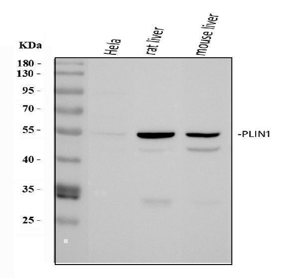
Western blot analysis of Perilipin A using anti-Perilipin A antibody (A03918-1).
Electrophoresis was performed on a 5-20% SDS-PAGE gel at 70V (Stacking gel) / 90V (Resolving gel) for 2-3 hours. The sample well of each lane was loaded with 30 ug of sample under reducing conditions.
Lane 1: human Hela whole cell lysates,
Lane 2: rat liver tissue lysates,
Lane 3: mouse liver tissue lysates.
After electrophoresis, proteins were transferred to a nitrocellulose membrane at 150 mA for 50-90 minutes. Blocked the membrane with 5% non-fat milk/TBS for 1.5 hour at RT. The membrane was incubated with rabbit anti-Perilipin A antigen affinity purified polyclonal antibody (Catalog # A03918-1) at 0.5 μg/mL overnight at 4°C, then washed with TBS-0.1%Tween 3 times with 5 minutes each and probed with a goat anti-rabbit IgG-HRP secondary antibody at a dilution of 1:5000 for 1.5 hour at RT. The signal is developed using an Enhanced Chemiluminescent detection (ECL) kit (Catalog # EK1002) with Tanon 5200 system. A specific band was detected for Perilipin A at approximately 56 kDa. The expected band size for Perilipin A is at 56 kDa.

Click image to see more details
Antioxidants preserve ADSC cell stemness and multidirectional differentiation potential during long-term in vitro expansion. After treatment with 10 μM GSH or melatonin, the ADSCs cultured for passage 3 (P3), passage 6 (P6), and passage 9 (P9) were used in the following analysis. a Osteogenesis differentiation of passaged ADSCs (Alizarin Red S staining; scale bar, 50 μm). b Adipogenesis differentiation of passaged ADSCs (Oil Red O staining; scale bar, 50 μm). c Western blot analysis for RUNX-2 in osteogenic cells. d Western blot analysis for perilipin A in adipogenic cells. e Chondrogenesis differentiation of passaged ADSCs (Alcian blue staining; scale bar, 50 μm). f Western blot analysis for SOX-9 in chondrogenic cells. g Western blot analysis for SOX-2, OCT-4, and β-actin in ADSCs. ADSCs, adipose tissue-derived stem cells; GSH, reduced glutathione
Index in PubMed under a CC BY license. PMID: 31623678
Specific Publications For Anti-Perilipin A/PLIN1 Antibody Picoband® (A03918-1)
Loading publications
Recommended Resources
Here are featured tools and databases that you might find useful.
- Boster's Pathways Library
- Protein Databases
- Bioscience Research Protocol Resources
- Data Processing & Analysis Software
- Photo Editing Software
- Scientific Literature Resources
- Research Paper Management Tools
- Molecular Biology Software
- Primer Design Tools
- Bioinformatics Tools
- Phylogenetic Tree Analysis
Customer Reviews
Have you used Anti-Perilipin A/PLIN1 Antibody Picoband®?
Share your experimental results or join a short interview to earn up to $1,000 in product credits or other rewards.
0 Reviews For Anti-Perilipin A/PLIN1 Antibody Picoband®
Customer Q&As
Have a question?
Find answers in Q&As, reviews.
Can't find your answer?
Submit your question
4 Customer Q&As for Anti-Perilipin A/PLIN1 Antibody Picoband®
Question
We purchased anti-Perilipin A/PLIN1 antibody for Flow Cytometry on liver a few years ago. I am using rat, and We are going to use the antibody for ICC next. We want examining liver as well as adipocyte in our next experiment. Do you have any suggestion on which antibody would work the best for ICC?
Verified Customer
Verified customer
Asked: 2019-10-30
Answer
I have checked the website and datasheets of our anti-Perilipin A/PLIN1 antibody and it seems that A03918-1 has been tested on rat in both Flow Cytometry and ICC. Thus A03918-1 should work for your application. Our Boster satisfaction guarantee will cover this product for ICC in rat even if the specific tissue type has not been validated. We do have a comprehensive range of products for ICC detection and you can check out our website bosterbio.com to find out more information about them.
Boster Scientific Support
Answered: 2019-10-30
Question
We are currently using anti-Perilipin A/PLIN1 antibody A03918-1 for human tissue, and we are happy with the Flow Cytometry results. The species of reactivity given in the datasheet says human, mouse, rat. Is it possible that the antibody can work on pig tissues as well?
Verified Customer
Verified customer
Asked: 2019-10-28
Answer
The anti-Perilipin A/PLIN1 antibody (A03918-1) has not been tested for cross reactivity specifically with pig tissues, but there is a good chance of cross reactivity. We have an innovator award program that if you test this antibody and show it works in pig you can get your next antibody for free. Please contact me if I can help you with anything.
Boster Scientific Support
Answered: 2019-10-28
Question
We were satisfied with the WB result of your anti-Perilipin A/PLIN1 antibody. However we have observed positive staining in adipose tissue endoplasmic reticulum using this antibody. Is that expected? Could you tell me where is PLIN1 supposed to be expressed?
Verified Customer
Verified customer
Asked: 2018-01-11
Answer
According to literature, adipose tissue does express PLIN1. Generally PLIN1 expresses in endoplasmic reticulum. Regarding which tissues have PLIN1 expression, here are a few articles citing expression in various tissues:
Adipocyte, Pubmed ID: 9521880
Brain, Pubmed ID: 15489334
Liver, Pubmed ID: 24275569
Boster Scientific Support
Answered: 2018-01-11
Question
We have observed staining in mouse liver. Are there any suggestions? Is anti-Perilipin A/PLIN1 antibody supposed to stain liver positively?
Verified Customer
Verified customer
Asked: 2017-11-21
Answer
From literature liver does express PLIN1. From Uniprot.org, PLIN1 is expressed in adipose tissue, adipocyte, brain, liver, among other tissues. Regarding which tissues have PLIN1 expression, here are a few articles citing expression in various tissues:
Adipocyte, Pubmed ID: 9521880
Brain, Pubmed ID: 15489334
Liver, Pubmed ID: 24275569
Boster Scientific Support
Answered: 2017-11-21
