Click image to see more details
Product Info Summary
| SKU: | PB9314 |
|---|---|
| Size: | 100μg/vial |
| Reactive Species: | Human |
| Host: | Rabbit |
| Application: | Flow Cytometry, WB |
Customers Who Bought This Also Bought

Product info
Product Name
Anti-PI 3 Kinase catalytic subunit alpha/PIK3CA Antibody Picoband®
SKU/Catalog Number
PB9314
Size
100μg/vial
Form
Lyophilized
Description
Boster Bio Anti-PI 3 Kinase catalytic subunit alpha/PIK3CA Antibody Picoband® catalog # PB9314. Tested in Flow Cytometry, WB applications. This antibody reacts with Human. The brand Picoband indicates this is a premium antibody that guarantees superior quality, high affinity, and strong signals with minimal background in Western blot applications. Only our best-performing antibodies are designated as Picoband, ensuring unmatched performance.
Storage & Handling
Store at -20˚C for one year from date of receipt. After reconstitution, at 4˚C for one month. It can also be aliquotted and stored frozen at -20˚C for six months. Avoid repeated freeze-thaw cycles.
Cite This Product
Anti-PI 3 Kinase catalytic subunit alpha/PIK3CA Antibody Picoband® (Boster Biological Technology, Pleasanton CA, USA, Catalog # PB9314)
Host
Rabbit
Contents
Each vial contains 4mg Trehalose, 0.9mg NaCl and 0.2mg Na2HPO4.
Clonality
Polyclonal
Clone Number
WH-3
Immunogen
E.coli-derived human PIK3CA recombinant protein (Position: H936-N1068). Human PIK3CA shares 98% amino acid (aa) sequence identity with mouse PIK3CA.
Cross-reactivity
No cross reactivity with other proteins
Reactive Species
PB9314 is reactive to PIK3CA in Human
Observed Molecular Weight
124 kDa
Calculated molecular weight
124.3 kDa
Background of PIK3CA
Phosphatidylinositol-4,5-bisphosphate 3-kinase, also called PIK3CA, is composed of an 85 kDa regulatory subunit and a 110 kDa catalytic subunit. PIK3CA gene is mapped to 3q26.32. The protein encoded by this gene represents the catalytic subunit, which uses ATP to phosphorylate phosphatidylinositols (PtdIns), PtdIns4P andPtdIns (4,5)P2. Recent evidence has shown that the PIK3CA gene is mutated in a range of human cancers. It has been found to be oncogenic and has been implicated in cervical cancers. PIK3CA mutations in breast cancer may be a predictive marker to guide the selection of patients who would benefit from mTOR inhibitor therapy. In addition to that, the presence of PIK3CA mutation may predict response to aspirin therapy for colorectal cancer, indicating power and promise of "Molecular Pathological Epidemiology (MPE)" approach as well as a complex interaction within the tumor microenvironment in this phenomenon.
Antibody Validation
Boster validates all antibodies on WB, IHC, ICC, Immunofluorescence, and ELISA with known positive control and negative samples to ensure specificity and high affinity, including thorough antibody incubations.
Application & Images
Applications
PB9314 is guaranteed for Flow Cytometry, WB Boster Guarantee
Recommend Dilution
| Application | Dilution | Species |
|---|---|---|
| Western blot | 0.1-0.5μg/ml | Human |
| Flow Cytometry (Fixed) | 1-3μg/1x106 cells | Human |
Tested application
Suggested blocking solution with 5% non-fat milk or BSA; (*)Recommended protein loading: 20-40 µg per lane
Validation Images & Assay Conditions
Click image to see more details
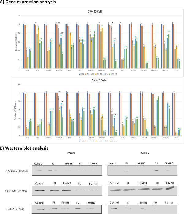
( A ) Gene expression analysis of substrates involved in the pathways related to the signaling downstream of insulin (INS) receptors by quantitative RT-PCR. Caco-2 and SW480 carcinoma cells were treated with 5-fluorouracil (FU) and irinotecan (IRI) in the concentration of 500 µM and IRI 50 µM, respectively for 48 h. The mRNA expression of INSR (insulin receptor), IRS 1 (insulin receptor substrate 1), PIK3CA (phosphatidylinositol 3-kinase catalytic subunit alpha), PIK3R1 (phosphatidylinositol 3-kinase regulatory subunit alpha), AKT1 and AKT2 (AKT Serine/Threonine Kinase 1 and 2), MAPK1 (Mitogen-Activated Protein Kinase 1), MAP2K2 (Mitogen-Activated Protein Kinase Kinase 2), GRB2 (Growth factor receptor-bound protein 2), glucose transporters (GLUT-1, GLUT-3, GLUT-4), SREBP-1c (sterol regulatory element-binding protein-1c), GSK3B (Glycogen synthase kinase 3 beta), antiapoptotic protein BCL-2, caspase 3 (CASP3) was determined by quantitative RT-PCR using gene-specific primers. Data are presented as mean ± SD. Experiments were run in triplicate and carried out once. *P < 0.05 compared with control group. ( B ) Western blotting analysis of expression of PIK3CA and GRB2 in Caco-2 and SW480 cancer cell lines.
Index in PubMed under a CC BY license. PMID: 31719636

Click image to see more details
Western blot analysis of PIK3CA using anti-PIK3CA antibody (PB9314).
Electrophoresis was performed on a 5-20% SDS-PAGE gel at 70V (Stacking gel) / 90V (Resolving gel) for 2-3 hours. The sample well of each lane was loaded with 30 ug of sample under reducing conditions.
Lane 1: human Jurkat whole cell lysates,
Lane 2: human MCF-7 whole cell lysates,
Lane 3: human K562 whole cell lysates,
Lane 4: human Raji whole cell lysates.
After electrophoresis, proteins were transferred to a nitrocellulose membrane at 150 mA for 50-90 minutes. Blocked the membrane with 5% non-fat milk/TBS for 1.5 hour at RT. The membrane was incubated with rabbit anti-PIK3CA antigen affinity purified polyclonal antibody (Catalog # PB9314) at 0.5 μg/mL overnight at 4°C, then washed with TBS-0.1%Tween 3 times with 5 minutes each and probed with a goat anti-rabbit IgG-HRP secondary antibody at a dilution of 1:5000 for 1.5 hour at RT. The signal is developed using an Enhanced Chemiluminescent detection (ECL) kit (Catalog # EK1002) with Tanon 5200 system. A specific band was detected for PIK3CA at approximately 124 kDa. The expected band size for PIK3CA is at 124 kDa.

Click image to see more details
Flow Cytometry analysis of Raji cells using anti-PIK3CA antibody (PB9314).
Overlay histogram showing Raji cells stained with PB9314 (Blue line). The cells were fixed with 4% paraformaldehyde and blocked with 10% normal goat serum. And then incubated with rabbit anti-PIK3CA Antibody (PB9314, 1 μg/1x106 cells) for 30 min at 20°C. DyLight®488 conjugated goat anti-rabbit IgG (BA1127, 5-10 μg/1x106 cells) was used as secondary antibody for 30 minutes at 20°C. Isotype control antibody (Green line) was rabbit IgG (1 μg/1x106) used under the same conditions. Unlabelled sample without incubation with primary antibody and secondary antibody (Red line) was used as a blank control.
Specific Publications For Anti-PI 3 Kinase catalytic subunit alpha/PIK3CA Antibody Picoband® (PB9314)
Loading publications
Recommended Resources
Here are featured tools and databases that you might find useful.
- Boster's Pathways Library
- Protein Databases
- Bioscience Research Protocol Resources
- Data Processing & Analysis Software
- Photo Editing Software
- Scientific Literature Resources
- Research Paper Management Tools
- Molecular Biology Software
- Primer Design Tools
- Bioinformatics Tools
- Phylogenetic Tree Analysis
Customer Reviews
Have you used Anti-PI 3 Kinase catalytic subunit alpha/PIK3CA Antibody Picoband®?
Share your experimental results or join a short interview to earn up to $1,000 in product credits or other rewards.
0 Reviews For Anti-PI 3 Kinase catalytic subunit alpha/PIK3CA Antibody Picoband®
Customer Q&As
Have a question?
Find answers in Q&As, reviews.
Can't find your answer?
Submit your question
